Retinal Vascular Density and Vessel Diameter in Sturge–Weber Syndrome Assessed by OCT-Angiography
Rosa Longo, Elena Gusson, Erika Lorenzetto, Luca Polinelli, Mariaelena Malvasi, Giacomo Panozzo, Giorgio Marchini

TL;DR
This study uses OCT-Angiography to find that retinal blood vessels in Sturge–Weber syndrome patients are denser and wider compared to healthy individuals.
Contribution
The study introduces OCTA as a novel tool to detect and monitor vascular changes specific to Sturge–Weber syndrome.
Findings
SWS patients showed significantly increased deep capillary plexus density compared to controls.
Vascular diameter was increased in multiple retinal plexuses in SWS patients.
OCTA can detect morphological vascular changes linked to disease progression.
Abstract
Background: Sturge–Weber syndrome (SWS) typically presents with a port-wine stain on the face, accompanied by leptomeningeal capillary malformations and ocular vascular anomalies. The aim of our study was to evaluate retinal vascular density and vessel diameter to better characterize the presence of vascular alterations. Methods: 15 patients diagnosed with Sturge–Weber syndrome and 15 healthy controls underwent comprehensive ophthalmologic evaluation, Optical Coherence Tomography (OCT) and Optical Coherence Tomography Angiography (OCTA), to evaluate the microvascular architecture of the retina and choroid. Results: Analysis of the processed images revealed a significant increase (p < 0.05 *) in the density of the deep capillary plexus in patients with SWS compared to healthy controls. Vascular diameter was found to be increased overall in several retinal vascular plexuses in SWS…
Genes, proteins, chemicals, diseases, species, mutations and cell lines named across the full text — each resolved to its canonical identifier and authoritative record.
Click any figure to enlarge with its caption.
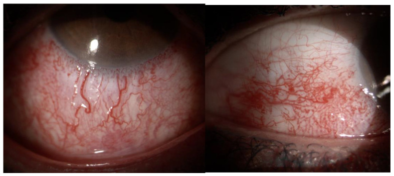 Figure 1
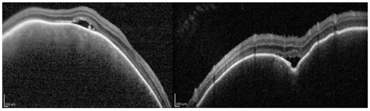 Figure 2
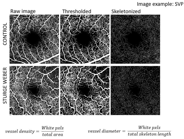 Figure 3
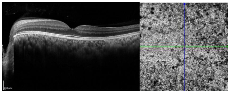 Figure 4
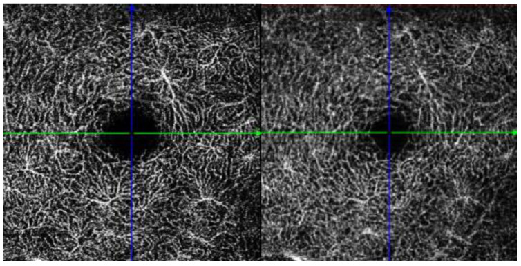 Figure 5
Peer Reviews
No public reviews on file for this paper yet. If you reviewed it on a platform where reviews are public (OpenReview, ICLR, NeurIPS, ICML), you can paste yours below so the community can read it here.
Videos
No videos yet. Explain this paper in a talk, walkthrough, or lecture? Add one.
Taxonomy
TopicsVascular Malformations and Hemangiomas · Vascular Malformations Diagnosis and Treatment · Histiocytic Disorders and Treatments