Prevalence, Diagnosis, and Treatment of Ovarian Cysts in Bitches and Queens: A Meta-Analysis
Kinga Domrazek, Katarzyna Kondratek, Filip Tobolewski, Piotr Jurka

TL;DR
This study finds that ovarian cysts are common in older female dogs and cats, but there's a lack of standard diagnostic methods and non-surgical treatments.
Contribution
The study provides a meta-analysis of ovarian cyst prevalence and treatment in bitches and queens, highlighting diagnostic and therapeutic gaps.
Findings
Ovarian cysts have a pooled prevalence of 41.7% in bitches and queens.
Follicular cysts are the most commonly reported subtype.
Dogs are more likely to develop cysts than cats, and older animals are at higher risk.
Abstract
Ovarian cysts—fluid-filled structures in the ovaries—are a common reproductive issue in female dogs and cats. These cysts can interfere with fertility and cause hormonal problems, yet little is known about how often they occur or how best to detect and treat them. In this study, we reviewed and analyzed data from 13 scientific articles, covering over 700 animals, to estimate how frequently these cysts occur and what diagnostic or treatment methods are being used. We found that ovarian cysts are quite common, especially in older animals, and are more frequently diagnosed in cats than in dogs. However, there is a lack of standard methods for diagnosing these cysts, and nearly all studies focused only on surgical removal of the ovaries. Non-surgical treatment options were rarely discussed, and important diagnostic tools such as hormone testing were underused. Our findings show a need for…
Genes, proteins, chemicals, diseases, species, mutations and cell lines named across the full text — each resolved to its canonical identifier and authoritative record.
Click any figure to enlarge with its caption.
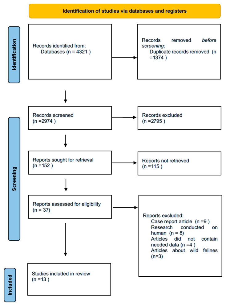 Figure 1
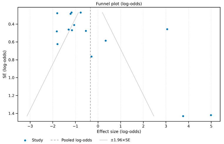 Figure 2
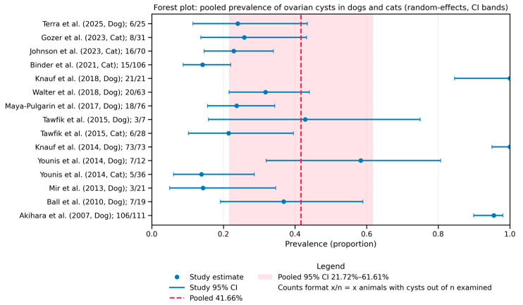 Figure 3
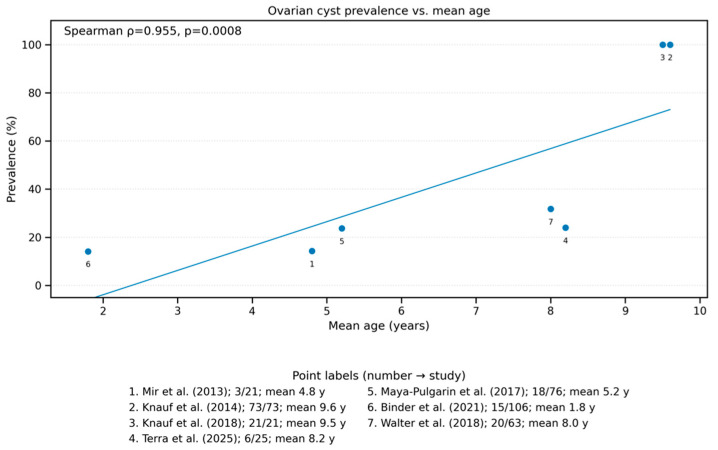 Figure 4
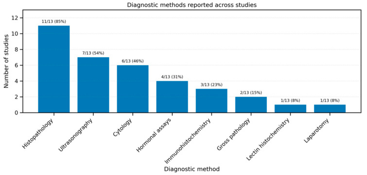 Figure 5
Peer Reviews
No public reviews on file for this paper yet. If you reviewed it on a platform where reviews are public (OpenReview, ICLR, NeurIPS, ICML), you can paste yours below so the community can read it here.
Videos
No videos yet. Explain this paper in a talk, walkthrough, or lecture? Add one.
Taxonomy
TopicsVeterinary Medicine and Surgery · Reproductive Physiology in Livestock · Veterinary Oncology Research
1. Introduction
Ovarian cysts are fluid-filled structures that develop within or on the surface of ovaries in female dogs (bitches) and cats (queens). They are distinguished into follicular cysts, lutein cysts, cysts of subsurface epithelial structure, cystic rete ovarii, and cystic corpus luteum [1,2,3]. Each type presents distinct histological characteristics and clinical implications. These cystic formations constitute a significant clinical entity in small animal reproduction, untreated often lead to infertility [1,2]. The prolonged presence of ovarian cysts that produce steroid hormones not only increases the risk of uterine disorders but also negatively impacts ovulation, resulting in reduced litter size. The most common uterine histopathologies observed were cystic endometrial hyperplasia (CEH), periglandular fibrosis, lymphoplasmacytic endometritis, and adenomyosis [2].
Ovarian cysts frequently disrupt normal reproductive physiology through hormonal imbalances [4,5]. Follicular cysts, the most clinically relevant type, produce sustained estradiol leading to persistent estrus, prolonged proestrus, and hyperestrogenism with associated complications including cystic endometrial hyperplasia-pyometra complex and bone marrow suppression [2,5]. Luteinized follicular cysts, luteal cysts and cystic corpus luteum, characterized by progesterone hypersecretion, induce mammary gland hyperplasia, and behavioral changes mimicking pregnancy, with associated risks of endometrial pathology and metabolic disturbances like insulin resistance [3,6,7].
Diagnostic challenges in ovarian cyst management arise from their often subclinical presentation and small size (most ≤0.5 cm) [4], often requiring both ultrasonography and hormonal assays for detection. Advanced diagnostic protocols may incorporate vaginal cytology additionally. Histopathological examination remains essential for definitive cyst classification and assessment of structural characteristics. It also enables assessment of endocrine activity through specialized staining techniques and immunohistochemical markers [1,8].
Therapeutic management presents clinical dilemmas between conservative and surgical intervention. While hormonal therapy using gonadotropin-releasing hormone analogs (GnRH) or human chorionic gonadotropin (hCG) has demonstrated success rates of approximately 63% in some studies [2,6], the potential for recurrence and the frequent concurrent presence of uterine pathology often necessitates surgical intervention. Ovariohysterectomy remains to be the most common used treatment [1]. Hormonal therapy challenges are further complicated by the need to exclude pre-existing uteropathies and estrogen-induced hematological changes before attempting conservative management, making comprehensive pre-treatment evaluation essential for optimal patient outcomes [4,7].
Although individual studies have addressed the presence of ovarian cysts in female dogs and cats, the findings remain fragmented and inconsistent. A systematic review and meta-analysis are needed to consolidate current knowledge, quantify the prevalence of ovarian cysts, and identify potential risk factors such as species, breed, and age. Such an analysis may also highlight diagnostic and therapeutic gaps and inform future clinical and research priorities.
The aim of this study was to estimate the prevalence of ovarian cysts in bitches and queens through a systematic review and meta-analysis. Additionally, we aimed to categorize the types of cysts identified, evaluate diagnostic methods and treatment approaches used in the literature, and assess the quality of available evidence.
Therefore, this systematic review and meta-analysis aimed to address the following research question: What is the prevalence of ovarian cysts in bitches and queens, and which diagnostic and therapeutic strategies have been most commonly reported? To answer this question, we applied the PICO framework: the population consisted of female dogs and cats described in clinical and pathological studies; the interventions included diagnostic methods such as ultrasonography, histopathology, and hormonal assays, as well as surgical and medical treatments; the comparisons focused on species differences (dogs versus cats) and diagnostic modalities; and the outcomes were the prevalence and types of ovarian cysts, the accuracy of diagnostic tools, and the reported treatment outcomes.
2. Materials and Methods
We followed PRISMA guidelines to perform a systematic review and meta-analysis, focusing on the prevalence, diagnostic approaches, and treatment strategies for ovarian cysts in bitches and queens. The systematic review and meta-analysis were registered in the PROSPERO database under the title: Prevalence, diagnosis, and treatment of ovarian cysts in bitches and queens: a systematic review and meta-analysis.
2.1. Study Selection
In the first stage, we conducted a systematic review of ovarian cysts in dogs and cats. After reviewing a large number of publications, we decided to perform a meta-analysis on the prevalence of various types of ovarian cysts in bitches and queens. We followed the PRISMA guidelines as a checklist while preparing the manuscript. The databases used included PubMed, Scopus, and Google Scholar. A comprehensive literature search was conducted in June and July 2025. The keywords used are presented in Table 1.
2.2. Inclusion and Exclusion Criteria
Eligible studies were included based on the following inclusion criteria: (a) original research articles reporting data on ovarian cysts in female dogs (bitches) and/or cats (queens); (b) studies published between January 2000 and June 2025; (c) articles written in English; (d) studies providing extractable numerical data (quantitative information allowing calculation of prevalence or proportions) on at least one of the following: prevalence, diagnostic method, treatment, treatment outcomes. The exclusion criteria were as follows: (a) duplicate publications or low-quality studies (e.g., missing raw data, inappropriate methodology); (b) conference abstracts, reviews, commentaries, editorials, or case reports; (c) studies involving other species or mixed-gender groups where data could not be isolated for female dogs or cats; (d) in vitro or experimental laboratory studies without in vivo animal outcomes.
2.3. Data Extraction
Screening and data extraction were carried out by one researcher who first conducted literature and data screening according to the criteria developed. Another researcher ensured that the collection of the material was consistent. The established literature screening criteria were as follows: initial screening to exclude articles that clearly did not meet the criteria on the basis of the title and the abstract, followed by a detailed reading of the literature to select the final articles for inclusion in this study on the basis of the inclusion and exclusion criteria. The following data were collected from the included articles: first author, country of study, year of publication, type of sample, detection methods, and results.
2.4. Assessment of Study Quality and Risk of Bias
The methodological quality of the included studies was assessed independently by two reviewers using appropriate tools based on study design. For observational studies, the Newcastle–Ottawa Scale (NOS) was applied, evaluating three domains: selection of participants, comparability of study groups, and outcome assessment. Each study could receive a maximum of 9 points, with ≥7 points considered high quality.
In the case of randomized controlled trials (RCTs) (if included), the Cochrane Risk of Bias 2 (RoB-2) tool was used, covering domains such as randomization process, deviations from intended interventions, missing outcome data, outcome measurement, and selection of reported results. Studies were categorized as low, moderate, or high risk of bias.
2.5. Statistical Analysis
From each eligible study we extracted the number of animals examined and the number diagnosed with ovarian (or para-ovarian) cysts. Study-specific prevalences were expressed as proportions with 95% Wilson score confidence intervals; a 0.5-animal continuity correction was applied to studies reporting 0% or 100%. Pooled prevalence and its 95% CI were calculated with a DerSimonian–Laird random-effects model; between-study heterogeneity was quantified with τ^2^ and I^2^ statistics. Publication-bias/small-study effects were explored visually with funnel plots (raw proportion and log-odds scales) and formally with Egger’s weighted regression. Species differences (dog vs. cat) were evaluated with Fisher’s exact test, Yates-corrected χ^2^, and by reporting risk and odds ratios. To assess age, we converted reported mean (or mid-range) cohort ages to years and tested the rank correlation between age and prevalence (Spearman ρ) plus a Mann–Whitney U comparison of cohorts ≤ 5 years versus >5 years. Random-effects sub-group meta-analyses were run for each diagnostic modality represented by at least two studies. All computations were performed in Python 3.11.9 (pandas 2.3.1, SciPy 1.16.0, statsmodels 0.14.5) visualizations were performed in matplotlib 3.8.4 and seaborn 0.13.2.
3. Results
3.1. Characteristics of Included Studies
Our systematic search across four databases (PubMed, Scopus, and Google Scholar) yielded a total of 4321 articles. After removing 1374 duplicate records retrieved from multiple sources, 2828 articles were excluded based on predefined inclusion and exclusion criteria after screening titles and abstracts. Following full-text assessment, an additional 92 studies were excluded due to insufficient primary data, lack of relevant outcomes, or inappropriate study design, resulting in a final inclusion of 13 articles for quantitative synthesis (Figure 1) [8]. These studies included data on female dogs and cats from various countries and covered different diagnostic approaches (e.g., ultrasound, histopathology). Data extracted from each eligible study were synthesized qualitatively and are summarized in Table 2. No studies addressing clinical aspects of ovarian cysts, such as treatment options or fertility implications, were identified. However, some relevant case reports are available and have been cited when they provide additional insights.
3.2. Characteristics of the Study Population
The analyzed studies included a total of 428 bitches and 273 queens. The age of the animals varied across studies. In dogs, the reported mean age ranged from 1.8 to 9.6 years, with individual age ranges extending from 0.3 to 16 years. For cats, age data were less frequently reported, but when available, it ranged from 0.6 to over 13 years. Regarding breed distribution, a wide variety of dog breeds were represented, with the most frequent being German Shepherds (n = 53), mixed-breed dogs (n = 25), and Labrador Retrievers (n = 14). Among cats, the majority were European Shorthairs (n = 105), mixed-breed (n = 9), and Persian cats (n = 20). In several studies, breed data were not specified (dogs: n = 194; cats: n = 129). The exact number of individuals for each breed is presented in Supplementary Table S1.
3.3. Risk of Bias and GRADE Evaluation
Assessment of publication bias using funnel plots (both log-odds and raw proportion scales) suggests some asymmetry (Figure 2), and Egger’s regression confirmed statistically significant small-study effects (intercept = 3.69, p = 0.008), indicating possible publication bias.
3.4. Ovarian Cysts
3.4.1. Prevalence of Ovarian Cysts
The pooled prevalence of ovarian cysts across the 13 included studies was 41.66% (95% CI: 21.72–61.61%), as shown in the forest plot (Figure 3). Heterogeneity between studies was high (I^2^ = 99.1%, τ^2^ = 0.149), indicating substantial variation across the data sources.
Each horizontal line represents a study with corresponding 95% confidence interval. The diamond at the bottom illustrates the overall pooled estimate using a random-effects model (DerSimonian–Laird).
3.4.2. Species Comparison: Dog vs. Cat
The analysis showed a statistically significant difference in the prevalence of ovarian cysts between dogs and cats. Dogs were more likely to develop ovarian cysts compared to cats. The risk ratio (RR) for dogs versus cats was 3.34, and the odds ratio (OR) was 7.12, both with p < 0.001 (Fisher exact test and Yates-corrected chi-square test). This suggests that the likelihood of ovarian cysts is significantly higher in dogs than in cats.
3.4.3. Association with Age
There was a strong positive correlation between the mean age of animals and cyst prevalence across studies (Spearman’s ρ = 0.955), indicating that older individuals were more likely to be diagnosed with ovarian cysts. Additionally, linear trend was observed between mean age and prevalence (Figure 4).
3.4.4. Most Common Cyst Types
For the analysis of ovarian cyst types, three studies were excluded—Knauf et al. [4], Gozer et al. [10], and Johnson et al. [11]—due to insufficient information on cyst classification. Based on the remaining studies, the most frequently reported ovarian cysts were follicular cysts, which accounted for 42.1% of all identified cysts (95% CI: 36.4–48.0%, 114/272). Subsurface epithelial cysts were the second most common, representing 37.9% (95% CI: 32.4–43.8%, 103/272), followed by rete ovarii cysts (13.1%, 95% CI: 9.6–17.6%, 35/272) and corpus luteum cysts (6.9%, 95% CI: 4.4–10.5%, 18/272).
3.5. Diagnosis
A variety of diagnostic methods were employed across the included studies to detect ovarian cysts (Figure 5). Histopathology was the most commonly used technique, applied in 11 out of 13 studies, followed by ultrasonography (8 studies) and cytology (7 studies). Immunohistochemistry and hormonal assays (such as AMH, estradiol, or progesterone) were used in 4 studies each, while gross pathology appeared in 2 studies. Laparotomy and lectin histochemistry were reported in individual studies only. These findings highlight the predominance of histopathological and imaging methods in confirming the presence and type of ovarian cysts.
Of the 13 articles, 12 did not describe the specificity or specificity of the diagnostic methods. Nor was there data in the cited literature from which these parameters could be calculated. Only Ball et al. reported that the sensitivity for ultrasound in the diagnosis of cysts was 75% [18].
3.6. Treatment of Ovarian Cysts
Systematic research on treatment options, their success rates, and associated side effects for various types of ovarian cysts remains limited. Ovariohysterectomy is generally considered a curative approach in most cases. Moreover, this procedure eliminates the risk of recurrence, particularly in non-breeding females or those affected by severe concurrent uterine pathologies.
Of the literature collected, in all the articles the treatment method was removal of the ovaries, ovarian remnants or the uterus and ovaries. None of the articles reviewed in the meta-analysis undertook drug treatment. Moreover, none of the articles reviewed had data on complications following the procedures.
4. Discussion
To the best of our knowledge, this is first meta-analysis performed on such large scale of data. Previously only one meta-analysis about canine cysts has been performed, but only on two studies [20]. This is probably due to the fact that there are few cross-sectional studies published in this area, and a lot of descriptions of individual clinical cases [21,22]. This situation indicates the need for more research on ovarian cysts in bitches and cats. Given the often incidental discovery of cysts during elective castration or necropsy, implementing routine screening protocols, especially in older or breeding animals, could improve early detection and provide more reliable prevalence estimates. In addition, the predominance of retrospective and cross-sectional study designs limits the ability to infer causality. Future prospective cohort studies would allow for a better understanding of the natural history and progression of ovarian cysts in dogs and cats. In clinical practice, ovaries are often not analyzed in detail during OVH. The introduction of systematic macroscopic and histopathological examination of removed ovaries may increase recognition and generate new data in this field.
Another aspect is that more studies on ovarian cysts are conducted on dogs than on cats. This is probably due to the peculiarities of the female cat’s estrous cycle, which influences the faster decision to castrate the female [23]. This situation may make it extremely difficult to collect a large group of mature cats for testing. The ovarian cysts occurs more frequently in older animals than in young ones [2]. In our meta-analysis, while age was significantly associated with higher prevalence of cysts, other potential risk factors such as breed, reproductive status, hormonal treatments, or environmental conditions were not systematically explored across studies. It is important to note that only 7 studies contained data where age-based analysis could be computed, therefore the computed correlation would be subject to change if more data on the subject was available (Figure 4). These areas represent critical gaps that warrant further investigation especially in bitches and cats intended for breeding, cysts can lead to infertility, which is why early diagnosis is particularly important for breeding line planning [1,2].
A notable limitation of the available literature is the lack of data on the sensitivity and specificity of diagnostic methods used to identify ovarian cysts. Although a range of diagnostic techniques were used, including ultrasonography, histopathology, cytology, and hormonal assays, there was no consistent protocol across studies. This heterogeneity highlights the urgent need for standardized diagnostic criteria and protocols for identifying ovarian cysts in companion animals as it takes place in human medicine [24]. There is a lack of clearly defined steps to follow in cases of suspected cysts—it is advisable to develop clinical diagnostic and therapeutic algorithms. In 13 out of 14 studies included in this review, these diagnostic performance parameters were either not reported or not calculable from the available data. Only one study (Ball et al.) provided information on sensitivity, reporting a value of 75% for ultrasonography [18]. This significant gap in the literature highlights the urgent need for well-designed studies evaluating and validating diagnostic tools for ovarian cysts in small animals. An overlooked aspect in many studies on ovarian cysts is the lack of hormonal testing of peripheral blood and cyst fluid. This could be of great value. In animals that are not scheduled for immediate castration, periodic hormone testing (estradiol, progesterone) could be a tool for early detection of hormonally active cysts and further decision-making regarding treatment.
In addition, there is a lack of current research on non-surgical treatment methods probably due to fact that the pathophysiology of ovarian cysts remains unclear. This is particularly important because the problem affects breeding animals, in which surgery to remove the ovaries is associated with loss of fertility and thus the possibility of further reproduction [2].
The overall methodological quality of the included studies was moderate, with the majority rated as having a moderate risk of bias. Only one study met criteria for a low risk of bias, while two were considered at high risk. These findings indicate that caution is needed when interpreting the results of this meta-analysis, as methodological limitations may affect the reliability of reported outcomes.
Future research should focus on prospective, standardized studies assessing both diagnostic accuracy and treatment efficacy. Comparative studies of diagnostic modalities, particularly ultrasonography versus histopathology, could provide valuable insights for clinical decision-making.
5. Conclusions
These findings highlight the urgent need for prospective, standardized research focusing on diagnostic accuracy, non-surgical treatment options, and the long-term clinical implications of ovarian cysts in small animals. The most important seems to be that the lack of knowledge of physiopathology as a major limitation for effective medical treatments. Future studies should also consider stratifying data by breed, reproductive status, and hormonal exposure to better understand risk factors and inform evidence-based clinical decisions. This meta-analysis confirms that ovarian cysts are a relatively common finding in bitches and queens, particularly in older animals, but more research in this field is needed.
The reference list from the paper itself. Each links out to its DOI / PubMed record.
- 1Knauf Y. Köhler K. Knauf S. Wehrend A. Histological Classification of Canine Ovarian Cyst Types with Reference to Medical History J. Vet. Sci.20181972510.4142/jvs.2018.19.6.72530304884 PMC 6265583 · doi ↗ · pubmed ↗
- 2Sasidharan J.K. Patra M.K. Singh L.K. Saxena A.C. De U.K. Singh V. Mathesh K. Kumar H. Krishnaswamy N. Ovarian Cysts in the Bitch: An Update Top. Companion Anim. Med.20214310051110.1016/j.tcam.2021.10051133434678 · doi ↗ · pubmed ↗
- 3Johnston S.D. Root Kustritz M.V. Olson P.S. Canine and Feline Theriogenology Saunders Philadelphia, PA, USA 20010721656072
- 4Knauf Y. Bostedt H. Failing K. Knauf S. Wehrend A. Gross Pathology and Endocrinology of Ovarian Cysts in Bitches Reprod. Domest. Anim.20144946310.1111/rda.1231124698026 PMC 4235420 · doi ↗ · pubmed ↗
- 5Meftakh I.A. Rybin E.V. Proshkin V.M. Current Aspects of the Problem of Ovarian Cysts in Bitches of Reproductive Age Leg. Regul. Vet. Med.20243565910.52419/issn 2782-6252.2024.3.56 · doi ↗
- 6Knauf Y. Failing K. Knauf S. Wehrend A. Treatment of Bitches with Ovarian Cysts Using Human Chorionic Gonadotropin-Releasing Hormone Analogon Tierärztliche Praxis. Ausg. K Kleintiere/Heimtiere 2013419310023608964 · pubmed ↗
- 7Pires M.A. Payan-Carreira R. Ovarian Cysts in Dogs’ Practice Advances in Medicine and Biology. Chapter 4 Berhardt L.V. Nova Science Publishers New York, NY, USA 2016 Volume 949781634841856
- 8Page M.J. Mc Kenzie J.E. Bossuyt P.M. Boutron I. Hoffmann T.C. Mulrow C.D. Shamseer L. Tetzlaff J.M. Akl E.A. Brennan S.E. The PRISMA 2020 Statement: An Updated Guideline for Reporting Systematic Reviews BMJ 2021372 n 7110.1136/bmj.n 7133782057 PMC 8005924 · doi ↗ · pubmed ↗