Investigation of the Prevalence and Characteristics of the Retromolar Canal Using Cone-Beam Computed Tomography in a Turkish Sample
Fatoş Can, Fahrettin Kalabalık, Emre Aytuğar

TL;DR
This study used 3D imaging to find that a canal behind the molars is common in a Turkish population and may be important for dental procedures.
Contribution
The study provides new prevalence data and anatomical insights on retromolar canals in a Turkish sample using CBCT.
Findings
57% of subjects had retromolar canals, with 32.4% being bilateral.
Type A1 was the most common canal type, with differences observed between sexes.
CBCT is effective for identifying and analyzing retromolar structures.
Abstract
Background: The aim of this study is to investigate the prevalence of the retromolar canal (RMC) and retromolar foramen (RMF) using cone-beam computed tomography (CBCT), and to evaluate the course and anatomical structure of the RMC. Methods: The study group consisted of CBCT images of 1008 subjects (541 females and 467 males). The prevalence and types of the RMC, as well as the frequency of the RMF, were analyzed according to age and sex. A significance level of 0.05 was accepted for all statistical analyses. Results: According to the findings, 575 (57.0%) RMCs and 298 (29.5%) RMFs were identified in 1008 subjects. Bilateral RMCs were observed in 327 subjects (32.4%), while unilateral RMCs were present in 248 subjects (24.6%). When 2016 retromolar regions were examined, a total of 902 RMCs and 400 RMFs were identified. No statistically significant difference was observed between the…
Genes, proteins, chemicals, diseases, species, mutations and cell lines named across the full text — each resolved to its canonical identifier and authoritative record.
Click any figure to enlarge with its caption.
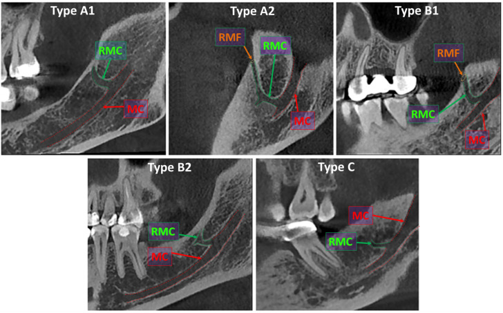 Figure 1
Peer Reviews
No public reviews on file for this paper yet. If you reviewed it on a platform where reviews are public (OpenReview, ICLR, NeurIPS, ICML), you can paste yours below so the community can read it here.
Videos
No videos yet. Explain this paper in a talk, walkthrough, or lecture? Add one.
Taxonomy
TopicsDental Radiography and Imaging · Oral and Maxillofacial Pathology · Endodontics and Root Canal Treatments