Proximal Pulmonary Fat Embolism on Non-Contrast Chest CT
Romain L’Huillier, Alexandra Braillon

TL;DR
A case report shows how a non-contrast chest CT scan helped detect a fat embolism causing cardiac arrest after hip surgery.
Contribution
Highlights the diagnostic benefit of non-contrast chest CT in identifying fat embolisms post-orthopedic surgery.
Findings
Proximal pulmonary fat embolism was detected using non-contrast chest CT.
Non-contrast CT aids in identifying fatty thrombi, improving diagnostic accuracy.
The case emphasizes the importance of non-contrast imaging in postoperative differential diagnosis.
Abstract
We report in this clinical case a proximal pulmonary fat embolism detected on unenhanced chest computed tomography (CT) responsible for a recovered cardiac arrest during a left total hip arthroplasty for a femoral neck fracture. This observation underscores the diagnostic value of integrating a non-contrast phase in chest CT in the postoperative context of orthopedic surgery, as it ensures accurate identification of the fatty nature of pulmonary arterial thrombi and thereby contributes to improved diagnostic accuracy and differential diagnosis.
Genes, proteins, chemicals, diseases, species, mutations and cell lines named across the full text — each resolved to its canonical identifier and authoritative record.
Click any figure to enlarge with its caption.
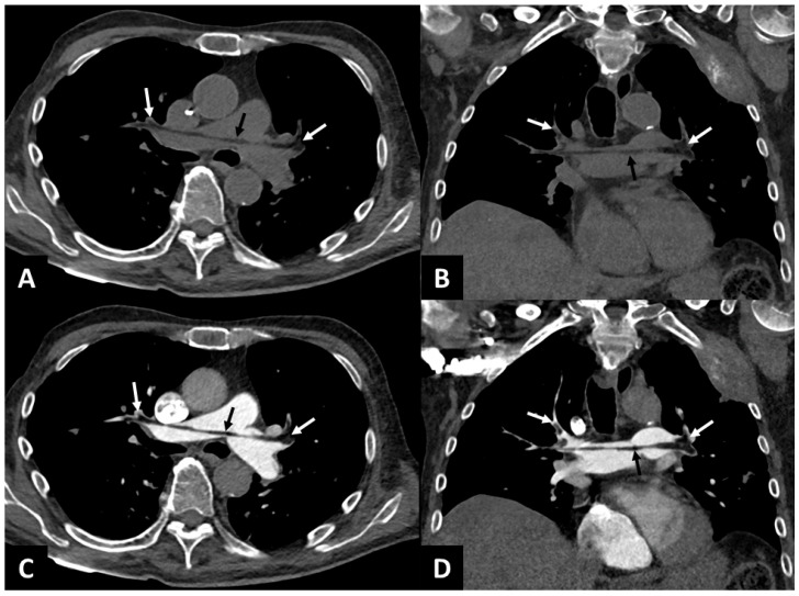 Figure 1
Peer Reviews
No public reviews on file for this paper yet. If you reviewed it on a platform where reviews are public (OpenReview, ICLR, NeurIPS, ICML), you can paste yours below so the community can read it here.
Videos
No videos yet. Explain this paper in a talk, walkthrough, or lecture? Add one.
Taxonomy
TopicsVenous Thromboembolism Diagnosis and Management · Hip and Femur Fractures · Orthopedic Infections and Treatments