Quantitative Evaluation of Gastrorenal Shunt Morphology Using Three-Dimensional Computed Tomography Portography: Comparison With Intraoperative Venography
Yoshimi Fujii, Masato Tanikake, Yurie Nishimura, Kazuma Yasui

TL;DR
This study compares 3DCT portography with intraoperative venography for evaluating gastrorenal shunt anatomy, finding that 3DCT is reliable for preprocedural planning in gastric variceal treatment.
Contribution
The study provides quantitative validation of 3DCT portography as a reliable alternative to intraoperative venography for preprocedural planning in BRTO.
Findings
3DCT portography showed high agreement with venography for vessel diameters and stenosis rates.
The vertebral level of stenosis was more caudal on 3DCT portography compared to venography.
Key anatomical features like angulation and confluence with the left adrenal vein were consistently identified by 3DCT.
Abstract
Background: Gastric varices are a serious complication of portal hypertension and may cause life-threatening bleeding when ruptured. Balloon-occluded retrograde transvenous obliteration (BRTO) is a standard endovascular treatment performed via the gastrorenal shunt (GRS). The present study examined concordance between three-dimensional computed tomography (3DCT) portography and intraoperative venography at the GRS outflow and evaluated the utility of 3DCT portography for preprocedural planning. Methods: Nineteen patients who underwent BRTO between 2017 and 2024 were retrospectively analyzed. Preoperative 3DCT portography and intraoperative venography were used to assess the following parameters: the diameter of the stenosis, the diameter of the common trunk, the stenosis rate, the distance from the left renal vein to the stenosis, and the vertebral level of the stenosis. Paired…
Genes, proteins, chemicals, diseases, species, mutations and cell lines named across the full text — each resolved to its canonical identifier and authoritative record.
Click any figure to enlarge with its caption.
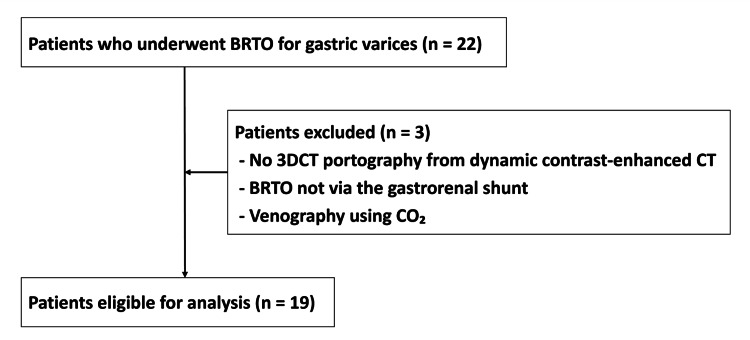 Figure 1
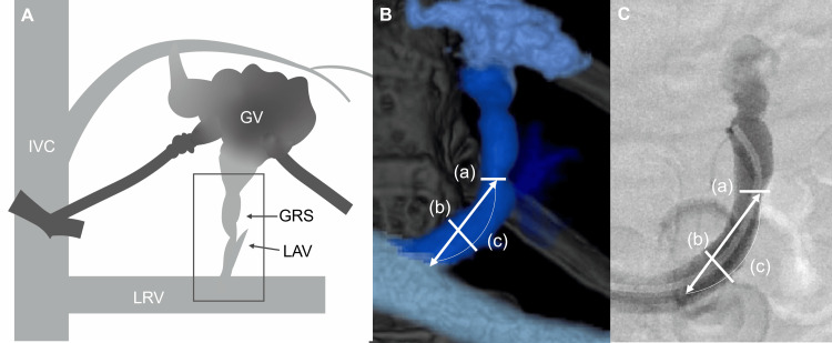 Figure 2
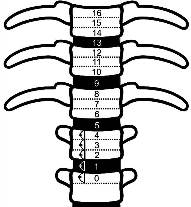 Figure 3
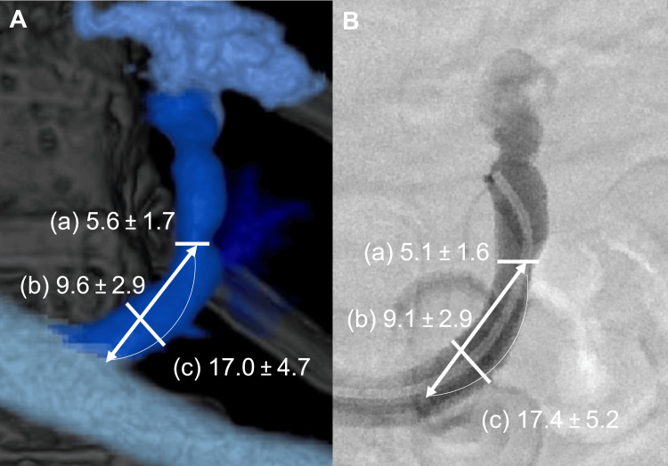 Figure 4
 Figure 5
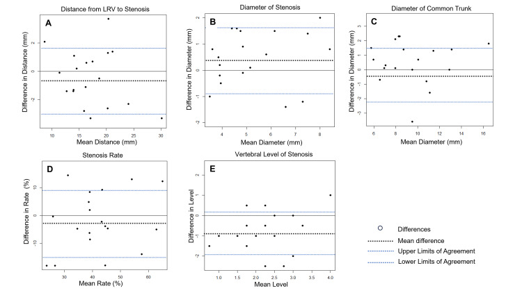 Figure 6
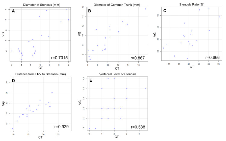 Figure 7
 Figure 8
Peer Reviews
No public reviews on file for this paper yet. If you reviewed it on a platform where reviews are public (OpenReview, ICLR, NeurIPS, ICML), you can paste yours below so the community can read it here.
Videos
No videos yet. Explain this paper in a talk, walkthrough, or lecture? Add one.
Taxonomy
TopicsLiver Disease and Transplantation · Organ Transplantation Techniques and Outcomes · Renal and Vascular Pathologies