A deep learning-based framework for standardized analysis of trabecular bone compartments from micro-CT imaging data in the mouse tibia
Amine Lagzouli, Lucinda Evans, Mark Hopkinson, Aikta Sharma, Natalia M. Castoldi, Davide Fontanarosa, Maria Antico, David M. L. Cooper, Alice Othmani, Vittorio Sansalone, Phil Salmon, Andrew A. Pitsillides, Peter Pivonka

TL;DR
This paper introduces a deep learning framework for standardized analysis of mouse tibia trabecular bone compartments using micro-CT scans, improving reproducibility in skeletal research.
Contribution
A novel deep learning-based framework for automated and standardized analysis of mouse tibia trabecular bone compartments using micro-CT data.
Findings
The classification model achieved high F1-scores (0.96-0.99) across four trabecular compartments.
The framework demonstrated strong generalizability on an external dataset with consistent performance.
Automated segmentation and analysis of trabecular compartments enable reliable comparisons across experimental groups.
Abstract
Understanding bone remodeling and disease progression is crucial in preclinical skeletal research, particularly for assessing pharmacological and mechanical interventions in the long bones of murine models. High-resolution micro-computed tomography (micro-CT) imaging enables detailed trabecular bone analysis; however, inconsistent and non-standardized definitions of the volumes of interest (VOIs) across the different trabecular compartments compromise reproducibility and may lead to misleading statistical interpretations. In this study, we introduce a deep learning framework for automated trabecular bone analysis from micro-CT scans (5 µm voxel size) of the epiphyseal-metaphyseal region in the mouse tibia. The epiphyseal-metaphyseal region is classified into four anatomical compartments, epiphyseal bone, growth plate, primary spongiosa, and secondary spongiosa, using a 2D slice-wise…
Genes, proteins, chemicals, diseases, species, mutations and cell lines named across the full text — each resolved to its canonical identifier and authoritative record.
Click any figure to enlarge with its caption.
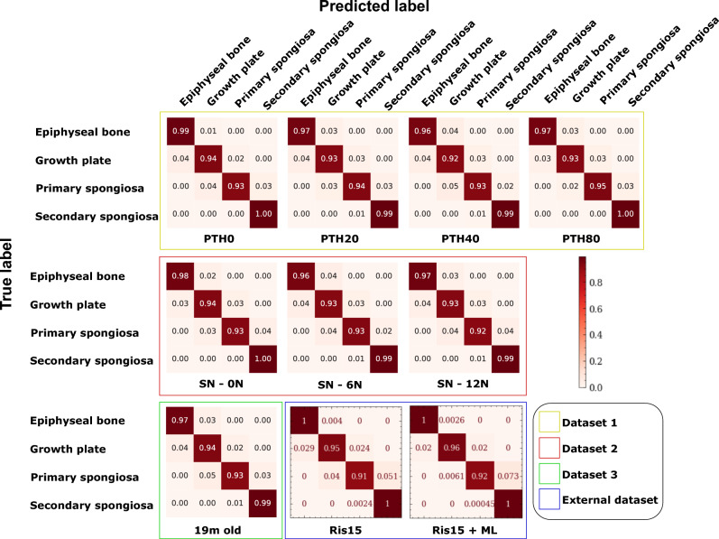 Figure 1
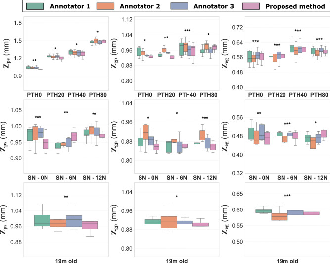 Figure 2
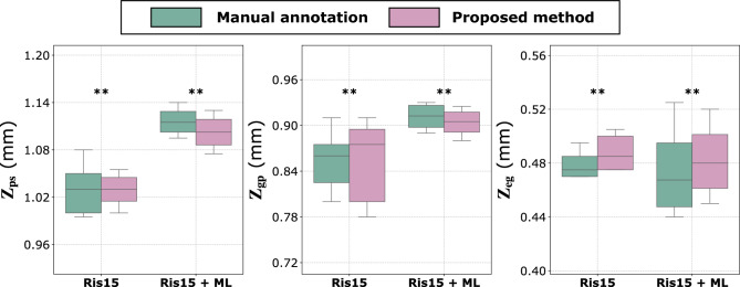 Figure 3
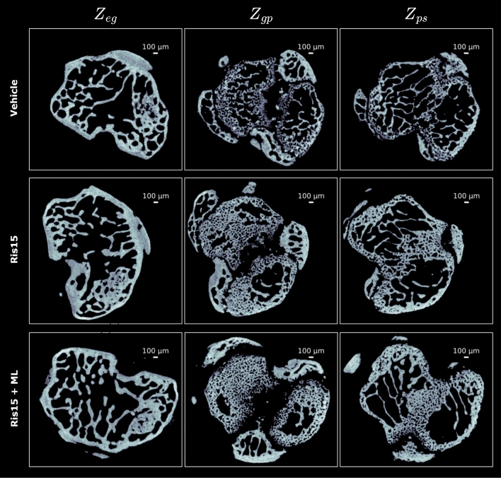 Figure 4
 Figure 5
 Figure 6
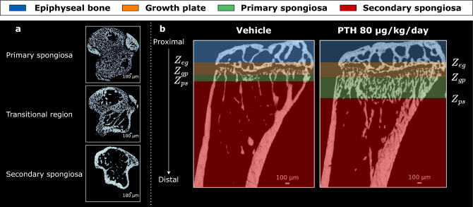 Figure 7
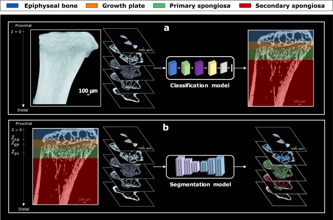 Figure 8
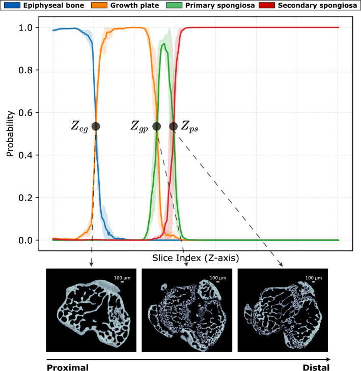 Figure 9
Peer Reviews
No public reviews on file for this paper yet. If you reviewed it on a platform where reviews are public (OpenReview, ICLR, NeurIPS, ICML), you can paste yours below so the community can read it here.
Videos
No videos yet. Explain this paper in a talk, walkthrough, or lecture? Add one.
Taxonomy
TopicsBone health and osteoporosis research · Radiomics and Machine Learning in Medical Imaging · Bone and Joint Diseases