Artificial Intelligence-Enhanced Cone-Beam Computed Tomography for the Diagnostic Evaluation of Chronic Rhinosinusitis: A Systematic Review
Muhammad H Chaudhary, Nada Dahan

TL;DR
This review assesses how artificial intelligence improves cone-beam CT for diagnosing chronic rhinosinusitis, finding it accurate but needing more validation.
Contribution
The study systematically evaluates AI-enhanced CBCT for CRS diagnosis, highlighting its potential and current limitations.
Findings
AI-enhanced CBCT shows high sensitivity and specificity for maxillary sinusitis and mucosal thickening detection.
Current AI methods for CRS have moderate methodological quality and limited external validation.
Applications include anatomical variant detection and classification, but evidence certainty varies by outcome.
Abstract
Chronic rhinosinusitis (CRS) is one of the most common chronic inflammatory conditions globally, affecting a significant proportion of adults. It is associated with reduced quality of life, recurrent healthcare utilization, and substantial economic burden. Imaging plays a pivotal role in its diagnosis and management. Cone-beam computed tomography (CBCT) has emerged as a valuable imaging modality that provides high-resolution, three-dimensional reconstructions with lower radiation exposure compared with conventional CT. However, CBCT interpretation requires expert knowledge, remains time-intensive, and is subject to inter-observer variability. Artificial intelligence (AI), particularly deep learning, has recently shown promise in enhancing diagnostic precision, standardizing interpretation, and improving efficiency. This systematic review evaluates the diagnostic performance,…
Genes, proteins, chemicals, diseases, species, mutations and cell lines named across the full text — each resolved to its canonical identifier and authoritative record.
Click any figure to enlarge with its caption.
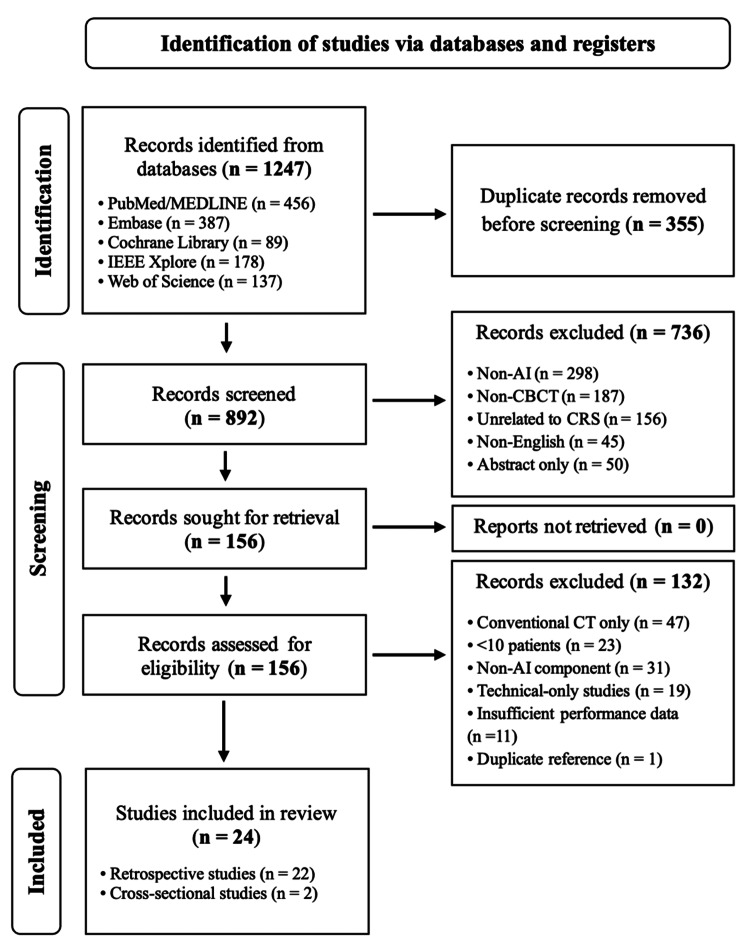 Figure 1
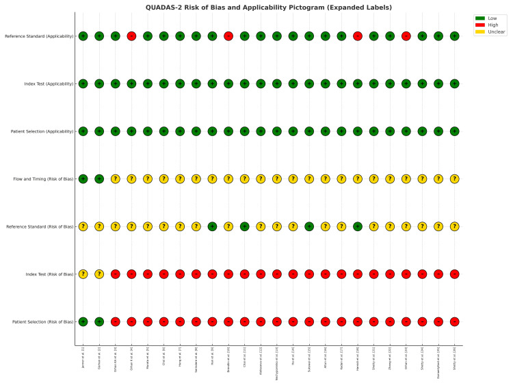 Figure 2
| Study | Year | Country | Design | Sample Size | AI Architecture | Primary Application | Key Findings |
| Janner et al. [ | 2020 | Switzerland | Cross-sectional | 156 | - | Sinus assessment comparison | ENT specialists are more accurate than dentists in sinus evaluation on CBCT; referral recommended for complex cases. |
| Gürhan et al. [ | 2020 | Turkey | Cross-sectional | 387 | - | Mucosal thickening assessment | Mucosal thickening associated with periapical lesions; CBCT useful for dental-sinus relationships. |
| Orhan et al. [ | 2020 | Turkey | Retrospective | 678 | AI algorithm | Periapical pathosis detection | AI achieved high accuracy in detecting periapical pathosis on CBCT; supports diagnostic use. |
| Orhan et al. [ | 2021 | Turkey | Retrospective | 534 | AI model | Third molar impaction detection | AI reliably detected impacted third molars; useful for clinical screening and planning. |
| Murata et al. [ | 2019 | Japan | Retrospective | 201 | CNN | Maxillary sinusitis on panoramic radiography | CNN showed good diagnostic performance for sinusitis; it outperformed conventional interpretation. |
| Choi et al. [ | 2022 | South Korea | Retrospective | 567 | Deep learning | Maxillary sinus segmentation | Deep learning enabled fully automatic and accurate segmentation of the maxillary sinus on CBCT. |
| Hung et al. [ | 2022 | Hong Kong | Retrospective | 678 | 3D CNN | Mucosal segmentation | 3D CNN successfully segmented mucosal changes; strong diagnostic potential. |
| Serindere et al. [ | 2022 | Turkey | Retrospective | 400 | CNN | Maxillary sinusitis detection | CNN effectively detected sinusitis on radiographs and CBCT; comparable to expert review. |
| Han et al. [ | 2022 | South Korea | Retrospective | 145 | - | CBCT vs MDCT comparison | CBCT provided comparable accuracy to MDCT with lower radiation exposure. |
| Brendlin et al. [ | 2022 | Germany | Retrospective | 89 | AI denoising | Image quality enhancement | AI denoising improved CBCT image quality and diagnostic confidence intraoperatively. |
| Chai et al. [ | 2021 | China | Retrospective | 298 | AI algorithm | Ameloblastoma and keratocyst diagnosis | AI improved differentiation of ameloblastoma vs. odontogenic keratocyst with higher accuracy. |
| Alekseeva et al. [ | 2023 | Ukraine | Retrospective | 234 | U-Net | Chronic odontogenic rhinosinusitis | U-Net decision support improved diagnosis of chronic odontogenic rhinosinusitis. |
| Nechyporenko et al. [ | 2023 | Ukraine | Retrospective | 198 | U-Net | Chronic odontogenic rhinosinusitis | U-Net segmentation accurately classified chronic odontogenic rhinosinusitis. |
| Ha et al. [ | 2023 | South Korea | Retrospective | 423 | CNN | Retention pseudocyst diagnosis | CNN reliably detected retentivity on pseudocysts on panoramic radiographs. |
| Sukswai et al. [ | 2024 | Thailand | Retrospective | 156 | Deep learning | Fungal ball rhinosinusitis | Deep learning achieved high accuracy for diagnosing fungal ball rhinosinusitis. |
| Altun et al. [ | 2024 | Turkey | Retrospective | 456 | Deep learning | Sinus segmentation and pathology classification | Deep learning enabled accurate sinus segmentation and pathology classification. |
| Kadan et al. [ | 2024 | Turkey | Retrospective | 234 | Deep learning | Molar-sinus relationship evaluation | Deep learning combined with CBCT improved molar-sinus relationship assessment. |
| Hamidi et al. [ | 2024 | Iran | Retrospective | 543 | GADNN | Age and sex determination | Hybrid GADNN accurately predicted age and sex using CBCT sinus images. |
| Shetty et al. [ | 2024 | UAE | Retrospective | 289 | Mask R-CNN | Nasal septal deviation | Mask R-CNN successfully detected nasal septal deviation; proof of feasibility. |
| Zhang et al. [ | 2025 | China | Retrospective | 1,245 | Deep CNN | Sinusitis diagnosisa | Deep CNN model achieved high accuracy for sinusitis diagnosis. |
| Orhan et al. [ | 2022 | Turkey | Retrospective | 867 | AI-based segmentation | Pharyngeal airway evaluation | AI segmentation accurately evaluated the pharyngeal airway in OSA patients. |
| Shetty et al. [ | 2025 | UAE | Retrospective | 278 | Deep learning | Accessory ostium detection | Deep learning models consistently identified accessory ostia on CBCT. |
| Esmaeilyfard et al. [ | 2025 | Iran | Retrospective | 312 | Deep learning | Cystic lesion detection | Deep learning accurately detected cystic lesions on CBCT scans. |
| Shetty et al. [ | 2025 | UAE | Retrospective | 345 | Deep learning | Concha bullosa detection | Deep learning reliably detected concha bullosa on CBCT; feasible for automated use. |
| Application Area | Number of Studies | Study | Sensitivity Range | Specificity Range | Accuracy Range | AUC Range |
| Maxillary sinusitis detection | 7 | [ | 82.3-96.7% | 78.9-94.2% | 85.1-95.4% | 0.887-0.976 |
| Mucosal thickening assessment | 6 | [ | 79.4-91.8% | 83.2-89.6% | 81.7-90.3% | 0.856-0.934 |
| Anatomical variant detection | 5 | [ | 85.6-93.2% | 80.1-92.4% | 83.9-91.7% | 0.879-0.951 |
| Chronic rhinosinusitis classification | 4 | [ | 77.8-88.9% | 82.1-91.3% | 80.2-89.8% | 0.834-0.912 |
| Fungal sinusitis detection | 2 | [ | 84.2-89.7% | 86.3-91.8% | 85.9-90.1% | 0.901-0.943 |
| Outcome | Number of Studies | Study | Study Design | Risk of Bias | Inconsistency | Indirectness | Imprecision | Overall Quality |
| Sinusitis detection accuracy | 7 | [ | Observational | Seriousa | Not serious | Not serious | Not serious | ⊕⊕⊕⊝ Moderate |
| Mucosal assessment | 6 | [ | Observational | Seriousa | Seriousb | Not serious | Not serious | ⊕⊕⊝⊝ Low |
| Anatomical variant detection | 5 | [ | Observational | Not serious | Not serious | Not serious | Seriousc | ⊕⊕⊕⊝ Moderate |
| Clinical implementation | 6 | [ | Observational | Very seriousd | Seriousb | Seriouse | Very seriousf | ⊕⊝⊝⊝ Very low |
| Assessment Domain | Kappa Coefficient | 95% Confidence Interval | Agreement Level |
| Study selection | 0.84 | 0.78-0.90 | Almost perfect |
| Data extraction | 0.78 | 0.71-0.85 | Substantial |
| Quality assessment | 0.82 | 0.76-0.88 | Almost perfect |
| GRADE evaluation | 0.76 | 0.69-0.83 | Substantial |
Peer Reviews
No public reviews on file for this paper yet. If you reviewed it on a platform where reviews are public (OpenReview, ICLR, NeurIPS, ICML), you can paste yours below so the community can read it here.
Videos
No videos yet. Explain this paper in a talk, walkthrough, or lecture? Add one.
Taxonomy
TopicsSinusitis and nasal conditions · Dental Radiography and Imaging · Nasal Surgery and Airway Studies
Introduction and background
Chronic rhinosinusitis (CRS) is a highly prevalent inflammatory disorder, affecting nearly 12% of adults worldwide. Its clinical manifestations - nasal obstruction, mucopurulent drainage, facial pressure, and olfactory dysfunction - result in a profound impact on quality of life and impose a significant healthcare burden [1]. In addition to impaired productivity, CRS is associated with recurrent physician visits, pharmacotherapy, and surgical interventions, all of which contribute to escalating healthcare expenditures [2,3].
Imaging is a cornerstone of CRS diagnosis and surgical planning. While conventional CT has been the standard modality, it is associated with relatively high radiation exposure and dependence on specialist interpretation. Inter-observer variability is a frequent challenge, leading to inconsistent diagnoses [4]. Cone-beam computed tomography (CBCT) represents a technological advancement that delivers superior spatial resolution at lower radiation doses, compared with conventional CT [5]. CBCT allows high-quality, three-dimensional reconstructions of sinonasal anatomy, aiding in the detection of anatomical variations and surgical planning [6-8]. However, CBCT interpretation is time-consuming and requires significant expertise, limiting accessibility and reproducibility [9].
Artificial intelligence (AI), particularly deep learning, is reshaping diagnostic imaging. Convolutional neural networks (CNNs) have demonstrated expert-level or superior performance across domains such as radiology, dermatology, and pathology [10-13]. In CRS, AI applied to CBCT has shown potential to automate segmentation, identify mucosal thickening, classify disease subtypes, and detect rare etiologies such as fungal or odontogenic sinusitis [14-18]. By reducing inter-observer variability and standardizing interpretation, AI-enhanced CBCT could transform CRS imaging into a more precise and efficient diagnostic tool.
Despite promising results, studies remain heterogeneous in methodology, AI architectures, and reporting. No prior synthesis has comprehensively evaluated diagnostic accuracy, methodological rigor, and clinical utility. This systematic review aims to critically appraise available evidence on AI-enhanced CBCT for CRS, focusing on diagnostic performance, quality of evidence, and implications for clinical practice.
Review
Methods
Study Design and Framework
This systematic review was conducted following PRISMA 2020 guidelines. The research question was structured using the PICO framework: Population (P): Patients with suspected or confirmed CRS, Intervention (I): AI-enhanced CBCT interpretation, Comparison (C): Conventional CBCT or CT interpretation by radiologists or ENT specialists, and Outcomes (O): Sensitivity, specificity, accuracy, AUC, observer agreement, efficiency, and clinical applicability.
Search Strategy
Database searches of PubMed, Embase, Cochrane Library, IEEE Xplore, and Web of Science were performed from inception through March 2025. Keywords included: (“artificial intelligence” OR “machine learning” OR “deep learning” OR “convolutional neural network”) AND (“cone beam CT” OR “CBCT” OR “cone beam computed tomography”) AND (“rhinosinusitis” OR “sinusitis” OR “sinonasal”). Reference lists of included articles and relevant reviews were hand-searched for additional studies.
Eligibility Criteria
To be included in the analysis, studies must apply AI to CBCT imaging for the diagnosis of CRS or other sinonasal evaluations. The included publications must be full-text, peer-reviewed, and written in English, with quantitative performance outcomes reported (e.g., sensitivity, specificity, accuracy, or AUC). The study must also involve a minimum of 10 patients.
Conversely, studies were excluded if they utilized only conventional CT rather than CBCT. Case reports, series with fewer than 10 patients, and publications consisting only of abstracts were also ineligible. Furthermore, purely technical model descriptions without any clinical validation were grounds for exclusion.
Study Selection and Data Extraction
Two reviewers independently screened titles and abstracts, then reviewed full texts. Discrepancies were resolved through consensus. Data extracted included study design, country, sample size, AI model type, training/validation approach, clinical application, and diagnostic metrics.
Quality Assessment
The QUADAS-2 tool was used to assess quality [19]. Domains included patient selection, index test, reference standard, and flow/timing. Certainty of evidence was graded using the GRADE methodology [20].
Inter-reviewer Agreement
Cohen’s kappa coefficient was calculated for study selection, data extraction, and quality appraisal. Agreement was classified as poor (<0.20), fair (0.21-0.40), moderate (0.41-0.60), substantial (0.61-0.80), or almost perfect (0.81-1.00). Statistical analyses were conducted with IBM SPSS Statistics for Windows, Version 29 (Released 2022; IBM Corp., Armonk, NY, USA).
Results
Study Selection
Database searches yielded 1,247 records. After the removal of 355 duplicates, 892 records were screened. Of these, 736 were excluded at title/abstract review. Of the total, 156 full texts were assessed, with 132 excluded (conventional CT only, n = 47; <10 patients, n = 23; no AI component, n = 31; technical-only studies, n = 19; insufficient diagnostic outcomes, n = 11; duplicate reference, n = 1). Ultimately, 24 studies were included. Figure 1 summarizes the study selection process. Inter-reviewer agreement for selection was excellent (κ = 0.84).
PRISMA flow diagram of the article selection process
Study Characteristics
The included studies, published between 2019 and 2025, were primarily retrospective (22 studies), with two cross-sectional. Sample sizes ranged from 89 to 1,245 patients. Geographically, most studies originated from Asia, Europe, or the Middle East. CNNs were the most frequently employed architecture (68%), followed by U-Net, Mask R-CNN, and hybrid networks. Table 1 summarizes the study characteristics.
Clinical Applications and Diagnostic Performance
The systematic review identified five major clinical applications of AI-enhanced CBCT in assessing CRS.
The most extensively studied application was maxillary sinusitis detection, examined in seven studies [6-8,14,16,21,22]. These consistently demonstrated high diagnostic performance, with sensitivity ranging from 82.3% to 96.7% and specificity between 78.9% and 94.2%. Accuracy values ranged from 85.1% to 95.4%. The robustness of these results can be explained by the clear anatomical features and imaging patterns of sinus opacification, which allow deep learning algorithms to reliably identify disease.
The evaluation of mucosal thickening, a hallmark feature of CRS, was investigated in six studies [2,7,12,13,16,17]. AI systems performed well in quantifying mucosal changes, with sensitivity between 79.4% and 91.8% and specificity between 83.2% and 89.6%. Importantly, U-Net and advanced segmentation architectures were particularly effective, as they enabled semantic interpretation of subtle variations in mucosa often overlooked in routine clinical assessments. Automated mucosal quantification may thus serve as a tool for standardizing diagnostic thresholds across centers.
Anatomical variants, including nasal septal deviation and concha bullosa, were studied in five papers [4,21,23-25]. These achieved sensitivities ranging from 85.6% to 93.2% and specificities from 80.1% to 92.4%. Automated detection of such variants has significant surgical implications, as these structural alterations can contribute to CRS pathophysiology by impairing sinus drainage. AI’s ability to capture three-dimensional relationships among sinonasal structures underscores its utility in surgical planning and preoperative risk assessment.
CRS classification was addressed in four studies [11-13,25]. These systems incorporated radiomics features and multiple imaging markers to distinguish CRS subtypes, such as allergic fungal rhinosinusitis, eosinophilic CRS, and CRS with nasal polyps. Reported sensitivities ranged from 77.8% to 88.9% and specificities from 82.1% to 91.3%. Although performance was somewhat lower compared with sinusitis detection, these findings suggest promise for personalized phenotyping of CRS, especially when radiomics and deep learning are combined.
Special applications included fungal sinusitis detection, reported in two studies [15,25]. These achieved excellent diagnostic performance, with sensitivity between 84.2% and 89.7% and specificity between 86.3% and 91.8%. Unique radiographic features, such as hyperdense calcifications and density heterogeneity, were readily recognized by AI systems. Additionally, the detection of odontogenic CRS was explored in two studies [12,13], reflecting growing awareness of dentally induced sinonasal disease. AI systems effectively identified characteristic inflammatory changes originating from dental pathology, highlighting another clinically relevant extension of CBCT-based diagnostics.
Overall, these findings confirm that AI-enhanced CBCT demonstrates strong diagnostic capability across diverse CRS applications, with particularly robust performance for maxillary sinusitis, mucosal assessment, and anatomical variant detection (Table 2).
Quality Assessment and Evidence Grading
QUADAS-2 demonstrated low risk of bias for index tests and reference standards, but moderate risk for patient selection (retrospective, single-center cohorts). Flow/timing reporting was variable. GRADE appraisals indicated moderate certainty for sinusitis detection and anatomical variants, low certainty for mucosal thickening and CRS classification, and very low certainty for clinical implementation outcomes. Figure 2 and Table 3 summarize the quality assessment results.
QUADAS-2 risk of bias and applicability assessment of the included 24 studiesGreen (+) indicates low risk, yellow (?) indicates unclear risk, and red (-) indicates high risk.
Inter-reviewer Agreement
Cohen’s kappa values indicated strong reproducibility across domains: study selection (κ = 0.84; 95% CI: 0.78-0.90), data extraction (κ = 0.78; 95% CI: 0.71-0.85), and quality appraisal (κ = 0.82; 95% CI: 0.76-0.88). Table 4 summarizes the inter-reviewer agreement results.
Discussion
This systematic review synthesizes evidence from 24 studies evaluating AI applications to CBCT for the diagnosis and characterization of CRS. Across applications, AI-enhanced CBCT demonstrated consistently high diagnostic accuracy, particularly in maxillary sinusitis detection and mucosal thickening assessment.
Diagnostic Accuracy and Applications
Maxillary sinusitis detection consistently demonstrated the highest performance, reflecting the distinct radiological patterns that AI algorithms can reliably recognize. Mucosal thickening quantification, central to CRS diagnosis, benefited particularly from U-Net architectures, which were able to detect subtle mucosal changes that may escape human interpretation. Anatomical variant detection - including septal deviation and concha bullosa - showed promise in preoperative planning and surgical risk assessment. Emerging applications, such as fungal sinusitis detection and CRS subtype classification, further underscore the adaptability of AI to rare or complex diagnostic challenges.
Comparison With Human Experts
Several studies demonstrated that AI-enhanced CBCT achieves diagnostic performance comparable to, and in some cases exceeding, conventional radiologist interpretation. For example, one study reported CNN models achieving sensitivity and specificity above 90% for maxillary sinusitis detection, matching expert ENT assessments on CBCT and outperforming general dental practitioners [8]. Similarly, other studies found U-Net and 3D CNN models provided fully automated mucosal segmentation with accuracy equivalent to senior radiologists, reducing inter-observer variability and interpretation time [6,7].
When benchmarked against conventional CT (multi-detector CT, or MDCT), it was observed that CBCT interpretation, whether manual or AI-assisted, provided comparable diagnostic accuracy for sinonasal anatomy and pathology, while offering markedly lower radiation exposure [9]. Moreover, AI applied to MDCT datasets in prior radiology literature typically reports AUC values between 0.85 and 0.95, consistent with the 0.88-0.98 range observed in AI-enhanced CBCT in this review. This suggests that CBCT, when augmented by AI, can deliver diagnostic performance on par with MDCT-based AI models, while offering improved accessibility and safety.
Collectively, these findings highlight that AI-enhanced CBCT can match or surpass conventional radiologist interpretation, provide consistency across observers, and approach the diagnostic capability of AI systems applied to higher-dose MDCT imaging, supporting its role as a practical and reliable alternative in CRS evaluation.
Methodological Strengths and Weaknesses
Although most studies were of moderate to high quality, methodological weaknesses persisted. Retrospective, single-center designs dominated, raising concerns about selection bias and limiting generalizability. Validation methods were inconsistent, ranging from internal split datasets to cross-validation, with few external validations. Reporting standards were heterogeneous, complicating direct comparison and quantitative synthesis.
Heterogeneity and Generalizability
Substantial heterogeneity was observed in sample sizes, ranging from under 100 to more than 1,200 participants. Geographic concentration of studies in Asia and the Middle East raises questions about generalizability across different populations and imaging protocols. Variability in CBCT acquisition further complicates external validation of algorithms.
Clinical Applicability and Workflow Integration
While diagnostic accuracy was high, studies rarely evaluated patient-centered outcomes, such as impact on surgery, recurrence, or quality of life. Similarly, workflow efficiency, cost-effectiveness, and clinician-AI interaction models remain untested. Translation into real-world settings will require regulatory oversight, standardized quality assurance, clinician training, and demonstration of cost-effectiveness.
Future directions
Looking forward, prospective multicenter trials are needed to validate AI models across diverse populations and CBCT protocols. These studies should incorporate patient-centered outcomes - including surgical success and quality-of-life measures - to demonstrate clinical value beyond technical performance. Economic analyses are equally important to determine cost-effectiveness and guide health policy. Standardized evaluation metrics and reporting protocols would improve comparability and facilitate evidence synthesis. Finally, research must examine integration into real-world workflows, addressing human-AI collaboration, training needs, and regulatory frameworks to ensure safe and effective adoption.
Conclusions
This systematic review indicates that AI-enhanced CBCT holds significant promise for CRS diagnosis. Across 24 studies, AI demonstrated high diagnostic accuracy in maxillary sinusitis detection, mucosal thickening quantification, anatomical variant identification, CRS classification, and fungal sinusitis detection. These findings highlight AI’s potential to improve diagnostic consistency, reduce variability, and provide automated, standardized assessments.
However, the evidence base is constrained by methodological heterogeneity, limited external validation, and a lack of patient-centered outcomes or cost-effectiveness analyses. While diagnostic performance is encouraging, readiness for clinical adoption remains low. To bridge this gap, rigorous prospective multicenter validation, standardized protocols, and integration of patient outcomes are urgently needed. Economic evaluations and regulatory frameworks will be essential to ensure sustainable and safe implementation. With these steps, AI-enhanced CBCT could transform CRS imaging into a more accurate, efficient, and equitable diagnostic tool.
The reference list from the paper itself. Each links out to its DOI / PubMed record.
- 1Sinus floor elevation or referral for further diagnosis and therapy: a comparison of maxillary sinus assessment by ENT specialists and dentists using cone beam computed tomography Clin Oral Implants Res Janner SF Dubach P Suter VG Caversaccio MD Buser D Bornstein MM 4634753120203199101010.1111/clr.13582 · doi ↗ · pubmed ↗
- 2Evaluation of factors affecting the association between thickening of sinus mucosa and the presence of periapical lesions using cone beam CT Int Endod J Gürhan C Şener E Mert A Şen GB 133913475320203262720410.1111/iej.13362 · doi ↗ · pubmed ↗
- 3Evaluation of artificial intelligence for detecting periapical pathosis on cone-beam computed tomography scans Int Endod J Orhan K Bayrakdar IS Ezhov M Kravtsov A Özyürek T 6806895320203192261210.1111/iej.13265 · doi ↗ · pubmed ↗
- 4Evaluation of artificial intelligence for detecting impacted third molars on cone-beam computed tomography scans J Stomatol Oral Maxillofac Surg Orhan K Bilgir E Bayrakdar IS Ezhov M Gusarev M Shumilov E 33333712220213334614510.1016/j.jormas.2020.12.006 · doi ↗ · pubmed ↗
- 5Deep-learning classification using convolutional neural network for evaluation of maxillary sinusitis on panoramic radiography Oral Radiol Murata M Ariji Y Ohashi Y 3013073520193053934210.1007/s 11282-018-0363-7 · doi ↗ · pubmed ↗
- 6Deep learning-based fully automatic segmentation of the maxillary sinus on cone-beam computed tomographic images Sci Rep Choi H Jeon KJ Kim YH Ha EG Lee C Han SS 140091220223597808610.1038/s 41598-022-18436-w PMC 9385721 · doi ↗ · pubmed ↗
- 7Automatic detection and segmentation of morphological changes of the maxillary sinus mucosa on cone-beam computed tomography images using a three-dimensional convolutional neural network Clin Oral Investig Hung KF Ai QY King AD Bornstein MM Wong LM Leung YY 3987399826202210.1007/s 00784-021-04365-x 35032193 · doi ↗ · pubmed ↗
- 8Evaluation of maxillary sinusitis from panoramic radiographs and cone-beam computed tomographic images using a convolutional neural network Imaging Sci Dent Serindere G Bilgili E Yesil C Ozveren N 1871955220223579996110.5624/isd.20210263 PMC 9226235 · doi ↗ · pubmed ↗