Nanobody Immunolabelling and three-dimensional imaging reveals spatially restricted LYVE1 expression by kidney lymphatic vessels in mice
Eva Maria Funk, Daniyal J. Jafree, Nils Rouven Hansmeier, Clàudia Abad Baucells, Rose Yinghan Behncke, Gideon Pomeranz, Maria Kolatsi-Joannou, William J. Mason, Dale Moulding, Lauren G. Russell, Ayshwarya Subramanian, Sascha Ulferts, Laura Wilson, David A. Long, René Hägerling

TL;DR
The study uses nanobodies to better visualize lymphatic vessels in mouse kidneys, revealing unique patterns of LYVE1 expression.
Contribution
Nanobodies enable improved 3D imaging of lymphatic vessels in intact organs, particularly in the kidney.
Findings
Nanobodies penetrate intact mouse organs more effectively than conventional antibodies.
Kidney lymphatic vessels show spatially restricted LYVE1 expression compared to other organs.
LYVE1- kidney lymphatics appear during the early postnatal period and have a distinct transcriptome.
Abstract
Lymphatic vessels are complex three-dimensional (3D) structures that facilitate tissue fluid clearance and regulate immune responses during health and inflammation. Recent advances in wholemount immunolabelling and 3D imaging have provided insights into organ-specific heterogeneity of lymphatic vessel structure and function. However, the visualisation of lymphatic vessels deep within an intact organ remains a challenge. We hypothesised that nanobodies, single-domain antibodies raised in camelid species, would result in improved labelling of lymphatics in intact mouse organs, without loss of information due to tissue sectioning or inadequate penetration of conventional antibodies into intact tissues. We generated and characterised nanobody clones targeting lymphatic vessel endothelial hyaluronan receptor 1 (LYVE1), a marker of lymphatic vessels. Compared with a conventional anti-LYVE1…
Genes, proteins, chemicals, diseases, species, mutations and cell lines named across the full text — each resolved to its canonical identifier and authoritative record.
Click any figure to enlarge with its caption.
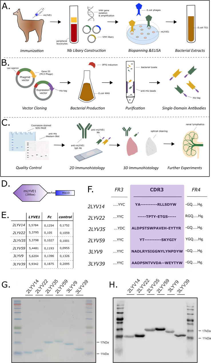 Figure 1
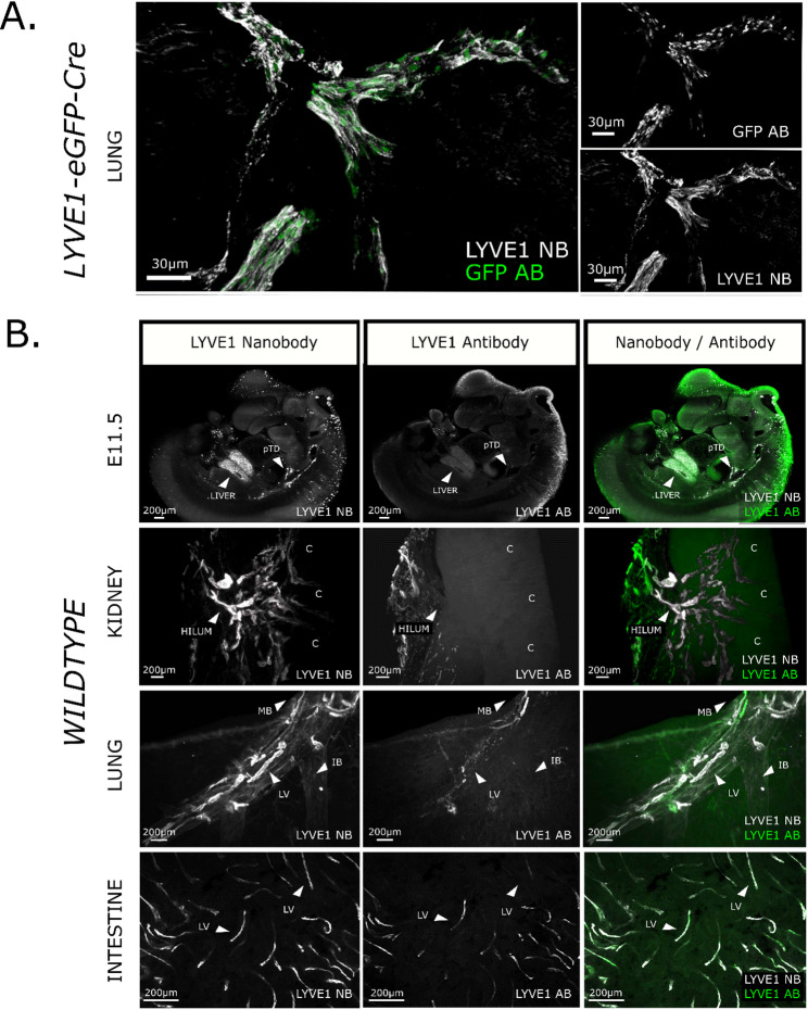 Figure 2
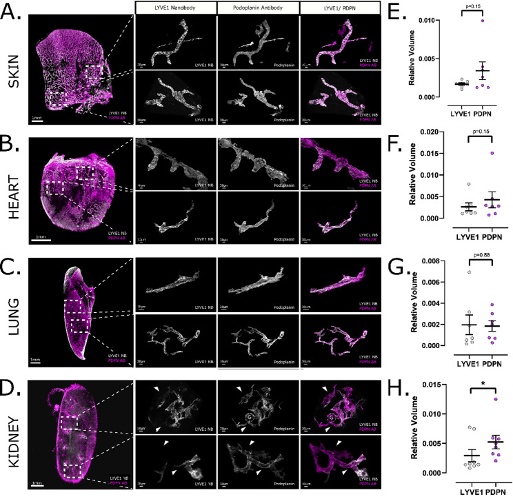 Figure 3
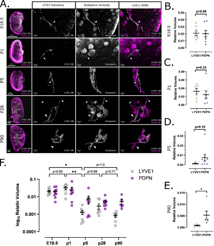 Figure 4
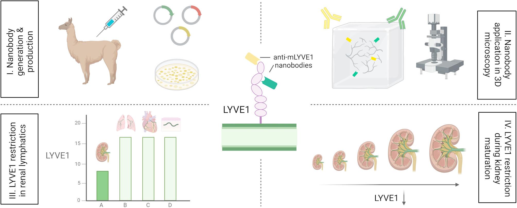 Figure 5
Peer Reviews
No public reviews on file for this paper yet. If you reviewed it on a platform where reviews are public (OpenReview, ICLR, NeurIPS, ICML), you can paste yours below so the community can read it here.
Videos
No videos yet. Explain this paper in a talk, walkthrough, or lecture? Add one.
Taxonomy
TopicsLymphatic System and Diseases · Single-cell and spatial transcriptomics · Neonatal Respiratory Health Research