Coronary intravascular imaging: a piece of history
Waiel Abusnina, Hector M. Garcia-Garcia, Pablo M. Rubio, Gary S. Mintz, Ron Waksman

TL;DR
This paper reviews the history and advancements of coronary imaging technologies that have improved heart disease diagnosis and treatment.
Contribution
The paper provides a historical overview and highlights key developments in intracoronary imaging technologies.
Findings
Intravascular ultrasound, optical coherence tomography, and near-infrared spectroscopy have evolved to enhance coronary artery disease management.
These imaging technologies have significantly improved diagnostic accuracy and outcomes in percutaneous coronary interventions.
Abstract
Since its early exploration in the 1950s, intracoronary imaging, including intravascular ultrasound (IVUS), optical coherence tomography (OCT), and near-infrared spectroscopy (NIRS), has revolutionized our understanding of coronary artery disease and improved percutaneous coronary intervention outcomes. These technologies have continuously evolved, enhancing our ability to diagnose and treat CAD and ultimately leading to better patient outcomes. This review focuses on the history and key developments of IVUS, OCT, and NIRS.
Genes, proteins, chemicals, diseases, species, mutations and cell lines named across the full text — each resolved to its canonical identifier and authoritative record.
Click any figure to enlarge with its caption.
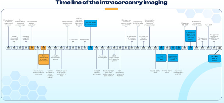 Figure 1
Peer Reviews
No public reviews on file for this paper yet. If you reviewed it on a platform where reviews are public (OpenReview, ICLR, NeurIPS, ICML), you can paste yours below so the community can read it here.
Videos
No videos yet. Explain this paper in a talk, walkthrough, or lecture? Add one.
Taxonomy
TopicsCoronary Interventions and Diagnostics · Optical Coherence Tomography Applications · Cerebrovascular and Carotid Artery Diseases