Hypomethylating agents increase L1 retroelement expression without inducing novel insertions in myeloid malignancies
Šárka Pavlová, Hana Svozilová, Marcela Krzyžánková, Radim Sonnek, Anastasiya Volakhava, Anastasia Smirnova, Tatiana Grigoreva, Zuzana Jašková, Hana Synáčková, Dennis Wahl, Michaela Bilčíková, Libor Červinek, Šárka Pospíšilová, Ilgar Mamedov, Karla Plevová

TL;DR
This study finds that hypomethylating agents used in blood cancers increase retroelement protein expression but do not cause new genetic insertions.
Contribution
The study is the first to investigate retroelement activity in leukemia and myeloid malignancies under hypomethylating agent treatment.
Findings
Hypomethylating agents increase LINE-1-encoded protein expression in myeloid cell lines.
No de novo retrotransposition events were detected in cell lines or patient samples after long-term HMA treatment.
The results suggest that HMAs do not promote new insertional mutagenesis in myeloid malignancies.
Abstract
Retroelements in the human genome are silenced via multiple mechanisms, including DNA methylation, to prevent their potential mutagenic effect. Retroelement activity, demonstrated by their expression and somatic retrotransposition events, was shown to be deregulated in multiple tumors but not yet in leukemia. We hypothesized that treatment with hypomethylating agents, commonly used in myelodysplastic syndromes and acute myeloid leukemia, could lead to increased retroelement activity and somatic retrotranspositions, thus contributing to disease progression. To address this hypothesis, we induced the expression of ORF1p protein with hypomethylating agents in DAMI and HL‐60 myeloid cell lines. To study whether long‐term hypomethylating agent therapy induces somatic retrotranspositions, we analyzed (i) both cell lines treated for 4 weeks, and (ii) sequential samples from 17 patients with…
Genes, proteins, chemicals, diseases, species, mutations and cell lines named across the full text — each resolved to its canonical identifier and authoritative record.
Click any figure to enlarge with its caption.
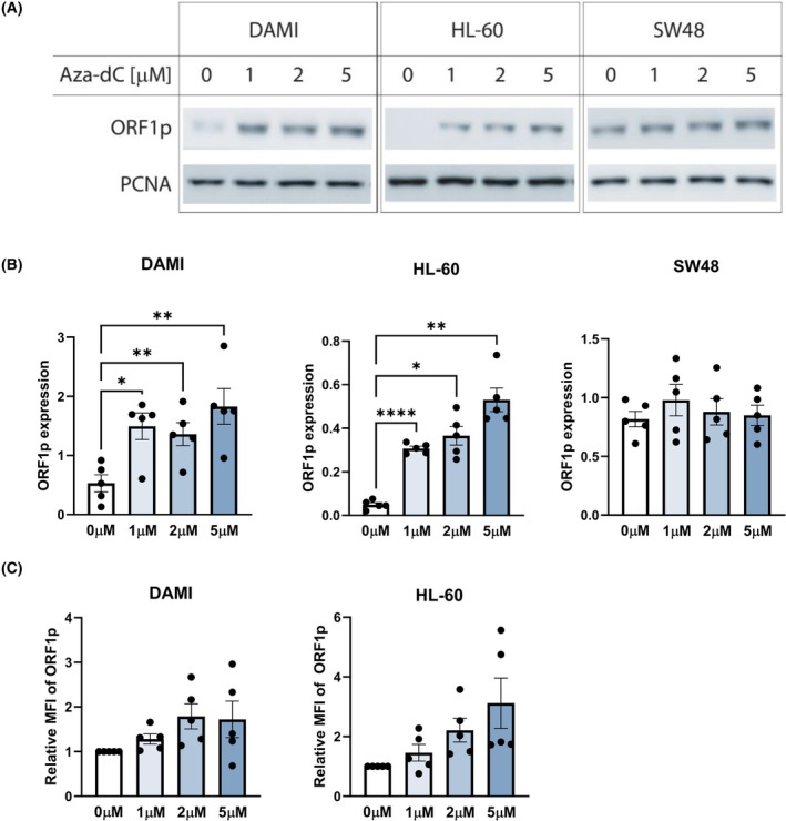 Fig. 1
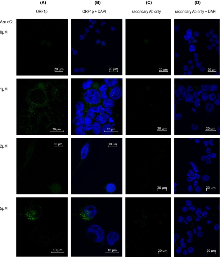 Fig. 2
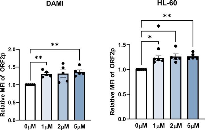 Fig. 3
- —The Ministry of Education, Youth and Sports of the Czech Republic10.13039/501100001823
- —The Ministry of Health of the Czech Republic10.13039/501100003243
- —The Czech Science Foundation10.13039/501100001824
Peer Reviews
No public reviews on file for this paper yet. If you reviewed it on a platform where reviews are public (OpenReview, ICLR, NeurIPS, ICML), you can paste yours below so the community can read it here.
Videos
No videos yet. Explain this paper in a talk, walkthrough, or lecture? Add one.
Taxonomy
TopicsAcute Myeloid Leukemia Research · RNA modifications and cancer · Epigenetics and DNA Methylation
Introduction
1
Long interspersed nuclear elements (LINEs), a non‐LTR subfamily of retrotransposons, occupy approximately 17% of the human genome [1]. Out of around 500 000 copies, the majority are inactive due to genomic rearrangements, point mutations, and 5′‐truncation to prevent their harmful impact on genome stability. The ongoing transposition is primarily caused by type 1 (LINE‐1; also known as L1). Only around 100 copies of L1 elements per genome are responsible for the majority of retrotransposon activity [2, 3].
The L1 transposable element comprises two open reading frames, ORF1, encoding an RNA chaperone, and ORF2, encoding an enzyme with single‐strand endonuclease and reverse transcriptase activities. After L1 transcription, the polyadenylated bicistronic L1 mRNAs are transported to the cytoplasm and translated into ORF1p and ORF2p proteins. Multiple ORF1p trimers and one or two ORF2p molecules bind the mRNA in cis to form the L1 ribonucleoprotein (L1 RNP). The strong predominance of ORF1p proteins (~30×) explains their much easier detection compared with ORF2p [4]. Indeed, ORF2p is nearly undetectable in primary tumors [5]. L1 RNP penetrates the nucleus during mitosis [6]. Subsequently, target‐primed reverse transcription occurs during the S‐phase, initiated by the endonuclease nicking genomic DNA at A‐ and T‐rich sites, followed by annealing of the RNA poly(A) tail and its reverse transcription [7, 8]. Using host proteins involved in DNA repair and replication, the single‐stranded DNA gap at the L1 integration site is recognized, followed by the integration and ligation of a newly synthesized L1 copy into the DNA, being flanked by the target site duplications.
The process of retrotransposition is suppressed by multiple cellular mechanisms, including DNA methylation, histone acetylation, piwi RNA complexes, and p53 machinery, with a frequency of a new retrotransposition event in the human population being between 1/20 and 1/200 births [9, 10]. As such, many L1 elements are present in individual genomes but absent in the haploid human genome reference. Contrary to normal cells, the expression levels of transposable elements and corresponding proteins, including ORF1p, are increased in tumor cells [11, 12]. Consequently, L1 retrotranspositions have been documented in multiple cancer types [13, 14, 15]. Active TEs are considered highly mutagenic and are commonly linked to the multiple steps of cancer development and progression [13, 16, 17, 18]. Moreover, L1 hypomethylation has been revealed in multiple cancer types, including lung cancer [19], colorectal cancer [20], breast cancer [21], prostate cancer [22], hepatocellular carcinoma [23], ovarian cancers [24], and esophageal squamous cell carcinoma [25], and was typically associated with poor clinical outcomes, likely due to resulting genome instability and presumed TE activation.
While high levels of somatic retrotranspositions have been shown mainly in solid tumors, in hematological malignancies, the levels are considered to be generally low [15]. In line with this, we did not identify new tumor‐specific RE insertions in childhood and adult acute leukemia samples using a sensitive NGS approach allowing for the detection of Alu and L1 insertions in 1% of cells [26].
Hypomethylating agents (HMA), 5‐Azacytidine (Aza), and 5‐Aza‐2′‐deoxycytidine (decitabine, Aza‐dC), either alone or in combination with other drugs, have become standard therapy for patients with high‐risk myelodysplastic syndromes (MDS) [27, 28] and acute myeloid leukemia (AML) [27]. Despite the unequivocal benefit for overall survival as well as improved quality of life, some patients do not respond to these drugs, and the majority of responding patients relapse. Since their use leads to hypomethylation of silenced regions in the human genome, including L1, we studied whether HMA affect the activity of L1 retrotransposons in tumor myeloid cell lines and whether de novo L1 retrotransposition can be detected in MDS patients treated with and progressing after Aza.
Materials and methods
2
Cell lines and patient samples
2.1
A panel of tumor cell lines (Table S1) was used for the initial screening of ORF1p expression following cultivation instructions provided by the supplying collection (DSMZ or ATCC). For further experiments, human myeloid cell lines DAMI and HL60 were selected. The breast carcinoma MCF‐7 cell line was used as a positive control for ORF1p expression. The HEK293T/17 cell line was used for transient transfections. All experiments were performed with mycoplasma‐free cells. During the cultivation period, the cell lines were tested on a weekly basis for the absence of mycoplasma contamination using the MycoAlert Plus Detection Kit (Lonza, Basel, Switzerland). All cell lines were authenticated, comparing their STR profiles in 16 loci against the reference sets in the Cellosaurus STR Similarity Search Tool, DSMZCellDive, or ATCC STR Database.
Thirty‐seven serial bone marrow samples from 17 MDS patients treated with 5‐azacytidine (Aza) at the Department of Internal Medicine—Hematology and Oncology, University Hospital Brno were collected between March 2012 and May 2021. The experiments were undertaken with the understanding and written informed consent of each individual subject. The project was approved by the Ethical Committee of the University Hospital Brno and followed the Declaration of Helsinki (approval ID: 19‐11299S). The samples were taken before treatment initiation, after several lines of Aza treatment, and/or in relapse; if a relapse sample was unavailable, the closest available sample before progression was used (Table S2). DNA was isolated from whole bone marrow leukocytes after erythrolysis.
Treatment of cell lines with 5‐Aza‐2′‐deoxycytidine
2.2
To induce ORF1p and ORF2p expression, a panel of tumor cell lines (Table S1) was treated with 5‐Aza‐2′‐deoxycytidine (Aza‐dC; Sigma‐Aldrich, St. Louis, MO, USA) for 72 h (0.5, 1, 2, and 5 μm). Briefly, 2.5 × 10^5^ cells·mL^−1^ were seeded in 6‐well plates (TPP, Trasadingen, Switzerland) in 5 mL of cell culture medium (Table S1) + 10% fetal bovine serum (FBS). On the following day, medium was replaced with fresh medium containing Aza‐dC (0.5, 1, 2, and 5 μm) and cultured for 48 h; then, the treatment was repeated and followed by cultivation for an additional 24 h. As a control, the cell lines were processed in the same way without adding Aza‐dC.
For fluorescent microscopy and flow cytometry, DAMI and HL‐60 were seeded in 6‐well plates (TPP) with or without sterile microscopy coverslips at a density of 3.0 × 10^6^ cells·mL^−1^ in 3 mL of DMEM + 10% ultra‐low IgG FBS (PAN‐Biotech, Aidenbach, Germany). On the following day, the cells were treated with 2 μm and 5 μm Aza‐dC, with a media change after 48 h, as described above.
Long‐term treatment of DAMI and HL‐60 was performed with 0.5 and 2 μm of Aza‐dC, with untreated cells as controls. Cells were harvested on Days 0, 3, 7, 14, and 28.
Immunoblotting
2.3
Cells were washed with PBS and lysed in ice‐cold NP‐40 buffer (Thermo Fisher Scientific, Waltham, MA, USA) with protease and phosphatase inhibitors (1:100, P8340 and P0044; Sigma‐Aldrich) for 30 min. Protein concentrations were determined by Bicinchoninic Acid Kit for Protein Determination (BCA1‐1KT; Sigma‐Aldrich) or the Bradford Protein Assay (Bio‐Rad, Hercules, CA, USA). The protein lysates were run on 10% sodium dodecyl sulfate‐polyacrylamide gels (SDS/PAGE) and transferred to a nitrocellulose membrane (Bio‐Rad) using a wet tank blotting system. The membranes were blocked with 5% nonfat milk in TBS buffer containing 0.1% Tween on a rocking shaker for 2 h. The membranes were washed in TBS‐T alone and incubated in the appropriate primary antibody overnight on a rocking shaker at 4 °C. The following day, membranes were washed and incubated in the appropriate secondary antibodies at room temperature for 1 h. For the list of antibodies, see Table S3. The proteins were detected using a chemiluminescence system (Clarity Western ECL Substrate, 1 705 061; Bio‐Rad) and imaged using a UVITEC Documentation System (Uvitec Cambridge, Mini HD9, or Alliance Q9 Advanced). The image analysis was performed in Alliance Q9 image analysis software with default settings and linear background extraction.
Intracellular detection of ORF1p and ORF2p proteins using fluorescence microscopy and flow cytometry
2.4
For fluorescent microscopy, the sterilized coverslips (13 mm/0.17 mm Menzel) were inserted into the wells of the 24‐well plate (TPP, Trasadingen, Switzerland), coated with 0.01% air‐dried poly‐l‐lysine (Sigma‐Aldrich) and seeded with the targeted cell line at a volume of 200 μL per well.
After completion of the cell line culture, cells were washed with PBS, fixed with 4% formaldehyde (10 min; F8775; Sigma‐Aldrich), washed with PBS three times, and blocked with 3% IgG‐free BSA blocking buffer (001‐000‐161; Jackson ImmunoResearch, West Grove, PA, USA) with the addition of 0.25% Triton X‐100 (SIALX100; Sigma‐Aldrich) for 1 h. The cells were incubated in the corresponding primary antibody for 1 h (Anti‐LINE‐1 ORF1p; Abcam, Cambridge, UK, or Anti‐LINE‐1 ORF2; Rockland Immunochemicals, Pottstown, PA, USA) and the secondary antibody for 30 min (Goat Anti Rabbit IgG AF488; Abcam, or Goat Anti Chicken IgY AF488; Thermo Fisher Scientific) with PBS wash in between (Table S3). Following four rounds of PBS wash, nuclear DNA was stained with Hoechst 33342 (2 min; Thermo Fisher Scientific) and washed with PBS and water. All procedures were performed at room temperature. Finally, individual stained coverslips with cells were carefully transferred with tweezers to slides with prepared mounting medium (S3023, DAKO, Glostrup, Denmark). Fluorescence detection of ORF1p and ORF2p was performed the day after the mounting medium dried using a ZEISS 700LSM confocal microscope with plan‐apochromat 40x oil objective lens. Individual images were detected using two filters with 405 and 488 nm wavelengths and 1 AU pinhole and assessed in ZEN 2009 software. The quantification of the nuclear ORF1p signal was performed in imagej (version 1.54f) by measuring the proportion of ORF1p fluorescence overlapping with the DAPI signal. The images were thresholded using Otsu's method, and regions of interest (ROIs) were created for both ORF1p and DAPI signals. The overlap between these ROIs was determined, and the percentage of the ORF1p signal localized in the nuclei was calculated as the area of overlap relative to the total ORF1p signal.
Intracellular staining for flow cytometry using FACSVerse (BD Biosciences, Franklin Lakes, NJ, USA) was performed as follows: Cells were incubated with BD Horizon Fixable Viability Stain 780 (1:100, 565 388; BD Biosciences) for 10 min at room temperature and washed with 5% ultra‐low IgG FBS in PBS (FBS/PBS). Fixation was carried out with 4% formaldehyde for 15 min at room temperature, followed by permeabilization with 50% methanol for 10 min on ice. After washing with FBS/PBS, staining proceeded similarly to the microscopy protocol, except for the omission of DAPI staining. The protocol concluded with cells being resuspended in PBS for flow cytometric analysis. An isotype control antibody was used for ORF1p expression evaluation, whereas the secondary antibody only was used as a negative control for ORF2p (see Fig. S1). The data were analyzed in flowjo v.10.8.1.
Preparation of ORF1 and ORF2 plasmids and transient transfection
2.5
E. coli DH5alpha with pBudORF1 (Addgene) and pBudORF2 (Addgene) were transferred on LB plates with Bleocin 1 mg·mL^−1^ according to the manufacturer's protocol and cultured overnight at 37 °C. Individual positive colonies were inoculated into individual Erlenmeyer flasks with 200 mL of culture LB medium and 1 mg·mL^−1^ Bleocin using sterile loops. Culturing in flasks was carried out on a shaker at 300 rpm and 37 °C until the next day. Plasmid DNA was isolated using the EndoFree Plasmid Maxi Kit (Qiagen, Hilden, Germany) according to the manufacturer's protocol.
Transient transfection for fluorescence microscopy was performed directly on coverslips coated with poly‐L‐lysine placed in 24‐well plates using the transfection reagent Polyethylenimine (PEI; Polysciences, Warrington, PA, USA) according to the manufacturer's protocol. Briefly, the day before transfection, HEK293T/17 cells were seeded in individual wells of a 24‐well plate with coverslips at a cell density of 0.5 × 10^6^/mL with 1 mL of DMEM/F12 culture medium (PAN‐Biotech) and 10% ultra‐low IgG FBS (PAN‐Biotech, Aidenbach, Germany). On the day of transfection, the culture medium was replaced with DMEM/F12:H2O transfection medium in a 1:1 ratio with the addition of 300 μL serum‐free 2 mm Glutamine (PAN‐Biotech). The premix was added to the individual wells with prepared transfection media after being left in the box at RT for half an hour at a DNA : PEI ratio (3:9 μg) of 150 μL. The transfected cells were incubated at 37 °C and 5% CO_2_. After 4 h, the transfection medium was replaced with fresh 1 mL DMEM/F12 medium + 10% ultra‐low IgG FBS, and plates were incubated for an additional 72 h. Fluorescence staining of the slide was carried out after PBS wash.
Transient transfection for the detection of ORF proteins by flow cytometry and western blots was performed in 6‐well plates without coverslips with the identical transfection procedure, and the volumes increased accordingly, that is, 1.5 × 10^6^ in 3 mL of HEK293T/17 cells were used, and the volumes of culture medium and transfection medium were 1 mL and 500 μL, respectively. After 3 days of culturing in 3 mL of fresh medium, cells were washed out with PBS supplemented with 15 mm EDTA and transferred into 1.5‐mL Eppendorf tubes.
Detection of DNA retrotransposition insertions in cancer cell lines and primary patient cells
2.6
A next‐generation sequencing method for detecting new L1 insertions of the transpositionally active subfamily L1HS was used according to our previous reports [26, 29]. Briefly, gDNA was digested by a mixture of selected endonucleases TaqI and FspBI to generate fragments that consisted of a 3′ part of the L1 retroelement and its adjacent genomic region (i.e., flank). In the next step, the fragmented DNA was ligated to a stem‐loop adapter containing unique molecular identifiers (UMI) that were used to quantify the number of cells bearing each insertion after sequencing. Next, a primer specific to the transpositionally active L1 subfamily L1HS and a primer corresponding to the ligated adaptor were used to selectively amplify genomic flanks adjacent to the 3′ end of L1. A product of the first PCR was used in the second semi‐nested PCR. Finally, an indexing PCR was carried out to introduce the sample barcodes and oligonucleotides necessary for Illumina sequencing on the NextSeq 550 machine (paired‐end, 150 + 150). We used a custom computational pipeline [30] to map all the sequenced insertions' flanks to the human reference genome and remove various artifacts generated during library preparation and sequencing. The coordinates of the insertions in the serial samples were matched to identify insertions related to clonal propagation (i.e., compared with the corresponding pretreatment sample). Following our previous report [26], candidate somatic L1 insertions were validated by an independent locus‐specific PCR with initial gDNA as a template and Encyclo Polymerase Mix (Evrogen, Moscow, Russia) and/or Q5 High‐Fidelity 2× Master Mix (New England Biolabs, Ipswich, MA, USA).
Results
3
5‐Aza‐2′‐deoxycytidine treatment induces ORF1p expression in tumor myeloid cell lines
3.1
First, we performed initial western blot screening to reveal which cell lines express ORF1p (Table S1, primary results provided in Zenodo file https://doi.org/10.5281/zenodo.15739421). While ORF1p was clearly detected in three carcinoma cell lines (MCF‐7, SW‐48, H1299), it was absent in all seven lymphoid and all four myeloid leukemia cell lines. Since DNA methylation is a key transposon silencing mechanism, we attempted to induce ORF1p expression using Aza‐dC. As shown in Fig. 1A,B, Aza‐dC induced ORF1p expression in two myeloid cell lines, DAMI and HL‐60. For the remaining two cell lines derived from myeloid disorders, MOLM‐13 and BV‐173, the Aza‐dC dosage led to a prominent decrease in cell viability, and no ORF1p induction was noted. Thus, DAMI and HL‐60 cell lines were used for further experiments.
*Western blot and flow cytometry measurements of ORF1p after Aza‐dC treatment: The western blot analysis (A, B) was performed on a panel of tumor cell lines (Table S1). Here, only DAMI and HL‐60 (myeloid cell lines) and SW48 (colorectal carcinoma cell line) are shown to demonstrate the ORF1p induction in the myeloid cell lines (P = 0.0012 and P = 0.0014 for DAMI and HL‐60, respectively), while in SW48, the ORF1p expression was present already in the baseline sample without Aza‐dC treatment. Representative results of the western blot analysis are shown (A), together with the summary of biological pentaplicates of the experiments, where the ORF1p expression was plotted as a ratio of ORF1p against PCNA signal intensity (B). The ORF1p induction in DAMI and HL‐60 was further confirmed using flow cytometry (C), however, without statistical significance (P > 0.05). For each sample, the mean fluorescence signal intensity (MFI) of ORF1p‐stained cells was corrected by subtracting the MFI of the isotype control. Data are presented as relative MFI, with each sample normalized to the untreated control from the same experiment. All experiments were performed in N = 5 biological replicates. Aza‐dC concentrations applied for the western blot analysis and flow cytometry: 0, 1, 2, and 5 μm for all cell lines. Only statistically significant results are marked: *P < 0.05, **P < 0.01, ***P < 0.001, ***P < 0.0001; one‐way ANOVA test with Geisser–Greenhouse correction, Tukey's multiple comparisons test. Data are presented as mean ± standard error of the mean.
To validate our results with an independent method, we implemented a protocol for intracellular ORF1p staining and analyzed treated and untreated DAMI and HL‐60 cells with flow cytometry. We confirmed the ORF1p induction in both tested cell lines (Fig. 1C). Similarly to the ORF1p levels detected by the western blot analysis in bulk samples, we did not observe a dose‐dependent effect when increasing Aza‐dC concentrations from 1 to 5 μm.
Next, to explore the intracellular localization of ORF1p in myeloid cell lines, we stained the cells intracellularly on microscope coverslips for visualization using fluorescent microscopy. As positive controls, we used MCF‐7 with a high endogenous expression (Fig. S2) and HEK293T/17 cells transfected with the pBudORF1 expression plasmid (Fig. S3A). The optimized procedure was used in DAMI cells that could adhere to the surface (unlike HL‐60). Again, we confirmed ORF1p induction in Aza‐dC‐treated cells (Fig. 2). This result prompted us to quantify ORF1p nuclear versus cytoplasmic signal across different Aza‐dC concentrations. The nuclear localization varied widely (N = 21 quantified images), with a mean of 60.9% at 0 μm Aza‐dC, 50.7% at 1 μm, 59.3% at 2 μm, and 46.2% at 5 μm and substantial variability among individual cells, indicating no consistent trend in the subcellular localization of ORF1p upon HMA treatment (Fig. S4).
Confocal microscopy detection of ORF1p induction after Aza‐dC treatment in DAMI cell line: (A) the immunofluorescence signal of ORF1p (green) only, (B) ORF1p (green) combined with the nuclei signal (DAPI, blue), (C) cells incubated with the secondary antibody only, displayed in the green channel (negative control) and (D) merged in the green and blue channels. Ab, antibody. Scale bars indicate 10 μm for images of Aza‐dC 2 μm (A, B) and 5 μm (A, B), and 20 μm for all remaining panels.
Furthermore, we also aimed to study the expression of ORF2p, a 150 kDa protein with much lower cellular abundance. For this purpose, we treated the DAMI cell line with Aza‐dC and analyzed it using flow cytometry. Although we observed a protein induction after Aza‐dC treatment (Fig. 3), we were unable to confirm the induction with the fluorescence microscopy despite successfully detecting ORF2p in HEK293T/17 cells transiently transfected with the pBudORF2 expression plasmid (Fig. S3B), likely due to different sensitivity of the two methods.
*Flow cytometric analysis of ORF2p induction after the treatment with Aza‐dC in DAMI and HL‐60 cell lines. For each sample, the mean fluorescence signal intensity (MFI) of ORF2p‐stained cells was corrected by subtracting the MFI of the secondary antibody‐only control. Data are presented as relative MFI, with each sample normalized to the untreated control from the same experiment. ORF2p induction after Aza‐dC treatment was significant in both DAMI (P = 0.0444) and HL‐60 (P = 0.0174). The experiment was repeated in independent biological pentaplicates; only statistically significant results are shown: *P < 0.05, *P < 0.01 (one‐way ANOVA with Geisser–Greenhouse correction, Tukey's multiple comparisons test). Data are presented as mean ± standard error of the mean.
Long‐term treatment of tumor myeloid cell lines with 5‐Aza‐2′‐deoxycytidine does not increase the retrotransposition rate
3.2
Based on our findings, showing that HMA may induce the expression of proteins encoded by L1 retrotransposon, we studied whether the long‐term exposure of myeloid tumor cells to HMA also increases the retrotransposition rates. To mimic in vitro the therapy of hematooncology patients, we treated DAMI and HL‐60 cell lines with 0.5 and 2 μm Aza‐dC for a period of 4 weeks. On Days 3, 7, 14, and 28, we collected DNA, protein, and cells.
First, we assessed ORF1p induction during Aza treatment using western blot and flow cytometry and observed the level of induction for 2 μm Aza comparable with our initial experiments shown in Fig. 1. This result prompted us to proceed with detecting novel insertions using NGS. We applied a high‐sensitive NGS‐based protocol enabling us to detect L1 insertions down to 1% of cells [26]. Candidate de novo L1 insertions for each time point (3, 7, 14, and 28 days after Aza‐dC addition) were determined by comparing their genomic coordinates to the list of L1 insertion sites identified in the same cell line before treatment (day 0). We additionally filtered out the insertions whose coordinates matched the insertions found in other treatment conditions. Such insertions likely comprise false positives, as the probability of an independent insertion into the same genomic locus in different cells is very low, whereas the chance of false positive results due to methodical limitations is much higher. Following our pipeline, manual filtering against the genomic sequence, and excluding regions containing known population L1 copies, we identified 358 candidate insertions. Based on read quality (R1 and R2 matching) and number of reads (>10) and UMIs (≥2), we selected 14 candidate insertions (8 in DAMI and 6 in HL‐60), for which we designed locus‐specific primers for PCR validation (Table S4, Fig. S5A,B). None of the candidate insertions was confirmed.
Retrotransposition is uncommon in MDS patients treated with HMA
3.3
Still, we wanted to find out whether we could identify any retrotranspositions in leukocyte DNA of MDS patients treated with Aza and whether the clonal expansion of cells carrying de novo L1 insertions may contribute to disease relapse. The rationale behind that was that they were exposed to the hypomethylation effect for a much longer period than the cell lines in 4‐week lasting cultivation, and the tumor cells eventually escape from the cell death caused by Aza. For this purpose, we again used our high‐sensitive NGS‐based protocol to explore 37 serial bone marrow samples from 17 MDS patients treated with Aza. The cohort consisted of cases with high‐risk disease, with five patients bearing TP53 defects and nine progressing to AML. The samples were taken before treatment initiation, several months on treatment and/or in relapse (for details about samplings, see Table S2). The patients received a median of 11 Aza cycles (ranging from 3 to 46) between the baseline and last samples. The cohort included both responders and nonresponders to Aza therapy. L1 insertions of each follow‐up sample were compared with the respective baseline sample of the same patient.
Following the same manual filtering as for treated tumor cell lines, we identified three candidate insertions in the genomes of three MDS patients undergoing therapy, each bearing a single candidate insertion that was subjected to validation using locus‐specific PCR (Fig. S5C–E, Table S5). Out of these candidate insertions, we were able to validate none of them, suggesting that L1 retrotransposition activity in primary patient samples is low.
Discussion
4
Epigenetic silencing of transposable elements is one of the key mechanisms of cellular defense against their potentially mutagenic effect. As REs become active after demethylation of their promoters [11, 23], we aimed to explore whether they become active in MDS treated with HMA and whether de novo insertions contribute to disease progression, at least in some patients.
MDS is a heterogeneous disease comprised of hematopoietic stem cell disorders leading to ineffective hematopoiesis, demonstrated by blood cytopenias and progression to acute myeloid leukemia in approximately one‐third of patients [31, 32]. Besides cytogenetic changes and gene mutations, epigenetic changes contribute to disease pathophysiology and progression. Widespread hypermethylation was correlated to impaired overall survival and progression to AML [33, 34]. Although HMAs are highly efficient in MDS, the mechanism of their action is not fully understood. It includes activation of tumor suppressor genes commonly inactivated in MDS, incorporation of modified cytidine derivatives into RNA and DNA, and activation of tumor antigens and retroelements recognized by the immune system (Reviewed by [35, 36]).
Indeed, we demonstrated with several methods that ORF1p levels increase after Aza‐dC treatment of tumor myeloid cell lines DAMI and HL‐60, pointing to retroelement activation. Fluorescent microscopy showed that ORF1p induced by Aza‐dC in DAMI was localized both in the cytoplasm and the nucleus, varying widely among individual cells. We were unable to localize ORF2p after treatment and could only show its expression and localization in the ectopic expression model. This is in line with other studies [5] and can be partially explained by the predominance of ORF1p proteins in L1 RNP [4]. As Aza‐dC caused cell death in other myeloid cell lines tested, we could not study ORF1p and ORF2p induction and localization in other models.
Despite the apparently increased level of L1 proteins, we did not observe de novo retrotranspositions in cell lines and patients treated with HMA regardless of the TP53 mutation status and response to therapy. This may be explained by the absence of a strong selective advantage that would lead to clonal expansion of affected cells. However, as we expected that novel L1 insertions could be present in minor subpopulations and the whole genome sequencing methods would have limited sensitivity to detect them, we applied a targeted‐NGS‐based approach with the ability to capture retrotransposition events in 1% of cells [26]. This approach was previously used to demonstrate somatic retrotranspositions in brain tissue [37] and colorectal cancer [29] and the absence of RE activity in leukemia [26]. Thus, we assume that the sensitivity of the NGS method was not the reason for the negative result.
The alternative explanation for the absence of L1 retrotransposition can be that myeloid tumor cells are negatively selected for L1 expression and/or the retrotransposition process. Indeed, a comprehensive study showed that decreased L1HS expression is associated with short overall survival in AML; it has been suggested that L1 silencing is required for oncogene‐induced transformation and propagation of AML‐initiating cells [38]. In a model system, activation of endogenous L1s resulted in increased L1 retrotransposition and impaired leukemia cell growth in vitro and in xenotransplants. In line, a model proposed by [39] suggests that the impact of TEs activity evolves during myeloid transformation. While the increased activity of TEs and piRNAs [40] induces an immune response and leads to the elimination of leukemic cells in early‐stage MDS, the leukemic cells finally escape the control of the immune system via suppression of TE/piRNA expression, resulting in the progression to high‐risk MDS, accumulation of leukemic blasts, and AML.
The somatic retrotranspositions are absent not only in AML and high‐risk MDS but also in acute lymphoblastoid leukemia, as we showed previously [26]. Of note, in the report, we also analyzed a set of childhood AML that is biologically distinct from AML in adults and the elderly, and we did not detect somatic retrotranspositions either [26]. Whether the intolerance of RE activity is a feature of hematopoietic stem and early progenitor cells or is inherent to the hematopoietic lineage as a whole is currently unknown. It would be of interest to explore RE activity, for example, in mature B‐cell neoplasms that are not directly related to a hematopoietic stem cell.
Significantly increased levels of ORF1p proteins and L1Hs transcripts were detected in TP53‐mutated Wilms tumor and colorectal carcinoma samples, respectively [41]. It has been suggested that keeping genome integrity via preventing RE events is an ancient function of tumor suppressor p53 [42]. TP53‐mutated myeloid neoplasms have recently been recognized as an aggressive entity [43]. To see whether loss of p53 function enables RE events in MDS, we included five patients with TP53 mutations in the studied cohort. The lack of RE events in these patients and the TP53‐deficient cell lines HL‐60 and DAMI suggests that intolerance of RE events in leukemic cells is p53‐independent and TP53 inactivation is not sufficient to allow for RE events in myeloid leukemia cells, although it is to be confirmed by future studies.
Conclusions
5
Overall, we show that although HMA induce the expression of L1‐encoded proteins in tumor myeloid cell lines, de novo somatic retrotransposition events do not arise during the treatment of MDS patients with these agents. Thus, while somatic retrotranspositions occur in carcinomas, we show they are uncommon in myeloid cells.
Conflict of interest
The authors declare no financial conflicts of interest.
Author contributions
SPav, IM, and KP conceived the study, designed and supervised experiments, and interpreted data. MK and IM developed methods. HS, MK, RS, AV, TG, ZJ, HS, and DW performed experiments and analyzed data. AS carried out the bioinformatic analyses. MB collected clinical and laboratory diagnostic information. LC provided samples and clinical expertise; SPav, IM and KP drafted the manuscript. SPosp, IM, and KP obtained funding. All authors reviewed and approved the manuscript.
Peer review
The peer review history for this article is available at https://www.webofscience.com/api/gateway/wos/peer‐review/10.1002/1878‐0261.70111.
Supporting information
Fig. S1. Strategy for gating positive cells using an isotype control for ORF1p staining and secondary antibody only for ORF2p staining in flow cytometry experiments. Fig. S2. Confocal microscopy detection of ORF1p in MCF‐7 cell line used as a positive control. Fig. S3. HEK293T cell transfection with ORF1 and ORF2 constructs. Fig. S4. The quantification of ORF1p subcellular localization in DAMI cells treated with Aza‐dC. Fig. S5. PCR validation of candidate L1 insertions detected by NGS.
Table S1. Cell lines used for experiments. Table S2. A cohort of MDS patients tested for L1 de novo insertions during Aza treatment. Table S3. Antibodies used for western blot analysis, flow cytometry, and immunofluorescence staining. Table S4. A list of candidate insertions detected using L1‐targeted amplicon‐based NGS protocol and validated using site‐specific PCR in the long‐term DAMI and HL‐60 treatment. Table S5. A list of candidate insertions detected using L1‐targeted amplicon‐based NGS protocol and validated using site‐specific PCR in MDS patient samples after Aza treatment.
The reference list from the paper itself. Each links out to its DOI / PubMed record.
- 1Wicker T , Sabot F , Hua‐Van A , Bennetzen JL , Capy P , Chalhoub B , et al. A unified classification system for eukaryotic transposable elements. Nat Rev Genet. 2007;8:973–982.17984973 10.1038/nrg 2165 · doi ↗ · pubmed ↗
- 2Brouha B , Schustak J , Badge RM , Lutz‐Prigge S , Farley AH , Moran JV , et al. Hot L 1s account for the bulk of retrotransposition in the human population. Proc Natl Acad Sci USA. 2003;100:5280–5285.12682288 10.1073/pnas.0831042100 PMC 154336 · doi ↗ · pubmed ↗
- 3Streva VA , Jordan VE , Linker S , Hedges DJ , Batzer MA , Deininger PL . Sequencing, identification and mapping of primed L 1 elements (SIMPLE) reveals significant variation in full length L 1 elements between individuals. BMC Genomics. 2015;16:220.25887476 10.1186/s 12864-015-1374-y PMC 4381410 · doi ↗ · pubmed ↗
- 4Taylor MS , La Cava J , Mita P , Molloy KR , Huang CR , Li D , et al. Affinity proteomics reveals human host factors implicated in discrete stages of LINE‐1 retrotransposition. Cell. 2013;155:1034–1048.24267889 10.1016/j.cell.2013.10.021PMC 3904357 · doi ↗ · pubmed ↗
- 5Ardeljan D , Wang X , Oghbaie M , Taylor MS , Husband D , Deshpande V , et al. LINE‐1 ORF 2p expression is nearly imperceptible in human cancers. Mob DNA. 2020;11:1.31892958 10.1186/s 13100-019-0191-2PMC 6937734 · doi ↗ · pubmed ↗
- 6Mita P , Wudzinska A , Sun X , Andrade J , Nayak S , Kahler DJ , et al. LINE‐1 protein localization and functional dynamics during the cell cycle. e Life. 2018;7:e 30058.29309036 10.7554/e Life.30058 PMC 5821460 · doi ↗ · pubmed ↗
- 7Cost GJ , Feng Q , Jacquier A , Boeke JD . Human L 1 element target‐primed reverse transcription in vitro. EMBO J. 2002;21:5899–5910.12411507 10.1093/emboj/cdf 592PMC 131089 · doi ↗ · pubmed ↗
- 8Doucet AJ , Wilusz JE , Miyoshi T , Liu Y , Moran JV . A 3′ poly(a) tract is required for LINE‐1 retrotransposition. Mol Cell. 2015;60:728–741.26585388 10.1016/j.molcel.2015.10.012PMC 4671821 · doi ↗ · pubmed ↗