Central Hemangioma of Mandible: Rare Case
Rakshya Shrestha, Reena Kumari Shrestha, Dipti Shrestha, Alok Sagtani, Pranay Shakya

TL;DR
This paper discusses a rare case of central hemangioma in the mandible and highlights the importance of accurate diagnosis to avoid life-threatening complications during surgery.
Contribution
The paper presents a rare clinical case emphasizing the need for thorough radiographic assessment and biopsy for central hemangioma diagnosis.
Findings
Central hemangioma should be considered in the differential diagnosis of intraosseous radiolucent lesions in younger patients.
Missed diagnosis can lead to profuse intraoperative bleeding and life-threatening complications.
Confirmatory biopsy and comprehensive radiographic assessment are crucial for effective management.
Abstract
Central hemangioma, though it is a rare entity, should always be considered in the differential diagnosis of intraosseous radiolucent lesions, especially in younger patients, as missed diagnosis can lead to profuse intraoperative bleeding and pose life‐threatening complications. Comprehensive radiographic assessment and confirmatory biopsy are crucial for definitive diagnosis, surgical planning, and effective management.
Genes, proteins, chemicals, diseases, species, mutations and cell lines named across the full text — each resolved to its canonical identifier and authoritative record.
Click any figure to enlarge with its caption.
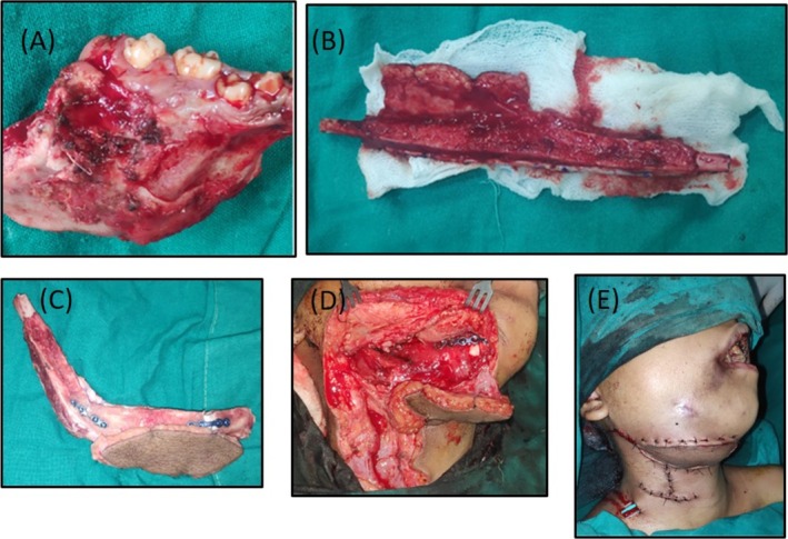 Figure 1
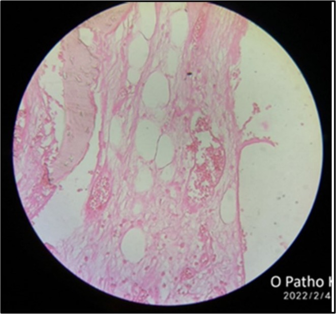 Figure 2
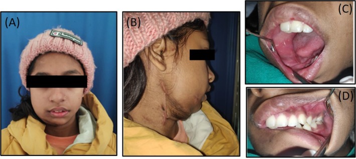 Figure 3
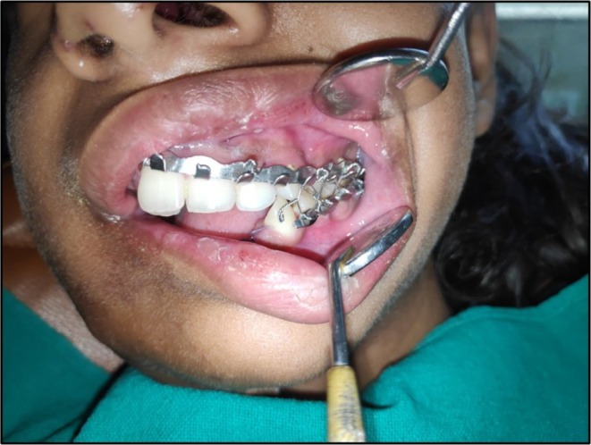 Figure 4
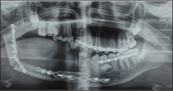 Figure 5
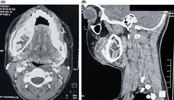 Figure 6
 Figure 7
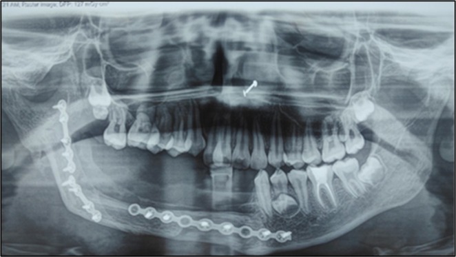 Figure 8
Peer Reviews
No public reviews on file for this paper yet. If you reviewed it on a platform where reviews are public (OpenReview, ICLR, NeurIPS, ICML), you can paste yours below so the community can read it here.
Videos
No videos yet. Explain this paper in a talk, walkthrough, or lecture? Add one.
Taxonomy
TopicsVascular Malformations and Hemangiomas · Bone Tumor Diagnosis and Treatments · Vascular Malformations Diagnosis and Treatment