Radiopathologic Characteristics of Invasive Mammary Carcinoma With Medullary Features: A Correlative Study
Sanjanika S, Pavithra V, Preetha Nethaji, Bhawna Dev

TL;DR
This study examines the radiologic and histopathologic features of medullary breast cancer to improve diagnosis accuracy and avoid misinterpretation due to its benign-like imaging appearance.
Contribution
The study provides a detailed radiopathologic correlation of medullary breast cancer, highlighting its diagnostic challenges and the necessity for histopathological confirmation.
Findings
Medullary carcinoma often appears as irregular masses with circumscribed margins on mammograms and hypoechoic masses with microlobulated margins on ultrasound.
Imaging features like posterior acoustic enhancement are common but not definitive for diagnosis, necessitating histopathological confirmation.
Histopathology reveals a syncytial growth pattern, high nuclear grade, and absence of glandular elements in most cases.
Abstract
Introduction Invasive mammary carcinoma with medullary features represents an uncommon subtype of breast cancer. Despite their high-grade histological appearances, they have a favourable prognosis. This study aims to correlate its radiologic and histopathologic characteristics. A comprehensive understanding of the radiopathologic profile is essential for enhancing the diagnosis precision and guiding patient treatment, particularly because of its typically benign imaging findings, which may result in misinterpretation and underdiagnosis. Materials and methods A retrospective observational study was conducted by reviewing cases of histologically confirmed invasive mammary carcinoma with medullary features and triple-negative basal-like carcinoma that met the WHO criteria over five years (2020-2025) at the Sri Ramachandra Institute of Higher Education and Research, Chennai, India. We…
Genes, proteins, chemicals, diseases, species, mutations and cell lines named across the full text — each resolved to its canonical identifier and authoritative record.
Click any figure to enlarge with its caption.
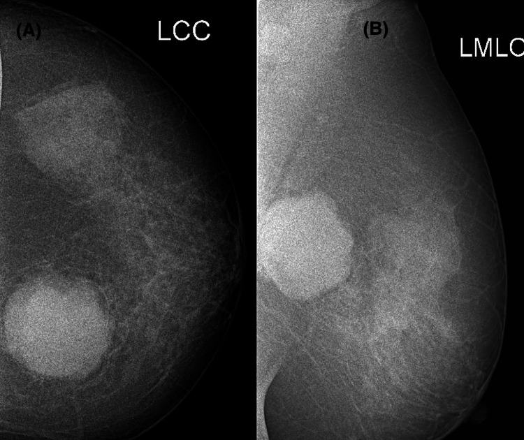 Figure 1
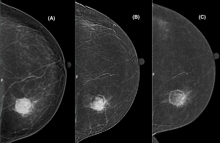 Figure 2
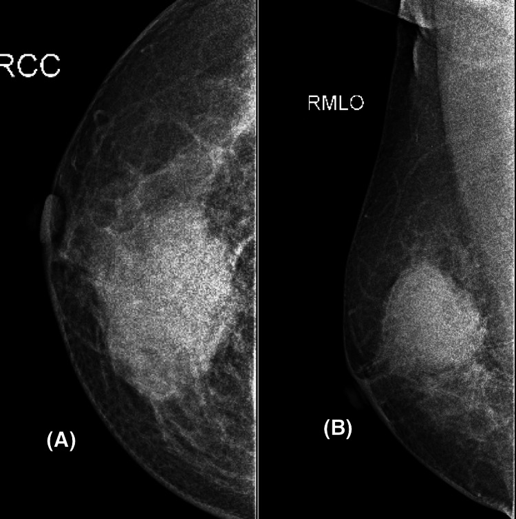 Figure 3
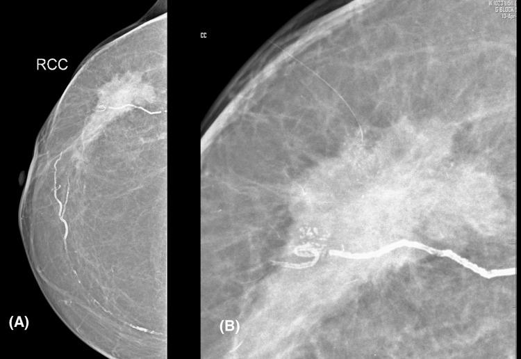 Figure 4
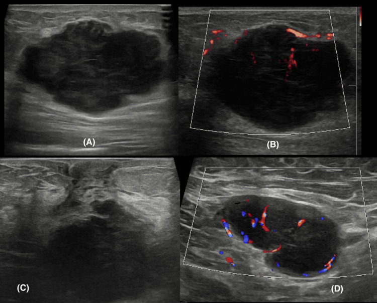 Figure 5
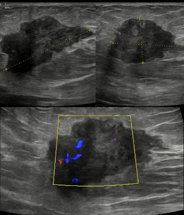 Figure 6
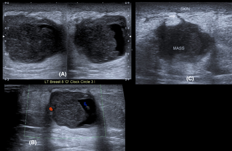 Figure 7
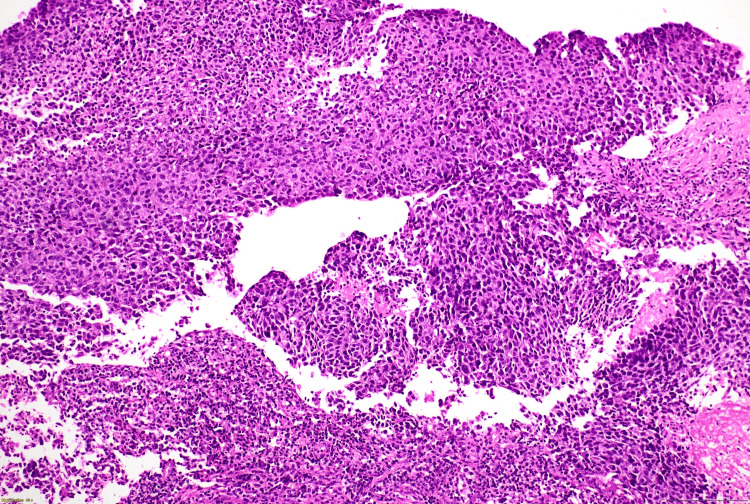 Figure 8
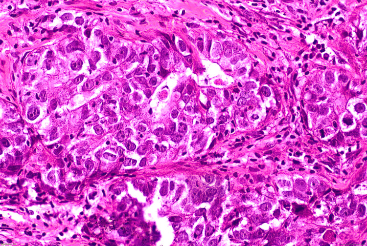 Figure 9
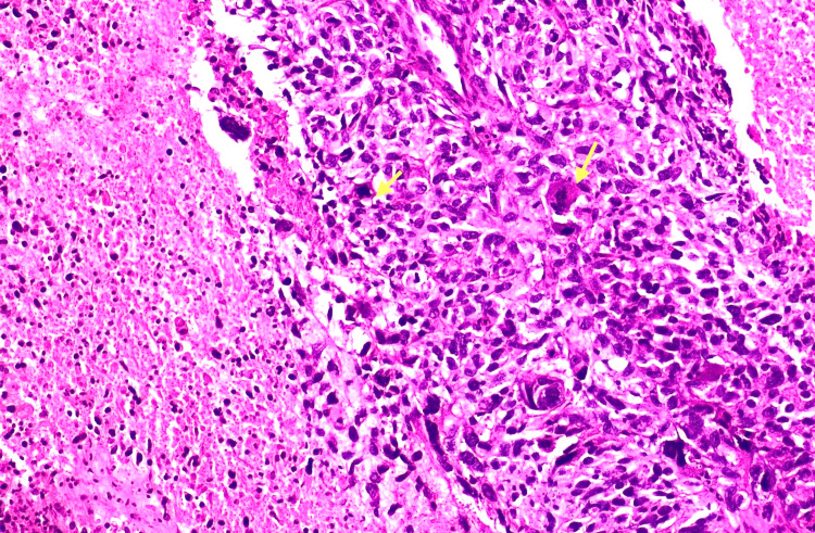 Figure 10
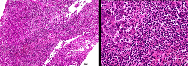 Figure 11
Peer Reviews
No public reviews on file for this paper yet. If you reviewed it on a platform where reviews are public (OpenReview, ICLR, NeurIPS, ICML), you can paste yours below so the community can read it here.
Videos
No videos yet. Explain this paper in a talk, walkthrough, or lecture? Add one.
Taxonomy
TopicsBreast Lesions and Carcinomas · Breast Cancer Treatment Studies · Metastasis and carcinoma case studies