Recurrent Aseptic Meningitis Without Identifiable Pathogens: A Case Report
Saima Nazish, Abdulhadi J Alotaibi, Obaid M Aljarbou, Abdulaziz Omair, Faisal H Aljamea

TL;DR
A 66-year-old man experienced multiple meningitis episodes without a clear cause, highlighting the challenges in diagnosing idiopathic aseptic meningitis.
Contribution
This case report adds to the understanding of idiopathic aseptic meningitis through a detailed clinical presentation and diagnostic journey.
Findings
The patient had five meningitic episodes with consistent CSF findings but no identifiable pathogen.
Extensive testing for infectious and autoimmune causes yielded negative results.
Symptoms resolved with supportive care and multidisciplinary management.
Abstract
Recurrent meningitis without an identifiable pathogen is an infrequent but taxing diagnostic dilemma, particularly when multiple systemic comorbidities obscure both the clinical picture and the laboratory data. A 66-year-old man with dyslipidemia, diabetes, hypertension, ischemic cardiomyopathy, and a left ventricular thrombus experienced five meningitic episodes over six years. Each event featured holocephalic headache radiating to the neck, neck stiffness, vomiting, transient dysarthria, and gait ataxia. The cerebrospinal fluid (CSF) consistently showed lymphocytic pleocytosis (6-48 cells/mm3), elevated protein (75-142 mg/dL), and sterile cultures. Serial viral polymerase chain reaction (PCR) panels, bacterial and mycobacterial studies, fungal cultures, extended autoimmune screens, complement levels, and paraneoplastic panels were negative. Brain magnetic resonance imaging (MRI)…
Genes, proteins, chemicals, diseases, species, mutations and cell lines named across the full text — each resolved to its canonical identifier and authoritative record.
Click any figure to enlarge with its caption.
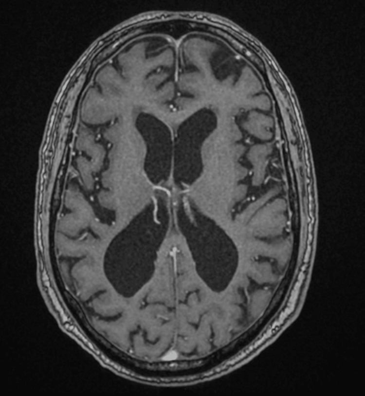 Figure 1
| Parameter | Reference range | March 2019 | September 2019 | June 2023 | December 2023 |
| Peripheral blood | |||||
| WBC (×10³/µL) | 4.0-11.0 | 6.2 | 7.1 | 5.6 | 6.4 |
| CRP (mg/L) | 0-5 | 5.6 | 14.7 | 8 | 5.4 |
| ESR (mm h⁻¹) | 0-15 | 60 | 99 | 44 | 51 |
| Procalcitonin (ng/mL) | <0.50 ng/mL (values ≥0.50 ng/mL raise concern for significant systemic bacterial infection) | 0.13 | 0.11 | 0.14 | 0.44 |
| CSF chemistry | |||||
| Protein (mg/dL) | 15-45 | 75 | 132 | 119 | 85 |
| Glucose (mg/dL)† | 45-80 | 227 | 127 | 95 | 120 |
| CSF cytology | |||||
| RBC (cells mm⁻³) | 0-5 | 205 | 1 | 2 | 1 |
| WBC (cells mm⁻³) | 0-5 | 15 (99 % L) | 22 (99 % L) | 48 (82 % L) | 6 (99 % L) |
| Test | March 2019 | September 2019 | June 2023 | December 2023 |
| Gram stain | No organisms | Rare WBC, none seen | No organisms | No organisms |
| Aerobic culture (48 hours) | No growth | No growth | No growth | No growth |
| India ink | Negative | Negative | Negative | Negative |
| AFB smear/culture | Negative (6 weeks) | Negative (6 weeks) | Negative (8 weeks) | Negative (6 weeks) |
|
| — | — | MTBC DNA not detected | — |
| HSV PCR | Negative | Negative | Negative | Negative |
| Fungal culture (4 weeks) | No growth | No growth | — | No growth |
| Peripheral blood culture (5 days) | No growth | No growth | No growth | No growth |
| Panel/assay | Reference range | Result |
| Autoimmune/inflammatory serology | ||
| ANA, anti‑dsDNA, anti‑Smith | N/A | Negative |
| SSA/SSB, anti‑β₂‑glycoprotein | N/A | Negative |
| ANCA (MPO, PR3) | N/A | Negative |
| Rheumatoid factor, anti‑CCP | N/A | Negative |
| Complement C3 | 90-180 mg/dL | Normal |
| Complement C4 | 10-40 mg/dL | Normal |
| Paraneoplastic antibody panel | N/A | Negative |
| Infectious disease workup | ||
| HIV, hepatitis panel | N/A | Non‑reactive |
| Brucella IgG/IgM, syphilis serology | N/A | Negative |
| Viral PCR (HSV‑1/2, VZV, CMV, EBV, enterovirus) | N/A | Negative |
| CSF indices (repeat tap, day 3) | ||
| Protein | 15-45 mg/dL | 142 mg/dL |
| Glucose† | 45- 80 mg/dL | 138 mg/dL |
| WBC | 0-5 cells/mm³ | 89 cells mm⁻³ (98 % L) |
| Gram stain/culture | N/A | No organisms/no growth |
| India ink, fungal culture | N/A | Negative/no growth |
| AFB smear/culture (8 weeks) | N/A | Negative |
| GeneXpert MTB/RIF | N/A | No MTB DNA detected |
| Toxicology and others | ||
| Drug screen | N/A | Negative |
| TSH | 0.4-4.0 mIU/L | Within reference limits |
| Vitamin B₁₂ | 200-900 pg/mL | Within reference limits |
| Folate (serum) | ≥4.0 ng/mL | Within reference limits |
Peer Reviews
No public reviews on file for this paper yet. If you reviewed it on a platform where reviews are public (OpenReview, ICLR, NeurIPS, ICML), you can paste yours below so the community can read it here.
Videos
No videos yet. Explain this paper in a talk, walkthrough, or lecture? Add one.
Taxonomy
TopicsPneumocystis jirovecii pneumonia detection and treatment · Bacterial Infections and Vaccines · Polyomavirus and related diseases
Introduction
Meningitis is a severe and sudden infection of the meninges, which can be triggered by bacteria, viruses, parasites, or fungi [1]; however, sometimes, there is inflammation of the brain meninges caused by various factors, leading to negative cerebrospinal fluid (CSF) bacterial cultures known as aseptic meningitis. Occasionally, these episodes are recurrent, leading to hospitalization and diagnostic workup. Recurrent meningitis is uncommon, affecting 2-9% of meningitis cases. It presents diagnostic challenges due to its various underlying causes. The primary reasons include anatomical defects (59%), immune system deficiencies (36%), and infections around the meninges (5%); other contributing factors may include head injuries, chronic inflammation within the brain, and issues with the complement system [2,3]. Recurrent episodes of aseptic meningitis are characterized by repeated attacks of headache, fever, and meningeal signs accompanied by CSF lymphocytic pleocytosis (white blood cell (WBC) >5 cells/mm³) and sterile CSF cultures; this clinical pattern is classically referred to as Mollaret's meningitis, first described by Pierre Mollaret in 1944 [4]. These episodes of meningitis are usually caused by herpes simplex virus type 2 (HSV-2) infection, although other viral and non-viral causes have also been reported [5,6]. The causes of aseptic meningitis can be broadly classified into infectious and non-infectious origins. Despite progress in diagnostic methods, only 30-65% of cases reveal a precise cause [7]. Cases without identified causes are termed idiopathic, though recently identified conditions, such as transient headache and neurologic deficits with CSF lymphocytosis (HaNDL) syndrome and meningitis-retention syndrome, may help reduce the proportion of cases classified as idiopathic aseptic meningitis [8]. The objective of writing this case report is to discuss diagnostic challenges physicians face for these rare causes of meningitis. By integrating this knowledge into their clinical practice, clinicians will be better equipped to deliver high-quality care, improving patient outcomes and safety. We also highlighted the importance of considering the timely adaptations of available treatment options in patients with recurrent meningitis without identifiable pathogens.
Case presentation
We present the case of a 66-year-old right-handed man with a complex medical history, including dyslipidemia, hypertension (HTN), diabetes mellitus (DM), ischemic heart disease, and left ventricular thrombus with heart failure, on apixaban 5 mg PO BID. He had a history of recurrent aseptic meningitis, 4-5 episodes in total over a six‑year period. Every time, he used to present with symptoms including headache, neck stiffness, vomiting, dysarthria, and ataxia with raised inflammatory markers and high CSF protein and white blood cell (WBC) counts with reactive atypical lymphocytes without any identifiable cause. In the first instance, antibiotics and antivirals were started. Meanwhile, we performed a workup, and the patient was discharged with normal CSF studies and inflammatory markers. Tables 1-2 show the record of the patient's CSF parameters and microbiological results over different periods.
This time, the patient again presented with a four-day history of a holocephalic, pressure-like headache, moderate in severity, radiating to the neck and aggravated by coughing. Associated symptoms included photophobia, neck stiffness, vomiting, unsteady gait, and generalized fatigability. Notably, there was no photophobia or fever. On examination, the patient was alert and oriented with stable vital signs. Pupils were small with sluggish light reaction; there were positive signs of meningeal irritation. Cogwheel rigidity, more pronounced in the right upper extremity, was present; rapid alternating movements were mildly slowed and irregular.** Sensory examination was normal with pyramidal signs. Considering his medical history and the complexity of recurrent meningitis without an identifiable pathogen, comprehensive microbiological testing of the CSF was performed to rule out infectious causes. This included screening for HSV-1 and HSV-2, human immunodeficiency virus (HIV), Epstein-Barr virus (EBV), cytomegalovirus (CMV), enteroviruses, mumps, measles, atypical bacteria such as Mycoplasma pneumoniae, Mycobacterium tuberculosis, Treponema pallidum, and Brucella, as well as parasites and fungi. CSF Gram stain, aerobic cultures, and multiplex viral polymerase chain reaction (PCR) panels were obtained, and all returned negative; extended fungal culture (four‑week incubation) was also negative. **Fungal culture, with a four-week incubation period, yielded no fungal growth. Brucella testing, including DNA PCR, isolation, and genetic sequencing, returned negative. Tuberculosis testing using the GeneXpert Mycobacterium tuberculosis (MTB)/rifampicin (RIF) assay and a six-week CSF culture showed no MTB DNA detection or growth. CSF cytology didn't reveal any malignant cells. In addition to CSF analysis, an extensive hematological, inflammatory, metabolic, and immunological workup was conducted. A broad range of non-infectious causes were considered, including systemic diseases such as Behçet's disease, Sjögren's syndrome, sarcoidosis, systemic lupus erythematosus, granulomatosis with polyangiitis, and paraneoplastic syndromes as summarized in Table 3.
Complement levels (C3, C4), soluble interleukin‑2 receptor, and a comprehensive toxicology drug screen were performed; all were within reference limits/negative.** **Drug-induced aseptic meningitis was also ruled out. HaNDL syndrome was also considered, but its criteria were not met. Thorough brain parenchymal and vascular imagings were performed to rule out potential inflammatory conditions, particularly vasculitis. Brain magnetic resonance imaging (MRI) showed mild ventricular enlargement, persistent pachymeningeal enhancement, and mild cerebral small vessel disease (Figure 1).
Axial T1-weighted brain MRIMRI: magnetic resonance imaging
Interdisciplinary care, with multiple teams, including infectious disease, neurology, and cardiology, was involved to work together to address the patient's clinical condition. The patient's clinical presentation, lack of identifiable pathogens, and exclusion of common causes led to a working diagnosis of aseptic meningitis. After excluding both infections and non-infectious causes of aseptic meningitis, ultimately, an idiopathic aseptic meningitis was considered. The patient was managed with a combination of supportive care and close monitoring for any changes in symptoms. The patient's clinical condition improved over a five-day period. His headache, unsteadiness, and fatigue completely resolved, and repeat CSF showed improvement. Inflammatory markers returned to normal, and the patient was discharged home.
Discussion
The presented case of recurrent aseptic meningitis posed a significant diagnostic challenge due to the presence of multiple comorbidities. The absence of classic meningitis symptoms, such as fever and photophobia, combined with negative microbiological findings, further complicated the clinical assessment. Recurrent aseptic meningitis requires a thorough and systematic diagnostic approach, with a broad differential diagnosis encompassing both infectious and non-infectious etiologies, including viral infections, autoimmune disorders, and paraneoplastic syndromes [9]. Given the patient's clinical presentation, alternative diagnoses such as Mollaret's meningitis and autoimmune etiologies were considered. Mollaret's meningitis, a benign recurrent form of aseptic meningitis, often presents with negative CSF PCR results for HSV. There were reports of Mollaret's meningitis caused by other virus infections, including varicella-zoster virus (VZV), West Nile virus, EBV, human herpesvirus-6 (HHV-6), influenza virus, and enterovirus [10]. Additionally, reports underscore the atypical presentation patterns and the diagnostic utility of CSF cytology in identifying this rare condition [11]. However, in this case, a comprehensive viral panel and CSF cytology did not reveal any abnormalities. Further investigations targeted atypical fastidious bacteria, parasites, fungi, as well as non-infectious causes. Despite multiple CSF analyses, advanced imaging studies, and extensive microbiological, immunological, and paraneoplastic evaluations, no definitive etiology was identified. This exhaustive diagnostic process ultimately led to a diagnosis of idiopathic aseptic meningitis. Advancements in diagnostic techniques, including PCR, next-generation sequencing, and enhanced identification of autoimmune and paraneoplastic syndromes, have significantly improved the detection of causative agents in recurrent aseptic meningitis. Nonetheless, up to two-thirds of cases remain idiopathic [8]. Some studies suggest that mutations resulting in glycosylphosphatidylinositol (GPI) anchoring protein deficiency could represent a novel pathogenic mechanism for recurrent meningitis of unknown origin. The absence of these proteins may lead to excessive complement system activation, triggering recurrent meningeal inflammation and related symptoms [12]. However, in our case, we did not identify any low complement levels. Management of aseptic meningitis cases requires careful evaluation of clinical presentations and diagnostic findings. Most patients benefit from symptomatic treatment alone. Indomethacin, for example, is thought to reduce fever and inflammation by inhibiting periodic eicosanoid acid abnormalities in the brain. In contrast, treatments such as steroids, colchicine, antihistamines, and butylamine have shown limited efficacy [13,14]. There is currently no consensus regarding the necessity for long-term pharmacological therapy or recommendations for preventive treatment. In this case, the decision to initiate antiviral therapy despite negative viral panels underscores the importance of clinical judgment amid diagnostic uncertainty. The de‑escalation after specialist review illustrates the importance of avoiding unnecessary antimicrobials and minimizing potential side effects. Effective management of this patient involved multidisciplinary collaboration among neurology, infectious disease, and internal medicine teams, which was pivotal in formulating a comprehensive treatment plan. This case emphasizes the value of a multidisciplinary approach when addressing complex cases of recurrent aseptic meningitis. Clinicians must maintain a high index of suspicion, consider a wide range of differential diagnoses, and carefully balance the risks and benefits of therapeutic interventions.
Conclusions
This case illustrates the complexities involved in diagnosing and managing recurrent aseptic meningitis, particularly in the absence of identifiable pathogens. A systematic, multidisciplinary strategy, coupled with vigilant monitoring, is essential for optimizing patient outcomes. Key insights include the necessity of considering viral etiologies in aseptic meningitis and acknowledging the diagnostic challenges posed by coexisting medical conditions. Further research into both infectious and non-infectious causes of recurrent meningitis is crucial for enhancing diagnostic accuracy and improving patient care.
The reference list from the paper itself. Each links out to its DOI / PubMed record.
- 1Magnetic resonance imaging of infectious meningitis and ventriculitis in adults Top Magn Reson Imaging Hazany S Go JL Law M 3153252320142529627610.1097/RMR.0000000000000034 · doi ↗ · pubmed ↗
- 2Recurrent meningitis--a review of current literature Przegl Epidemiol Janocha-Litwin J Simon K 4112941-5, 125-9672013 https://pubmed.ncbi.nlm.nih.gov/23745374/23745374 · pubmed ↗
- 3Epidemiology, etiology, pathogenesis, and diagnosis of recurrent bacterial meningitis Clin Microbiol Rev Tebruegge M Curtis N 5195372120081862568610.1128/CMR.00009-08PMC 2493086 · doi ↗ · pubmed ↗
- 4Mollaret's meningitis: a case report and review of the literature TAJ J Teach Assoc Hoque MA Chowdhury NM Hoque Z Siddiqui N Islam R Ahmad Q 4146172009
- 5Herpes simplex virus type 1 DNA in cerebrospinal fluid of a patient with Mollaret's meningitis N Engl J Med Yamamoto LJ Tedder DG Ashley R Levin MJ 108210853251991165390010.1056/NEJM 199110103251507 · doi ↗ · pubmed ↗
- 6Recurrent lymphocytic meningitis by herpes simplex virus type 2 [Article in Spanish]Medicina (B Aires) Benchetrit AG García JL Janota F Solari RM 513515792019 https://pubmed.ncbi.nlm.nih.gov/31829956/31829956 · pubmed ↗
- 7Etiologies and management of aseptic meningitis in patients admitted to an internal medicine department Medicine (Baltimore) Jarrin I Sellier P Lopes A 095201610.1097/MD.0000000000002372 PMC 471823726765411 · doi ↗ · pubmed ↗
- 8Aseptic meningitis Rev Neurol (Paris) Tattevin P TchamgouéS Belem A Bénézit F Pronier C Revest M 47548017520193137528610.1016/j.neurol.2019.07.005 · doi ↗ · pubmed ↗