Review of the wolf-spider genus Draposa Kronestedt, 2010 from China (Araneae, Lycosidae)

Abstract
Genes, proteins, chemicals, diseases, species, mutations and cell lines named across the full text — each resolved to its canonical identifier and authoritative record.
Click any figure to enlarge with its caption.
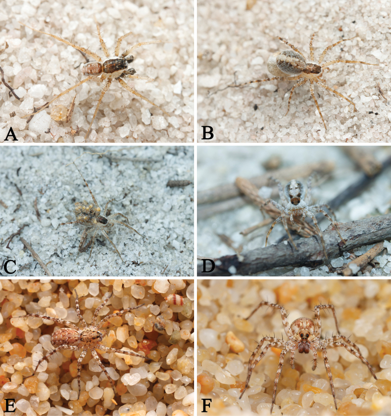 Figure 1
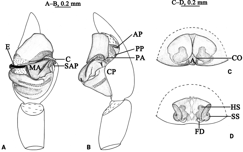 Figure 2
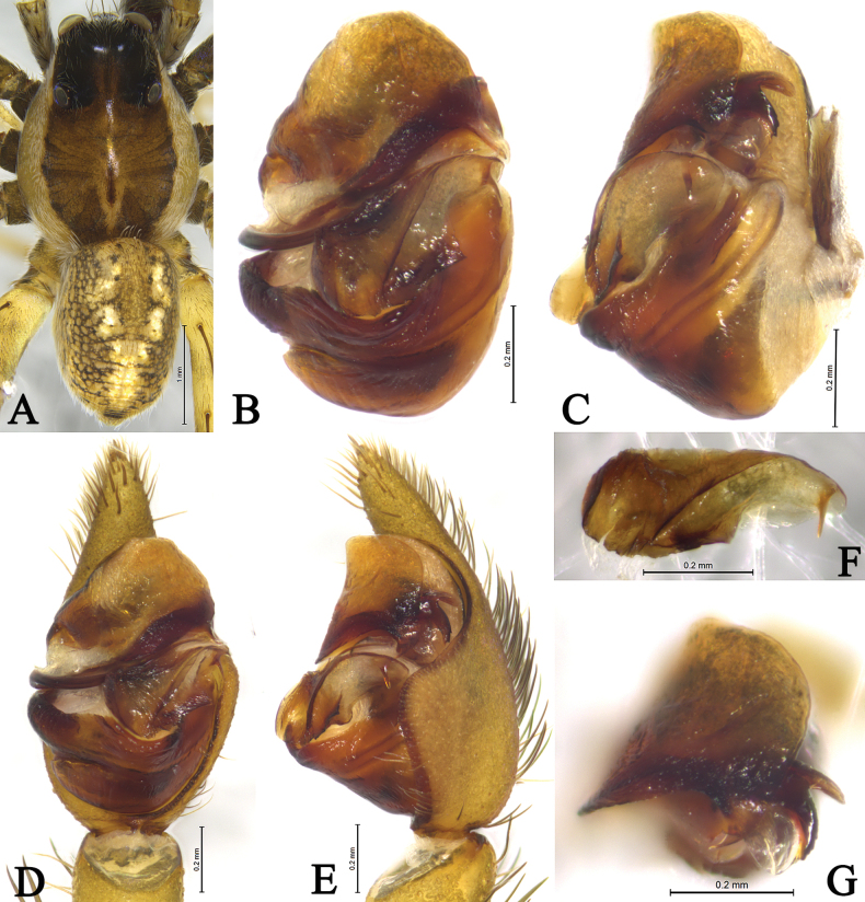 Figure 3
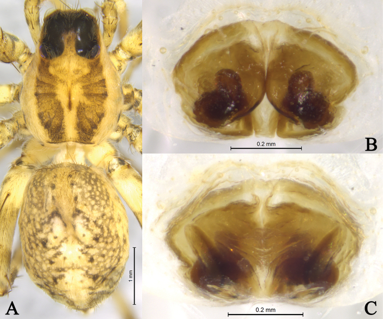 Figure 4
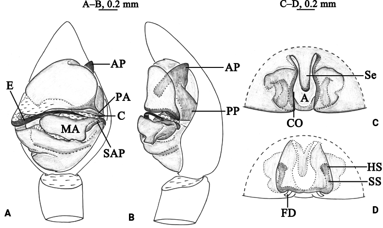 Figure 5
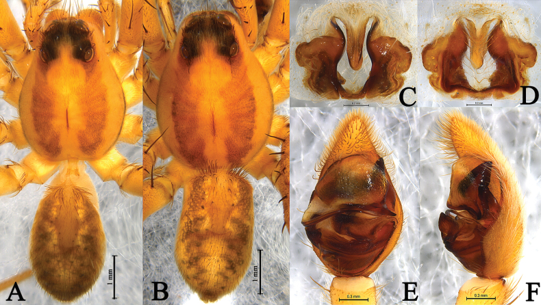 Figure 6
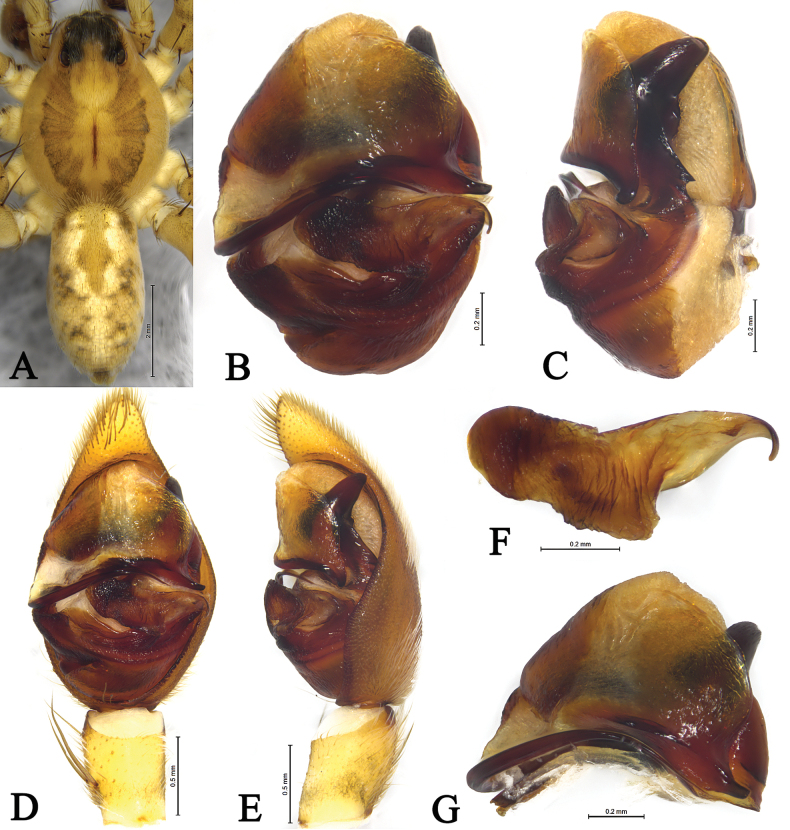 Figure 7
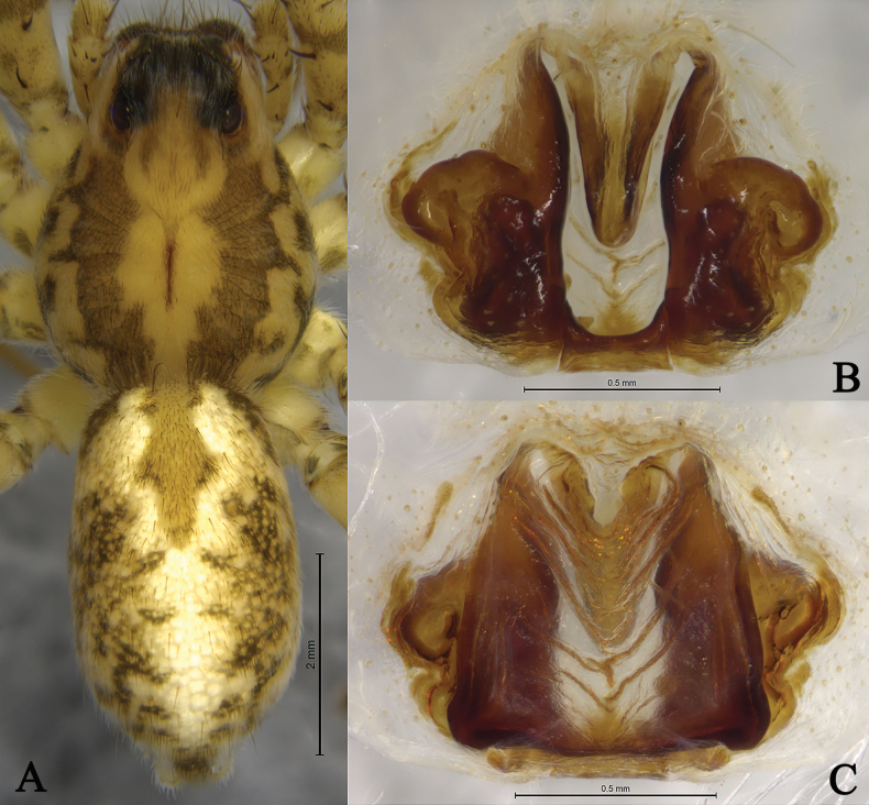 Figure 8
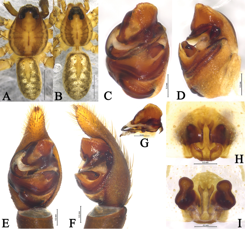 Figure 9
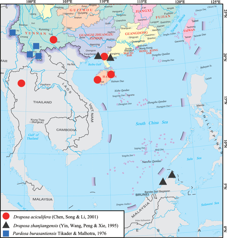 Figure 10
Peer Reviews
No public reviews on file for this paper yet. If you reviewed it on a platform where reviews are public (OpenReview, ICLR, NeurIPS, ICML), you can paste yours below so the community can read it here.
Videos
No videos yet. Explain this paper in a talk, walkthrough, or lecture? Add one.
Taxonomy
TopicsSpider Taxonomy and Behavior Studies · Lepidoptera: Biology and Taxonomy · Hymenoptera taxonomy and phylogeny
Introduction
Draposa Kronestedt, 2010 is a small genus with 12 named species distributed from the United Arab Emirates to the Indomalayan Realm (Alderweireldt and Jocqué 2017; WSC 2025). Draposa has been well studied in Bangladesh, Bhutan, India, Indonesia, Iran, Maldives, Myanmar, Pakistan, Sri Lanka, and the United Arab Emirates (Kronestedt 2010; Marusik and Omelko 2016; Alderweireldt and Jocqué 2017; Abhijith and Sudhikumar 2023). Currently, only two species of the genus have been recorded in China: D.burasantiensis (Tikader & Malhotra, 1976) and D.zhanjiangensis (Yin, Wang, Peng & Xie, 1995) (WSC 2025). Here, we review Draposa from China based on recently collected specimens, as well as the type specimens of D.zhanjiangensis, Pardosaaciculifera Chen, Song & Li, 2001, and P.shugangensis Yin, Bao & Peng, 1997. We propose that Pardosaaciculifera Chen, Song & Li, 2001 should be transferred to Draposa and that Pardosashugangensis Yin, Bao & Peng, 1997 is a junior synonym of Draposazhanjiangensis (Yin, Wang, Peng & Xie, 1995).
Materials and methods
All specimens are preserved in 75% ethanol and were examined, illustrated, photographed, and measured using a Leica M205A stereomicroscope equipped with a drawing tube, a Leica DFC450 Camera, and LAS v. 4.6 software. Male palps and epigynes were examined and illustrated after dissection. Epigynes were cleared by immersing them in a pancreatin solution (Álvarez-Padilla and Hormiga 2007). Eye sizes were measured as the maximum dorsal diameter. Leg measurements are shown as: total length (femur, patella and tibia, metatarsus, tarsus). All measurements are in millimetres. Specimens examined here are deposited in the Collection of Spiders, School of Life Sciences, Southwest University, Chongqing, China (SWUC); the Institute of Zoology, Chinese Academy of Sciences (IZCAS); and the Hunan Normal University (HNU).
Terminology follows Kronestedt (2010) and Zhang et al. (2025). Abbreviations used in the text: ALE, anterior lateral eye; AME, anterior median eye; MOA, median ocular area; PLE, posterior lateral eye; PME, posterior median eye.
Taxonomy
Family Lycosidae Sundevall, 1833 (狼蛛科)
Draposa
Taxon classificationAnimaliaAraneaeLycosidae
Genus
Kronestedt, 2010
F5420D6A-ACF4-54F5-8553-3BB92A76B388
Draposa Kronestedt, 2010: 33.
Type species.
Lycosanicobarica Thorell, 1891.
Diagnosis.
Males of Draposa differ from other Pardosinae by the presence of a subpaleal sclerite with two processes (AP and PP, Figs 2B, 3C, E, 5B, 6F, 7C, E) partly hidden by the palea, as well as by the median apophysis (= tegular apophysis in Kronestedt 2010) being transverse with wide basal part carrying variously shaped projections and narrow distal part carrying small subapical protrusion before evenly curved tip; females differ by the epigynal cavity being only partly divided by a tongue-shaped septum.
Description.
See Kronestedt (2010).
Composition and distribution.
Twelve species are known from the Arabian Peninsula to Indomalayan Region.
Draposa
aciculifera
Taxon classificationAnimaliaAraneaeLycosidae
(Chen, Song & Li, 2001) comb. nov.
8CF97FB7-5E57-577B-8924-526D78CEBF0F
Pardosa aciculifera Chen, Song & Li, 2001: 476, figs 1–7 (♂♀); Wang et al. 2021: 48, fig. 42A–H (♂♀).
Material examined.
China: • 1♂ 2♀, Hainan Prov., Jianfengling, 18.7°N, 108.8°E, 12.07.1990 (holotype and 2 paratypes, IZCAS-Ar-9487, IZCAS-Ar-9488, and IZCAS-Ar-9489) • 1♀, Hainan Prov., Qianghai Co., 20.5.1990 (paratype, IZCAS-Ar-9490) • 2♂ 1♀, Yunnan Prov., Kaiyuan City, 23°40'54"N, 103°20'27"E, elev. 1389 m, 14.06.2017, L.Y. Wang et al. leg. (SWUC, SWUC-LYDA-01–03) • 5♂ 14♀, Guangxi Prov., Beihai City, Hepu Co., Shankou Mangrove Nature Reserve, Dunzai Vill., 21°31'2"N, 109°45'36"E, 13.05.2024, Q.L. Lu leg. (SWUC, SWUC-LYDA-04–22) • Thailand: 6♂ 6♀, Chiang Mai, Amphoe Hot, 18°09'14"N, 98°25'51"E, elev. 782 m, 5.07.2014, Z.S. Zhang et al. leg. (SWUC, SWUC-LYDA-23–34).
Living Draposa spp. A–D. D.aciculifera (Chen, Song & Li, 2001) (A. Male, B–D. Female); E, F. D.zhanjiangensis (Yin, Wang, Peng & Xie, 1995) (female). Photographed by Qian-Le Lu.
Draposaaciculifera (Chen, Song & Li, 2001) male (A, B) and female (C, D). A. Left male palp, ventral view; B. Same, retrolateral view; C. Epigyne, ventral view; D. Same, dorsal view. Abbreviations: A = atrium; AP = anterior subpaleal process; C = conductor; CO = copulatory opening; CP = cymbium protruding; E = embolus; FD = fertilization duct; HS = head of spermathecae; MA = median apophysis; PA = paleal apophysis; PP = posterior subpaleal process; SAP = subapical protrusion on median apophysis; SS = stalk of spermathecae.
Comments.
Kronestedt (2010) has already suggested that this species might belong to Draposa.
Diagnosis.
Draposaaciculifera can be distinguished from all congeners by the acicular subapical protrusion (SAP) of the median apophysis (MA) in the male palp (vs triangular) and the absence of epigynal septum (vs present).
Description.
Male (Fig. 3A) total length 4.14. Carapace 2.41 long, 1.85 wide; opisthosoma 1.89 long, 1.29 wide. Carapace dark brown, with lateral margin with yellow bands, and covered with white setae; small, light, longitudinal stripe around fovea. Cervical groove and radial furrows indistinct. Eye sizes and interdistances: AME 0.11, ALE 0.07, PME 0.32, PLE 0.28; AME–AME 0.10, AME–ALE 0.04, PME–PME 0.37, PME–PLE 0.45. Clypeus height 0.25. Chelicerae brown. Endites and labium brown. Sternum black. Leg measurements: I 8.86 (1.90, 2.41, 1.76, 1.07); II 6.78 (1.74, 2.27, 1.77, 1.00); III 6.81 (1.76, 2.15, 1.94, 0.96); IV 10.02 (2.51, 2.96, 3.14, 1.41). Opisthosoma oval, yellow-brown and covered with numerous black spots dorsally. Heart mark distinct. Venter yellow-brown.
Draposaaciculifera (Chen, Song & Li, 2001) male from Kaiyuan City, Yunnan. A. Male habitus, dorsal view; B. Left bulb, ventral view; C. Same, retrolateral view; D. Left male palp, ventral view; E. Same, retrolateral view; F. Median apophysis, ventral view; G. Terminal part in obliquely retrolateral.
Palp (Figs 2A, B, 3B–G). Tibia yellow-brown, 1.7 times longer than wide. Cymbium yellow-brown, with strong protruding (CP) of retrolateral margin. Median apophysis (MA) moderately long, 2.4 times longer than wide, with lamellar projection in basal half, its subapical protrusion (SAP) acicular, tip of median apophysis semicircle. Paleal apophysis (PA) corniform; anterior subpaleal process narrow (AP), lamellar, with rounded tip, as long as posterior subpaleal process; posterior subpaleal process (PP) wider than long, half sclerotized and half membranous, with serrated edges. Embolus (E) narrow, originating at approximately 9-o’clock position, without membrane, evenly tapering to tip.
Female (Fig. 4A) total length 5.27. Carapace 2.41 long, 1.83 wide; opisthosoma 2.62 long, 1.89 wide. Eye sizes and interdistances: AME 0.10, ALE 0.05, PME 0.32, PLE 0.27; AME–AME 0.11, AME–ALE 0.04, PME–PME 0.37, PME–PLE 0.45. Clypeus height 0.22. Leg measurements: I 6.51 (1.85, 2.23, 1.53, 0.90); II 5.38 (1.48, 1.77, 1.21, 0.92); III 6.21 (1.62, 2.00, 1.68, 0.91); IV 9.69 (2.26, 2.87, 3.15, 1.41).
Draposaaciculifera (Chen, Song & Li, 2001) female from Kaiyuan City, Yunnan, China. A. Female habitus, dorsal view; B. Epigyne, ventral view; C. Same, dorsal view.
Epigyne (Figs 2C, D, 4B, C). Plate ~1.6 times wider than long. Atrium (A) narrow; septum absent; copulatory openings (CO) crack-shaped, located on both side of the atrium. Spermathecal heads (HS) spherical, visible through cuticle of lateral walls of atrium. Spermathecal stalks (SS) arc-shaped. Fertilization ducts (FD) crescent.
Note.
As described above, copulatory organs of this species are completely consistent with this genus except tongue-shaped septum (absent). Therefore, we transfer Pardosaaciculifera to Draposa.
Distribution.
China (Hainan, Guangxi, Yunnan), Thailand (Chiang Mai) (Fig. 10).
Draposa
zhanjiangensis
Taxon classificationAnimaliaAraneaeLycosidae
(Yin, Wang, Peng & Xie, 1995)
572F06BF-1E4F-5DDC-97B3-2C3B96DFD85B
Figs 1E, F , 5 , 6 , 7 , 8 , 10
Pardosa zhanjiangensis
Yin et al., 1995: 74, figs 18–22 (♂♀); Yin et al. 1997b: 281, fig. 133a–e (♂♀); Song et al. 1999: 335, figs 199Q, 200B (♂♀). Pardosa shugangensis Yin, Bao & Peng, 1997a: 24, figs 39–41 (♂); Yin et al. 1997b: 269, fig. 127a–c (♂); Song et al. 1999: 334, fig. 198M (♂). Syn. nov.
Material examined.
China: • 1♂ 2♀ (holotype ♀, paratype ♀ and allotype ♂ of P.zhanjiangensis), Guangdong Prov., Zhanjiang City, 25.06.1985, Y.J. Zhang leg. (HNU) • 2♂ (holotype and paratype of P.shugangensis), Guangxi Prov., Beihai City, Shuangang Dadao, 25.11.1995, C.M. Yin leg. (HNU) • Malaysia: Sabah: 6♂ 6♀, Kota Kinabalu, bank of Kawa Kawa R., 06°25'18"N, 116°24'19"E, elev. 50 m, 13.10.2015, L.Y. Wang et al. leg. (SWUC) • 8♂ 8♀, Pitas, 06°41'15"N, 116°57'44"E, elev. 24 m, 20.10.2015, L.Y. Wang et al. leg. (SWUC).
Diagnosis.
This species is similar to D.nicobarica (Thorell, 1891) (Kronestedt 2010: 44, figs 26, 51–52) and D.tenasserimensis (Thorell, 1895) (Kronestedt 2010: 51, figs 28, 54–55) in having strong posterior subpaleal process (PP), hook-like tip of median apophysis, conspicuous deep atrium and tongue-shaped septum, but it can be distinguished by the lamellar, rounded tip in basal half of median apophysis (MA) (vs acute tooth-like), the atrium widens both anteriorly and posteriorly (vs widens posteriorly).
Description.
Female holotype of P.zhanjiangensis (Fig. 6A) total length 6.69. Carapace 3.34 long, 2.45 wide; opisthosoma 2.66 long, 1.75 wide. Carapace yellow-brown. Cervical groove and radial furrows indistinct. Eye sizes and interdistances: AME 0.15, ALE 0.12, PME 0.40, PLE 0.35; AME–AME 0.10, AME–ALE 0.06, PME–PME 0.29, PME–PLE 0.35. Clypeus height 0.19. Chelicerae yellow-brown. Endites and labium yellowish brown, longer than wide. Sternum yellowish brown, covered with brown setae. Leg measurements: I 9.85 (2.60, 3.57, 2.23, 1.45); II 9.65 (2.70, 3.35, 2.15, 1.45); III 8.76 (2.37, 2.91, 2.22, 1.26); IV 12.63 (3.24, 4.05, 3.75, 1.59). Opisthosoma oval, yellow-brown, and covered with numerous black spots. Heart mark distinct. Venter yellow-brown.
Epigyne (Figs 5C, D, 6C, D, 8B, C). Plate ~1.2 times wider than long. Conspicuous deep atrium (A), atrium widens both anteriorly and posteriorlys; copulatory openings (CO) crack-shaped, as long as atrium. Spermathecal heads (HS) spherical, spermathecal stalks (SS) ~3 times longer than wide, both visible through cuticle of lateral walls of atrium. Fertilization ducts (FD) hook-shaped.
Draposazhanjiangensis (Yin, Wang, Peng & Xie, 1995) male allotype (A, B) and female holotype (C, D). A. Left male palp, ventral view; B. Same, retrolateral view; C. Epigyne, ventral view; D. Same, dorsal view. Abbreviations: A = atrium; AP = anterior subpaleal process; C = conductor; CO = copulatory opening; E = embolus; FD = fertilization duct; HS = head of spermathecae; MA = median apophysis; PA = paleal apophysis; PP = posterior subpaleal process; SAP = subapical protrusion on median apophysis; Se = Septum; SS = stalk of spermathecae.
Male allotype of P.zhanjiangensis (Fig. 6B) total length 6.62. Carapace 3.56 long, 2.68 wide; opisthosoma 2.92 long, 1.69 wide. Eye sizes and interdistances: AME 0.14, ALE 0.11, PME 0.41, PLE 0.34; AME–AME 0.10, AME–ALE 0.05, PME–PME 0.28, PME–PLE 0.36. Clypeus height 0.22. Leg measurements: I 10.81 (2.74, 3.85, 2.61, 1.61); II 10.19 (2.73, 3.47, 2.47, 1.52); III 9.83 (2.58, 3.31, 2.61, 1.33); IV 14.13 (3.40, 4.48, 4.40, 1.85).
Draposazhanjiangensis (Yin, Wang, Peng & Xie, 1995) female holotype (A, C, D) and male allotype (B, E, F). A. Female habitus, dorsal view; B. Male habitus, dorsal view; C. Epigyne, ventral view; D. Vulva, dorsal view. E. Left male palp, ventral view; F. Same, retrolateral view.
Draposazhanjiangensis (Yin, Wang, Peng & Xie, 1995) male from Pitas, Sabah, Malaysia. A. Male habitus, dorsal view; B. Left bulb, ventral view; C. Same, retrolateral view; D. Left male palp, ventral view; E. Same, retrolateral view; F. Median apophysis, ventral view; G. Terminal part in obliquely retrolateral.
Draposazhanjiangensis (Yin, Wang, Peng & Xie, 1995) female from Pitas, Sabah, Malaysia. A. Female habitus, dorsal view; B. Epigyne, ventral view; C. Same, dorsal view.
Palp (Figs 5A, B, 6E, F, 7B–G). Tibia yellow-brown, 1.4 times longer than wide. Cymbium yellow-brown, 1.7 times longer than wide. Paleal apophysis triangular (PA). Anterior subpaleal process (AP) strong and rodlike, with a rounded end. Posterior subpaleal process (PP) lamellar, with a notch. Median apophysis (MA) moderately long, with lamellar projection in basal half, subapical protrusion triangular (SAP), tip of median apophysis hook-like. Embolus (E) narrow, evenly tapering to tip.
Distribution.
China (Guangxi and Guangdong), Malaysia (Sabah) (Fig. 10). Records from Malaysia (Tioman Island, Sarawak) and Indonesia (Kronestedt 2010) are doubtful and require confirmation.
Pardosa
burasantiensis
Taxon classificationAnimaliaAraneaeLycosidae
Tikader & Malhotra, 1976 sensu Yin et al. (1997b)
711957A2-D0AE-53F7-82F1-F0B8BF8518C1
Pardosa burasantiensis Tikader & Malhotra, 1976: 130, figs 10–12 (♂♀); Yin et al. 1997b: 239, fig. 112a–g (♂♀, misidentified per Kronestedt 2010: 34); Song et al 1999: 330, figs 194C (♀, misidentified per Kronestedt 2010: 34); Yin et al. 2012: 833, fig. 416a–g (♂♀). Draposa burasantiensis : Dhali et al., 2012: 1202. For complete list of references see WSC (2025).
Comments.
This species was originally described based on a female holotype and male paratypes collected from Dehradun, Uttar Pradesh, India. Kronestedt (2010) proposed a potential taxonomic reclassification, suggesting that this species might belong to Draposa, based on the presence of a subpaleal sclerite in the illustrated male specimen. However, it should be noted that the figure of the paratype male is rather schematic. Morphological examination of the female holotype indicates characteristics consistent with the nebulosa species group within Pardosa C.L. Koch, 1847. Dhali et al. (2012) transferred two Pardosa species (P.amkhasensis Tikader & Malhotra, 1976 and P.burasantiensis Tikader & Malhotra, 1976) to Draposa, referring to Kronestedt (2010) as justification. However, their work lacked substantial morphological evidence to support this reclassification, providing neither detailed descriptions nor clear images of diagnostic characteristics. Study of the paratype female of this species by Dr Souvik Sen from the Zoological Survey of India (Kronestedt pers. comm.) reveals that it belongs to Pardosa and male paratype is not conspecific with female. Pardosaburasantiensis was first reported from China by Yin et al. (1997b) and figures provided in their publication refer to Pardosa and most likely not conspecific with the holotype of P.burasantiensis. Therefore, species reported from China does not belong to Draposa. In addition, we hereby reinstate the original taxonomic classification for P.burasantiensis Tikader & Malhotra, 1976.
Pardosaburasantiensis sensu Yin et al. (1997b) from Puer City, Yunnan, China. A. Male habitus, dorsal view; B. Female habitus, dorsal view; C. Left male palp, bulb, ventral view; D. Same, retrolateral view; E. Left male palp, ventral view; F. Same, retrolateral view; G. Terminal part in obliquely retrolateral; H. Epigyne, ventral view; I. Same, dorsal view.
Distribution records of Draposaaciculifera (Chen, Song & Li, 2001) and Draposazhanjiangensis (Yin, Wang, Peng & Xie, 1995) and Chinese records of Pardosaburasantiensis Tikader & Malhotra, 1976.
Supplementary Material
XML Treatment for Draposa
XML Treatment for Draposa aciculifera
XML Treatment for Draposa zhanjiangensis
XML Treatment for Pardosa burasantiensis
The reference list from the paper itself. Each links out to its DOI / PubMed record.
- 1Abhijith RS Sudhikumar AV (2023) Description of a new wolf spider species (Arachnida: Araneae: Lycosidae: Draposa) from Western Ghats, India.Taprobanica 12(1): 1–4. [pls 1–3.] 10.47605/tapro.v 12i 1.294 · doi ↗
- 2Alderweireldt M JocquéR (2017) Order Araneae, family Lycosidae.Arthropod Fauna of the UAE 6: 3–20.
- 3Álvarez-Padilla F Hormiga G (2007) A protocol for digesting internal soft tissues and mounting spiders for scanning electron microscopy.The Journal of Arachnology 35(3): 538–542. 10.1636/Sh 06-55.1 · doi ↗
- 4Dhali DC Roy TK Sen S Saha S Raychaudhuri D (2012) Wolf spiders (Araneae: Lycosidae) of the reserve forests of Dooars, West Bengal, India.Munis Entomology and Zoology 7(2): 1199–1213.
- 5Kronestedt T (2010) Draposa, a new wolf spider genus from South and Southeast Asia (Araneae: Lycosidae).Zootaxa 2637(1): 31–54. 10.11646/zootaxa.2637.1.2 · doi ↗
- 6Marusik YM Omelko MM (2016) Redescription of Draposasubhadrae (Patel & Reddy, 1993) (Araneae: Lycosidae, Pardosinae).Zootaxa 4107(1): 89–92. 10.11646/zootaxa.4107.1.627394808 · doi ↗ · pubmed ↗
- 7Song DX Zhu MS Chen J (1999) The Spiders of China.Hebei Science and Technology Publishing House, Shijiazhuang, 640 pp.
- 8Tikader BK Malhotra MS (1976) Studies on some spiders of the genus Pardosa Koch from India (family: Lycosidae).Proceedings of the Indian Academy of Sciences, Section B, Biological Sciences 83(3): 123–131. 10.1007/BF 03045698 · doi ↗