Microstructural alterations of the trigeminal ganglion in chronic ocular surface pain patients: A diffusion MRI study
Alpen Ortug, Nicholas Reyes, Anat Galor, David Valdes-Arias, Ema Karakoleva, Cameron Talbert, Nicholas J. Pondelis, Pradip Pattany, Elizabeth Felix, Scott Holmes, David Zurakowski, Barry Sessle, Emi Takahashi, Eric A. Moulton

TL;DR
This study uses MRI to find changes in the trigeminal ganglion of people with chronic eye surface pain, suggesting possible nerve damage.
Contribution
The study demonstrates the feasibility of using diffusion tractography to assess microstructural alterations in the trigeminal ganglion in chronic ocular surface pain.
Findings
Significant decreases in NQA, MD, and RD were observed in the left trigeminal ganglion of COSP patients.
These changes suggest axonal loss and fiber damage in the left TG of COSP patients.
No significant differences were found in the right TG between COSP patients and controls.
Abstract
Noxious stimuli to the ocular surface are encoded by sensory axons of trigeminal ganglion (TG) neurons and conveyed through the ophthalmic branch of the trigeminal nerve (CN V1). We hypothesized that chronic ocular surface pain (COSP) may be associated with microstructural alterations of the trigeminal nerve structures. Our objective was to demonstrate the feasibility of using diffusion tractography to identify and analyze diffusion properties to assess TG microstructure in individuals with and without COSP. Forty COSP patients (27 males and 13 females; mean age: 56.2 ± 11.9 yrs; range: 34–77 yrs) and 17 controls without pain (15 males and 2 females; mean age: 55.4 ± 8.9 yrs, range: 37–66 yrs) were included in the study. Using 3T diffusion MRI (dMRI), we performed tractography to reconstruct TG and CN V1 with a generalized q-sampling imaging (GQI) algorithm. dMRI-based indices such as…
Genes, proteins, chemicals, diseases, species, mutations and cell lines named across the full text — each resolved to its canonical identifier and authoritative record.
Click any figure to enlarge with its caption.
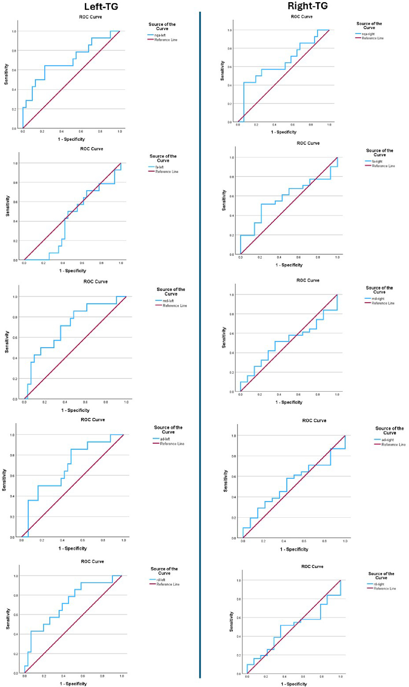 Figure 1
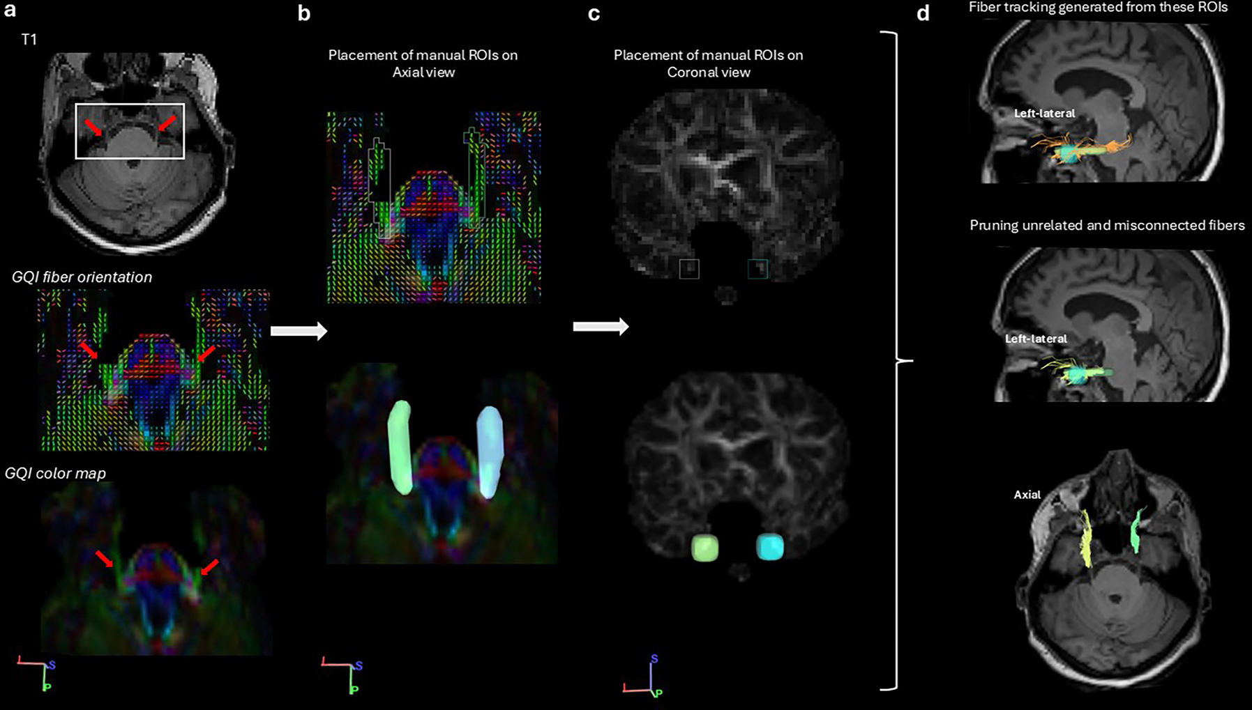 Figure 2
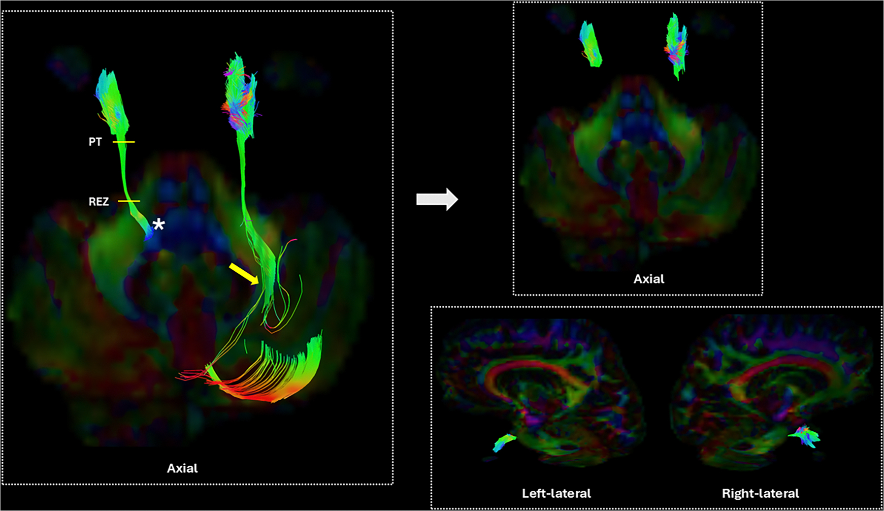 Figure 3
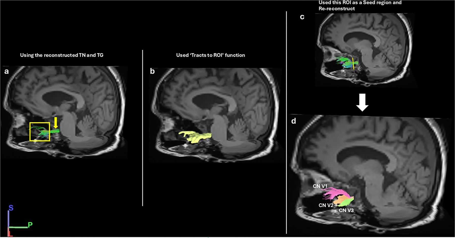 Figure 4
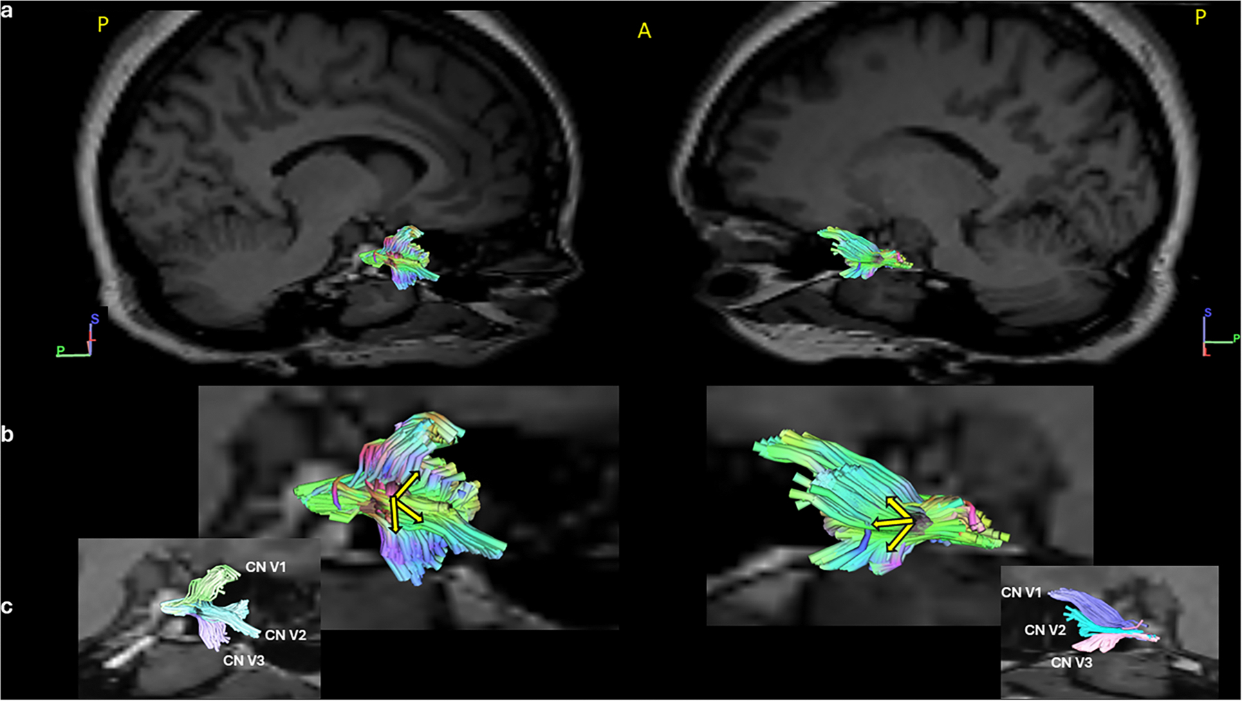 Figure 5
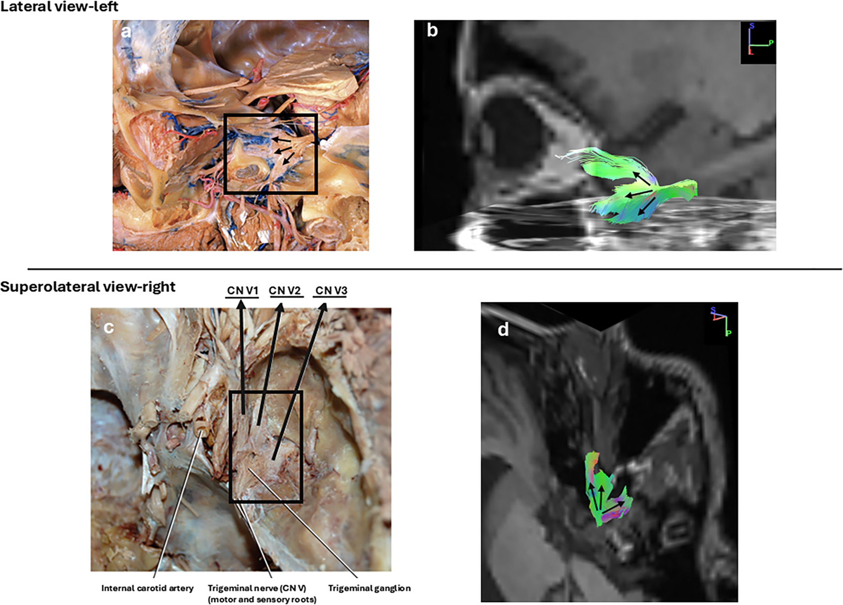 Figure 6
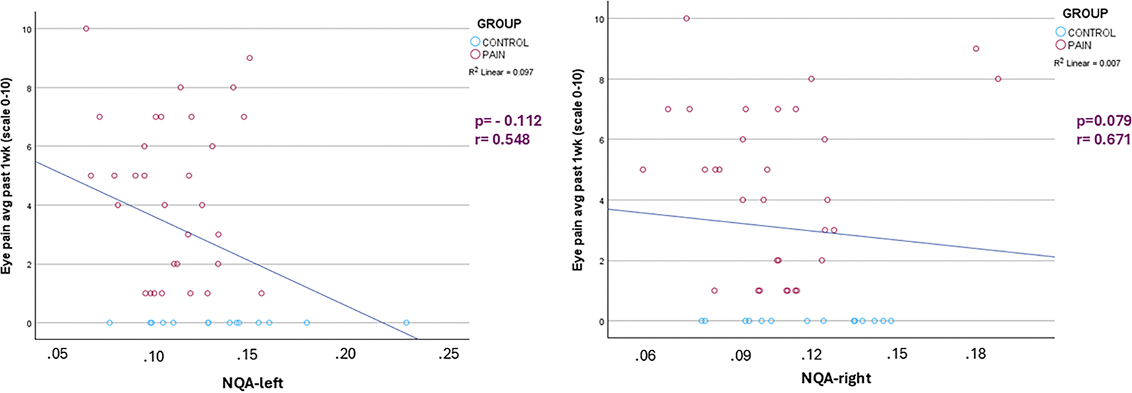 Figure 7
Peer Reviews
No public reviews on file for this paper yet. If you reviewed it on a platform where reviews are public (OpenReview, ICLR, NeurIPS, ICML), you can paste yours below so the community can read it here.
Videos
No videos yet. Explain this paper in a talk, walkthrough, or lecture? Add one.
Taxonomy
TopicsOphthalmology and Eye Disorders · Advanced Neuroimaging Techniques and Applications · Glaucoma and retinal disorders