Microsurgical Reconstruction of a Cranial Defect Using a Latissimus Dorsi Flap: A Case Report
Arym P Preza Estrada, Alexis A Granados Flores, José L Villarreal-Salgado, Damaris E Navarro Nuño, Cesar Oropeza Duarte

TL;DR
A 64-year-old woman with a complex cranial defect in the temporal region successfully underwent microsurgical reconstruction using a latissimus dorsi flap, achieving good functional and aesthetic results.
Contribution
This case report demonstrates the effectiveness of the free latissimus dorsi flap for reconstructing complex cranial defects in the temporal region.
Findings
The latissimus dorsi flap provided reliable coverage and restored cranial contour in a complex temporal region defect.
Microsurgical techniques enabled precise vascular anastomosis, promoting flap integration and reducing complications.
A multidisciplinary approach and thorough preoperative planning were key to successful reconstruction.
Abstract
The reconstruction of complex cranial defects, particularly in the temporal region, poses a significant surgical challenge due to both functional and aesthetic considerations, as well as the anatomical constraints of the area. We present the case of a 64-year-old female patient with an extensive cranial defect in the temporal region, secondary to a previous surgical resection. Owing to the complexity of the defect and the presence of bone exposure, a microsurgical reconstruction using a free latissimus dorsi (LD) flap was undertaken. The procedure involved harvesting the flap in the lateral decubitus position, ensuring a long, well-calibered vascular pedicle, followed by free tissue transfer and microvascular anastomosis to the superficial temporal vessels. The LD flap was chosen for its volume, versatility, and reliable vascular supply, which enabled effective coverage and restoration…
Genes, proteins, chemicals, diseases, species, mutations and cell lines named across the full text — each resolved to its canonical identifier and authoritative record.
Click any figure to enlarge with its caption.
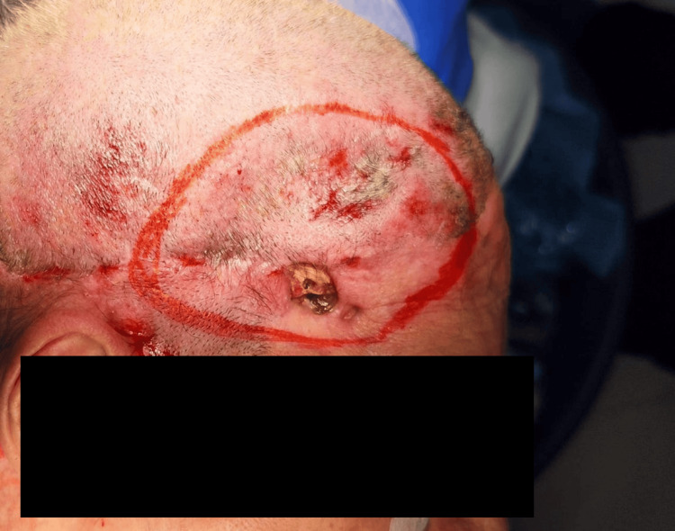 Figure 1
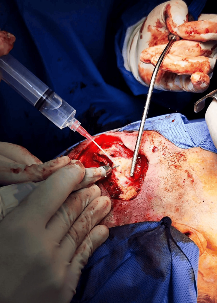 Figure 2
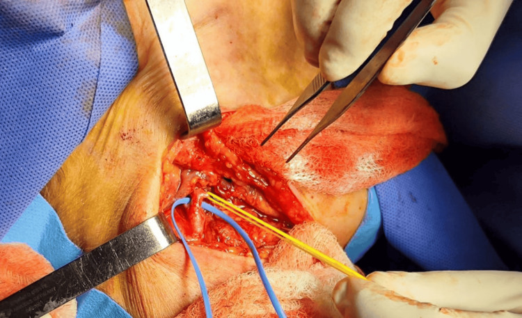 Figure 3
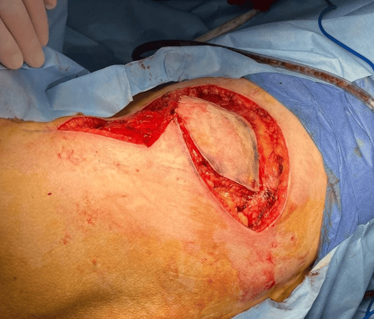 Figure 4
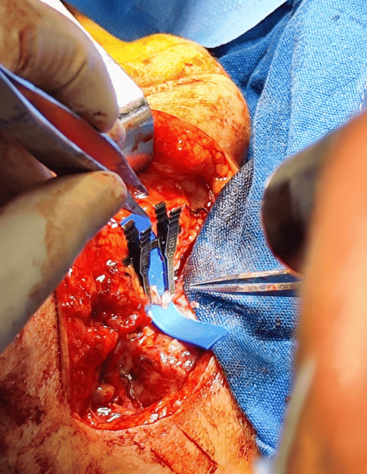 Figure 5
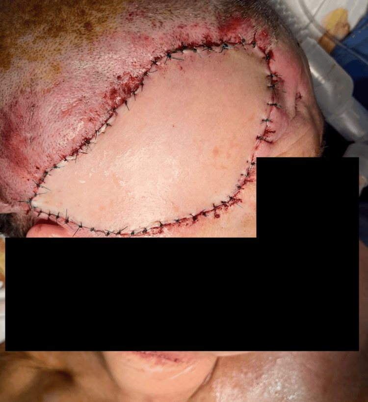 Figure 6
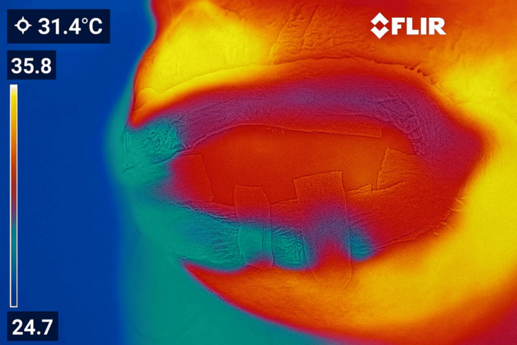 Figure 7
Peer Reviews
No public reviews on file for this paper yet. If you reviewed it on a platform where reviews are public (OpenReview, ICLR, NeurIPS, ICML), you can paste yours below so the community can read it here.
Videos
No videos yet. Explain this paper in a talk, walkthrough, or lecture? Add one.
Taxonomy
TopicsReconstructive Surgery and Microvascular Techniques · Head and Neck Surgical Oncology · Craniofacial Disorders and Treatments