Generating 2.5D pathology for enhanced viewing and AI diagnosis
Ekaterina Redekop, Mara Pleasure, Zichen Wang, Anthony Sisk, Yang Zong, Kimberly Flores, William Speier, Corey W. Arnold

TL;DR
This paper introduces a new method to create 2.5D pathology images from biopsy samples, improving both AI diagnosis and pathologist visualization.
Contribution
A novel morphology-preserving alignment framework to generate 2.5D biopsy cores for enhanced AI and pathologist use.
Findings
The framework was applied to over 12,000 biopsy samples across prostate, breast, and renal tissues.
A deep learning model trained on 2.5D cores showed improved cancer grading performance.
Pathologists confirmed the 2.5D cores improved their ability to evaluate tissue structures.
Abstract
Histological analysis of biopsy samples by pathologists can require the evaluation of complex three-dimensional (3D) tissue structures. This process involves studying the same tissue region across slides, which requires laborious zooming and panning for localization. Additionally, standard deep learning frameworks typically focus on cross-sections cut from biopsy specimens, limiting their ability to capture 3D tissue spatial information. We present a novel framework that constructs 2.5D biopsy cores via the extraction and co-alignment of serial tissue sections using a novel morphology-preserving alignment framework. These 2.5D cores can then be used for enhanced viewing by pathologists and as input to video transformer models that can capture depth-wide spatial dependencies. We used our framework to construct 2.5D cores for 10,210 prostate biopsies, 156 breast biopsies, and 1869 renal…
Genes, proteins, chemicals, diseases, species, mutations and cell lines named across the full text — each resolved to its canonical identifier and authoritative record.
Click any figure to enlarge with its caption.
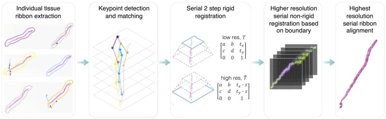 Figure 1
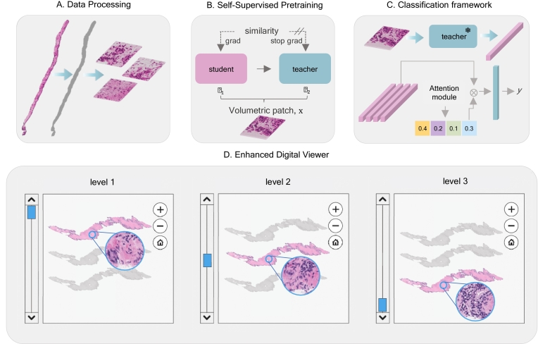 Figure 2
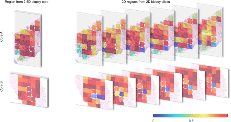 Figure 3
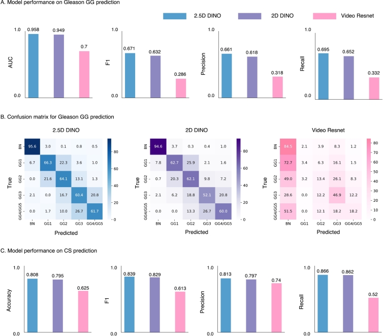 Figure 4
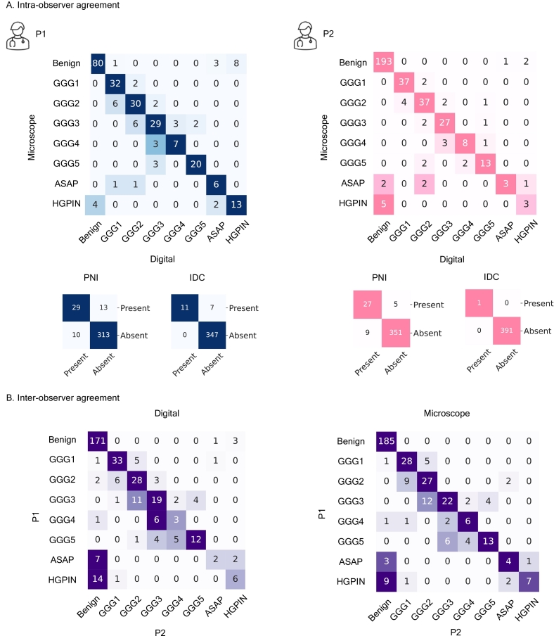 Figure 5
Peer Reviews
No public reviews on file for this paper yet. If you reviewed it on a platform where reviews are public (OpenReview, ICLR, NeurIPS, ICML), you can paste yours below so the community can read it here.
Videos
No videos yet. Explain this paper in a talk, walkthrough, or lecture? Add one.
Taxonomy
TopicsAI in cancer detection · Cell Image Analysis Techniques · Retinal Imaging and Analysis