Quantitative Evaluation of the Invasion Depth of Colorectal Cancer Located on a Colorectal Fold Through the Width of Colorectal-Fold Lateral Contour Using a Lateral Split-View Computed Tomographic Air-Contrast Enema Image
Mitsutoshi Miyasaka, Toshio Muraki, Yusuke Nishimuta, Eiji Oki, Kousei Ishigami, Daisuke Tsurumaru

TL;DR
This study shows that measuring the width of a specific contour in CT images can help determine how deep colorectal cancer has invaded when it's located on a fold in the colon.
Contribution
The first demonstration that quantifying the lateral contour width in CT enema images improves depth-of-invasion diagnosis for fold-located colorectal cancer.
Findings
T1b/more deeply invading CRCs had significantly larger lateral contour widths (12.1 mm) than intramucosal/T1a CRCs (3.3 mm).
A 6 mm cut-off value achieved 92.9% sensitivity and 87.5% specificity for differentiating invasion depths.
High inter-rater reliability (intraclass correlation coefficient of 0.949) was observed for the measurements.
Abstract
Purpose: The aim of the study was to investigate the usefulness of quantitatively evaluating the width of lateral contour on a lateral split-view computed tomographic air-contrast enema (CT enema) image to diagnose the invasion depth of colorectal cancer (CRC) located on a colorectal fold. Methods: The cases of 22 patients with 22 fold-located CRCs, that is, 12 (54.5%) early CRCs and 10 (45.5%) advanced CRCs, who underwent a pretherapeutic CT colonography, were retrospectively examined. T1-stage CRCs were classified into two categories according to the Japanese guideline: T1a-stage (carcinoma invading the superficial submucosa (<1000 μm)) and T1b-stage (carcinoma invading the deeper submucosa (≧1000 μm)). The maximum width of colorectal-fold lateral contour on which the CRC was located, i.e., the gap distance between the two adjacent haustrations, was calculated from the lateral…
Genes, proteins, chemicals, diseases, species, mutations and cell lines named across the full text — each resolved to its canonical identifier and authoritative record.
Click any figure to enlarge with its caption.
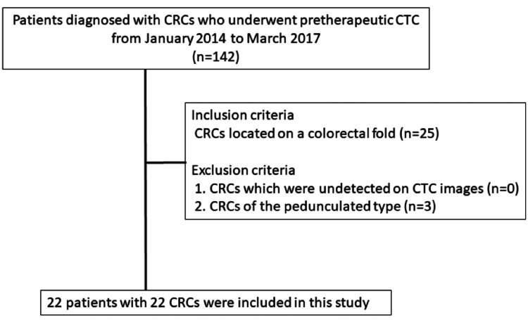 Figure 1
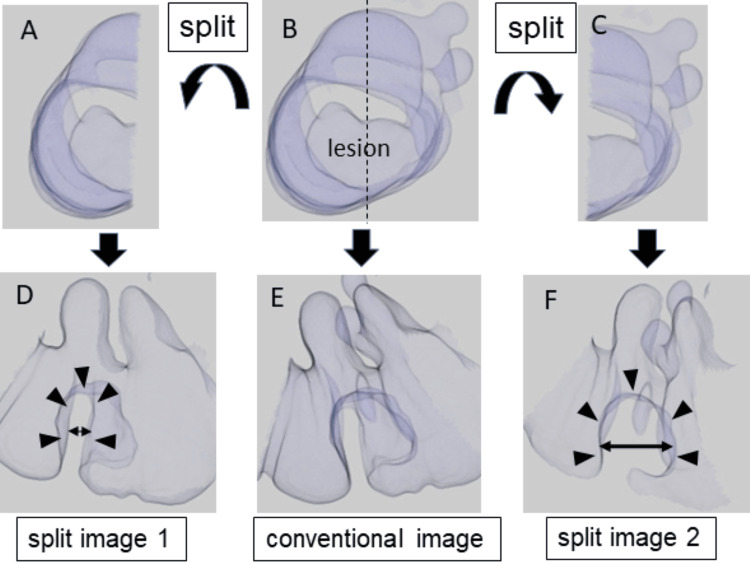 Figure 2
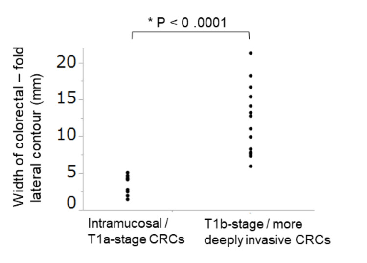 Figure 3
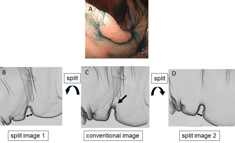 Figure 4
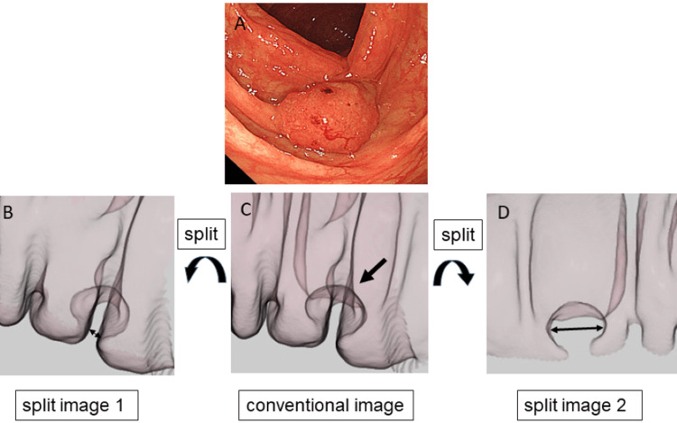 Figure 5
Peer Reviews
No public reviews on file for this paper yet. If you reviewed it on a platform where reviews are public (OpenReview, ICLR, NeurIPS, ICML), you can paste yours below so the community can read it here.
Videos
No videos yet. Explain this paper in a talk, walkthrough, or lecture? Add one.
Taxonomy
TopicsColorectal Cancer Surgical Treatments · Radiomics and Machine Learning in Medical Imaging · Colorectal Cancer Screening and Detection