The diagnostic value of confocal laser endomicroscopy in a cancer-mimicking lesion
Yujie Shi, Qin Zeng, Yuxuan Chen, Mi Zhou, Shuo Zhang

Abstract
Genes, proteins, chemicals, diseases, species, mutations and cell lines named across the full text — each resolved to its canonical identifier and authoritative record.
Click any figure to enlarge with its caption.
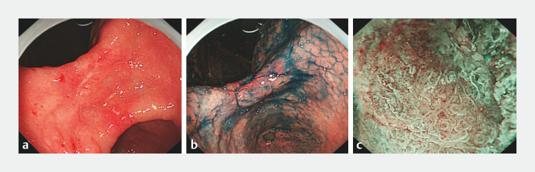 Fig. 1
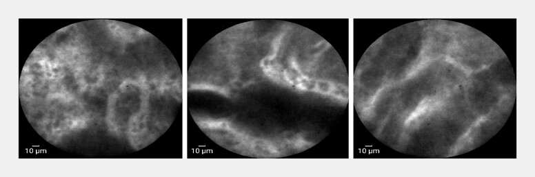 Fig. 2
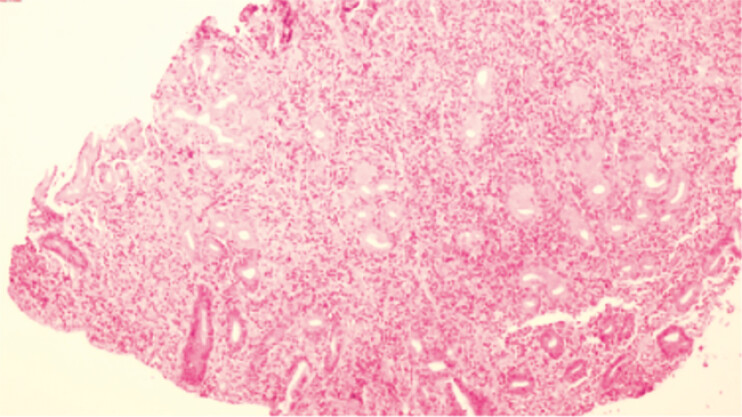 Fig. 3
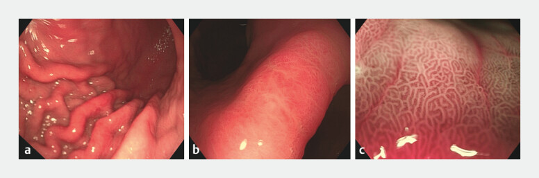 Fig. 4
- —Zhejiang Province Traditional Chinese Medicine Science and Technology Project Youth Talent Program
- —Research Fund of National Health Commission
- —The Chinese Medicine Clinical Research Program
- —The Fourth Batch of Zhejiang Province “Ten Thousand People Plan” Scientific and Technological Innovation Leaders
Peer Reviews
No public reviews on file for this paper yet. If you reviewed it on a platform where reviews are public (OpenReview, ICLR, NeurIPS, ICML), you can paste yours below so the community can read it here.
Videos
No videos yet. Explain this paper in a talk, walkthrough, or lecture? Add one.
Taxonomy
TopicsEsophageal Cancer Research and Treatment · Gastric Cancer Management and Outcomes · Metastasis and carcinoma case studies
A 25-year-old woman presented with epigastric pain for two months. Laboratory tests revealed mild anemia (hemoglobin: 114 g/L) and elevated C-reactive protein (8.9 mg/L). Treponema pallidum antibody and rapid plasma reagin (RPR) tests were positive (titer: 1:32), while tumor markers, fecal occult blood test, hepatitis B surface antigen, and HIV screening were all negative. Abdominal computed tomography showed only a left renal cyst. One month earlier, the patient had undergone gastroscopy at a local hospital, which revealed a large irregular ulcer at the gastric angle. Biopsy suggested chronic atrophic gastritis with erosion. Although immunohistochemistry excluded lymphoma, the lesion appeared highly suspicious, and the patient was referred to our center for further evaluation.
Repeat gastroscopy confirmed an irregular ulcer at the gastric angle, surrounded by nodular elevated mucosa ( Fig. 1 a ). After staining the lesion with acetic acid and indigo carmine ( Fig. 1 b ), magnifying narrow-band imaging (M-NBI) was performed, which revealed disrupted glandular architecture and irregular microvasculature ( Fig. 1 c ). Due to the highly suspicious cancer-like appearance, we proceeded with confocal laser endomicroscopy (CLE), which showed overall preserved glandular structures with focal mild irregularities and polygonal epithelial changes, dark-stained goblet cells between epithelial cells in some glands, and extensive fluorescein leakage along with abundant microvasculature in the stroma ( Fig. 2 , Video 1 ). Based on CLE imaging, inflammatory changes with intestinal metaplasia and focal atypia were suspected. Subsequent histopathology confirmed mild to moderate chronic active gastritis with prominent plasma cell infiltration, without evidence of malignancy ( Fig. 3 ). CLE findings and serological results were combined, and a final diagnosis of gastric syphilis was established. The patient received standard benzathine penicillin therapy for three weeks with symptom relief. Follow-up endoscopy one month later showed ulcer healing ( Fig. 4 a, b ), and M-NBI demonstrated normalization of glandular and vascular patterns ( Fig. 4 c ).
a Irregular ulcer at the gastric angle under white-light endoscopy. b Acetic acid-indigo carmine staining of the lesion. c Magnifying narrow-band imaging (M-NBI) shows disrupted glandular architecture and irregular microvasculature around the ulcer.
Confocal laser endomicroscopy reveals overall preserved glandular structures with mild focal irregularities. Dark-stained goblet cells visible between epithelial cells; extensive fluorescein leakage and abundant microvasculature in the stroma.
Hematoxylin and eosin staining (×40) shows marked plasma cell infiltration in the lamina propria and mild to moderate active inflammation; no cytological atypia observed.
a, b White-light endoscopic images of the gastric body and angle after treatment. c M-NBI shows normalized glandular and vascular architecture at the gastric angle.
Confocal laser endomicroscopy of the gastric lesion showing preserved glandular architecture, dark goblet cells, and extensive fluorescein leakage in the stroma.Video 1
This case highlights the utility of CLE in accurately distinguishing benign lesions from cancer-mimicking findings when conventional endoscopy results are suspicious but histology is inconclusive. Approximately 17% of gastric syphilis cases have reportedly undergone unnecessary surgery due to misdiagnosis 1 . CLE enables real-time visualization of cellular and microvascular structures, providing critical information for differentiating ulcer etiology and malignancy 2 . With its “optical biopsy” capability, CLE can reduce reliance on conventional tissue biopsies and significantly improve diagnostic efficiency and accuracy, showcasing its promising potential in precision endoscopic diagnostics.
Endoscopy_UCTN_Code_CCL_1AB_2AC_3AB
The reference list from the paper itself. Each links out to its DOI / PubMed record.
- 1Mylona EE Baraboutis IG Papastamopoulos V Gastric syphilis: A systematic review of published cases of the last 50 years Sex Transm Dis 20103717718310.1097/OLQ.0b 013e 3181 c 0d 51f 20023597 · doi ↗ · pubmed ↗
- 2Han W Kong R Wang N Confocal laser endomicroscopy for detection of early upper gastrointestinal cancer Cancers (Basel)20231577610.3390/cancers 1503077636765734 PMC 9913498 · doi ↗ · pubmed ↗