Anti-Helicobacter pylori and Anti-Inflammatory Sesquiterpenoids from the Rhizoma of Atractylodes macrocephala
So Yeong Jeong, Dong-Min Kang, Hyun-Jun Kim, Sang Won Yeon, Hak Hyun Lee, Min Hee Kim, Bang Yeon Hwang, Mi-Jeong Ahn, Mi Kyeong Lee

TL;DR
This study identifies compounds in Atractylodes macrocephala that effectively inhibit Helicobacter pylori and reduce inflammation, offering potential for treating related infections.
Contribution
The first report on anti-H. pylori efficacy of A. macrocephala constituents and their strain-specific inhibitory effects.
Findings
Atractylenolide I showed strong H. pylori inhibition comparable to metronidazole.
Atractylenolide III exhibited selective activity against strain 51 with moderate efficacy.
Both compounds demonstrated anti-inflammatory activity but had little effect on urease.
Abstract
Helicobacter pylori, a spiral-shaped bacterium found in the stomach, is associated with various gastrointestinal and systemic health conditions. Effective suppression of H. pylori is therefore critical for managing gastrointestinal diseases. In a search for natural products with anti-H. pylori activity, the extract of Atractylodes macrocephala rhizoma showed significant inhibitory effects. Chromatographic purification of A. macrocephala extract yielded thirteen compounds, which were identified as ten sesquiterpenes and three polyacetylenes by spectroscopic analysis. The sesquiterpene compounds belong to the eudesmane or eudesmane lactone types and exhibited structure-dependent efficacy. The major eudesmane lactone sesquiterpene, atractylenolide I (1), showed strong inhibitory activity comparable to metronidazole, a positive control, and atractylenolide III (3) also showed good efficacy.…
Genes, proteins, chemicals, diseases, species, mutations and cell lines named across the full text — each resolved to its canonical identifier and authoritative record.
Click any figure to enlarge with its caption.
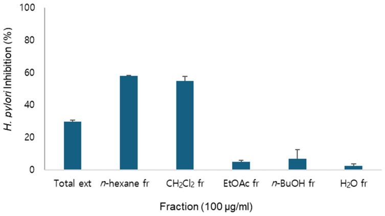 Figure 1
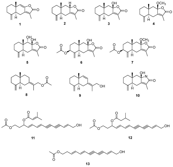 Figure 2
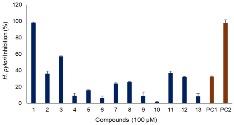 Figure 3
- —National Institute of Forest Science, Korea
- —National Research Foundation of Korea (NFR)
Peer Reviews
No public reviews on file for this paper yet. If you reviewed it on a platform where reviews are public (OpenReview, ICLR, NeurIPS, ICML), you can paste yours below so the community can read it here.
Videos
No videos yet. Explain this paper in a talk, walkthrough, or lecture? Add one.
Taxonomy
TopicsHelicobacter pylori-related gastroenterology studies · Phytochemical Studies and Bioactivities · Phytochemistry and Bioactive Compounds
1. Introduction
Gastric diseases, ranging from mild indigestion to stomach cancer, are prevalent worldwide and significantly impact health and quality of life. Helicobacter pylori, a spiral-shaped bacterium residing in the stomach, is a critical factor in the development and progression of various gastric diseases, including chronic gastritis, peptic ulcers, and stomach cancer [1,2,3]. Helicobacter pylori is uniquely adapted to survive in the harsh, acidic environment of the stomach. It colonizes the stomach’s mucus layer using its flagella and neutralizes gastric acid through the production of urease. These adaptations enable the bacterium to persist in the stomach, causing damage to the gastric mucosa and triggering inflammatory responses [4,5,6,7,8]. Therapies targeting H. pylori focus on disrupting these survival mechanisms [9,10,11]. Eradication of the bacterium remains the cornerstone of treatment, supplemented by anti-inflammatory and gastric acid-suppressive therapies. Antibiotics are the primary therapy for eradication, but antibiotic resistance poses significant challenges [12,13]. Consequently, there is an urgent need to develop new substances with anti-H. pylori efficacy to overcome antibiotic resistance.
Natural products, rich in diverse metabolites with unique structures, are considered valuable resources for drug discovery [14,15]. For the treatment of H. pylori infection, natural products hold promise for overcoming antibiotic resistance and providing synergistic effects, including anti-inflammatory benefits, through diverse mechanisms [16,17]. Efforts are also underway to enhance the effectiveness through combination of natural products with antibiotics.
Atractylodes macrocephala, a perennial herbaceous plant belonging to the Asteraceae family, is native to Korea, China, and Japan. It has traditionally been used as a tonic to strengthen the immune system and boost energy, as well as to treat gastrointestinal disorders such as indigestion, diarrhea, and loss of appetite [18]. A wide range of phytochemicals have been identified, including sesquiterpenoids, triterpenoids, polyacetylenes, coumarins, phenylpropanoids, flavonoids and polysaccharides [17]. Among the identified constituents, sesquiterpenes, polyacetylenes, and polysaccharides have been recognized as major components, and have been associated with a range of biological activities such as anti-inflammatory, anticancer, and antibacterial effects [19,20]. While Atractylodes macrocephala has been traditionally utilized for gastrointestinal ailments, its anti-H. pylori potential and the characterization of its active compounds have not been thoroughly investigated.
In the screening of natural products for anti-H. pylori activity, the extract of A. macrocephala rhizoma demonstrated inhibitory activity on the growth of H. pylori and fractionation revealed the increased inhibitory activity on n-hexane and CH_2_Cl_2_ fractions (Figure 1). Therefore, this study aimed to identify the active constituents of A. macrocephala and evaluate their antibacterial activity against H. pylori. Additionally, the anti-inflammatory properties and inhibitory effects on urease were assessed to determine its potential for treating gastric disorders.
2. Results and Discussions
2.1. Anti-H.pylori Activity of A. macrocephala Extract and Fractions
As the total extract of A. macrocephala exhibited anti-H. pylori activity, identification of its active constituents was attempted. Fractionation of the total extract yielded n-hexane, CH_2_Cl_2_, EtOAc, n-BuOH and H_2_O fractions.
Among these, the n-hexane and CH_2_Cl_2_ fractions exhibited anti-H. pylori activity (Figure 1) and exhibited similar HPLC chromatograms. Therefore, they were combined for further purification.
2.2. Isolation of Compounds from A. macrocephala
Chromatographic separation led to the isolation of thirteen compounds. The structures of these isolated compounds were identified as atractylenolide I (1), atractylenolide II (2), atractylenolide III (3), 8β-methoxyatractylenolide (4), 8β,9α-dihydroxyeudesman4(15),7(11)-dien-8,12-olide (5), atractylenolide IV (6), 9α-methoxy-3,8-dimethyl-5-methylene-2-oxo-2,4,4α,5,6,7,8,8α,9,9α-decahydronaphtho [2,3-β]furan-6-yl acetate (7), atractylenolide VII (8), atractylmacrols D (9), taenialactam B (10), 14-acetoxy-12-senecioyloxytetradeca-2E,8E,10E-trien-4,6-diyn-1-ol (11), 14-acetoxy-12-methylbutyl-2E,8E,10E-trien-4,6-diyn-1-ol (12) and 4,6,12-tetradecatriene-8,10-diyn-1-ol,1-acetate (13) by the comparison with previously published data (Figure 2) [21,22,23,24,25,26,27]. These compounds are ten sesquiterpenes (1–10) and three polyacetylenes (11–13), which have been reported previously from Atractylodes species.
2.3. Anti-H. pylori Activity of Compounds of A. macrocephala Rhizoma
2.3.1. H. pylori Inhibitory Activity
The anti-H. pylori activity of the isolated compounds was evaluated (Figure 3). Among the compounds derived from A. macrocephala, atractylenolide I (1) exhibited the best efficacy, with an inhibitory effect of 98% at a concentration of 100 μg/mL, similar to that of metronidazole, a positive control used as an antibiotic in clinical field. Atractylenolide III (3) also demonstrated an inhibitory efficacy of 57% at the same concentration. However, other sesquiterpenes exhibited weaker efficacy, with inhibition rates of less than 30%. Additionally, the polyacetylenes, 14-acetoxy-12-senecioyloxytetradeca-2E,8E,10E-trien-4,6-diyn-1-ol (11) and 14-acetoxy-12-methylbutyl-2E,8E,10E-trien-4,6-diyn-1-ol (12), showed anti-H. pylori activities of 37% and 32%, respectively, which were comparable to the activity of quercetin, a natural compound used as a positive control (Figure 3).
2.3.2. Structure Activity Relationship for H. pylori Inhibitory Activity
The compounds isolated from A. macrocephala in this study can be categorized into sesquiterpenes (1–10) and polyacetylenes (11–13). The anti-H. pylori activity exhibited by these compounds was highly dependent on their structural features. All the sesquiterpenes isolated in this study share eudesmane skeleton, which can be further divided into eudesmanes (8–10) and eudesmane lactones (1–7) based on the presence of lactone moiety in the structure. Each sesquiterpene differs in the substituents, such as double bonds, hydroxyl (OH), methoxyl (OCH_3_), and acetyl (CH_3_CO) groups, as well as their positions, which significantly affected their efficacy. Compounds 1–7 belong to eudesmane lactone category. Among them, atractylenolide I (1), which possesses an additional double bond, showed the best efficacy, whereas atractylenolide II (2), with the same structure as atractylenolide I (1) but lacking a double bond, exhibited relatively mild efficacy. Atractylenolide III (3), which showed excellent efficacy, has hydroxyl moiety at C-8. The efficacy decreased when there was no hydroxyl moiety, as in atractylenolide II (2), or when the hydroxyl moiety was substituted with methoxyl, as in 8β-methoxyatractylenolide (4). The addition of hydroxyl or acetyl moieties, as seen in 8β,9α-dihydroxyeudesman4(15),7(11)-dien-8,12-olide (5) and atractylenolide IV (6), reduced efficacy. Moreover, replacement of oxygen of lactone with nitrogen also reduced efficacy. Additionally, eudesmane lactone sesquiterpenes were more effective than eudesmane without a lactone moiety. Based on our structure-activity relationship, it was confirmed that eudesmane lactone sesquiterpenes hold the potential for anti-H. pylori efficacy, though there are considerable differences depending on their structures. In the case of polyacetylenes, compounds with acetyl and prenyl moieties exhibited mild efficacy. Therefore, it is presumed that these compounds may contribute synergistically to the overall anti-H. pylori activity of A. macrocephala.
2.3.3. Minimum Inhibitory Concentrations (MIC) Against Different H. pylori Strains
The MICs of atractylenolide I (1) and atractylenolide III (3), which exhibited excellent efficacy, were determined for several H. pylori strains 51, 26695, and 43504, as shown in Table 1. Atractylenolide I (1) showed excellent inhibitory efficacy against all strains. In particular, atractylenolide I (1) showed the most potent anti-H. pylori activity against strain 51 with MIC_50_, MIC_90_ and MBC values of 27.3, 45.8 and 54.9 μg/mL, respectively. These values were comparable to the antibiotic metronidazole, an antibiotic in clinical field, which showed MIC_50_, MIC_90_ and MBC values of 14.9, 49.8, and 59.7 μM, respectively. Atractylenolide I (1) also showed potent anti-H. pylori activity against strains 26695 and 43504, with MIC_50_ values of 43.3 and 48.6 μg/mL, MIC_90_ values of 79.1 and 97.4 μg/mL and MBC values of 91.9 and 94.3 μg/mL, respectively. The anti-H. pylori activity of atractylenolide III (3) was weaker compared to atractylenolide I (1) but still showed lower MIC values than quercetin, a positive control derived from natural product.
2.3.4. Urease Inhibitory Activity
The virulence factors of H. pylori include motility, exotoxin, mucinase, adhesion, and urease activity [28]. The acid tolerance of H. pylori is primarily attributed to its urease activity, an enzyme found in the cytoplasm. Urease produced by H. pylori initiates hydrolysis of urea, resulting in ammonia production, which creates an alkaline environment in the stomach. This alkaline condition supports the survival of H. pylori and further triggers inflammation and various diseases. Urease inhibitors are therefore used in clinical therapy [29]. The inhibitory effects of atractylenolide I (1) and atractylenolide III (3) on urease activity were evaluated, but no inhibition was observed in our assay system (Table 2).
2.4. Anti-Inflammatory Activity
Inflammation is a key pathological characteristic of H. pylori infection. Persistent infection with H. pylori leads to inflammation through the activation of macrophages, resulting in damage to the gastric mucosa and various gastric diseases. Therefore, anti-inflammatory therapy can help alleviate the damages caused by H. pylori infection. Therefore, anti-inflammatory activity of atractylenolide I (1) and atractylenolide III (3) were evaluated. Both compounds showed anti-inflammatory activity in LPS-stimulated RAW 264.7 macrophage cells (Table 3). The IC_50_ values of atractylenolide I (1) and atractylenolide III (3) were 23.3 and 31.2 μM, respectively, showing comparable activity to the positive control, aminoguanidine, which had an IC_50_ of 24.6 μM.
2.5. Potential as Treatment for H. pylori Infection
Atractylodes species are well-recognized for their immunomodulatory effects in traditional medicine [30,31]. Notably, their essential oil components have been traditionally utilized for the improvement and treatment of gastrointestinal disorders. These oils have demonstrated anti-inflammatory and antioxidant properties, contributing to the prevention and treatment of gastric ulcers and gastric cancer [19]. Additionally, essential oils from Atractylodes spp. exhibit antibacterial and antiviral activities, including reported efficacy against methicillin-resistant Staphylococcus aureus (MRSA) and suppression of SARS-CoV-2 replication [32,33]. However, their inhibitory activity against H. pylori has not yet been reported.
In this study, we evaluated the anti-H. pylori activity of A. macerophala extract and investigated the activity of their major bioactive components. Thirteen compounds, including ten sesquiterpenes and three polyacetylenes, were isolated. Although the compounds were previous reported, the anti-H. pylori activity of these compounds have not been previously reported. Therefore, their anti-H. pylori activity were comparatively analyzed and structure-activity relationship was suggested. The major compound of A. macerophala exhibited strong H. pylori inhibitory activity and our study also demonstrated anti-H. pylori activity against multiple H. pylori strains which support its broad spectrum efficacy for its potential therapeutic application. To better understand the mechanism of action, urease inhibition was examined. Interestingly, while the major constituents exhibited strong anti-H. pylori effects, they did not show urease inhibition activity, implying a non-urease-related mechanism of action such as the inhibition of H. pylori virulence factors including CagA and VacA. They also exhibited strong anti-inflammatory effects, which suggested the potential of A. macerophala as an effective treatment for H. pylori infection, potentially offering a synergistic effect due to its combined anti-H. pylori and anti-inflammatory properties. Taken together our present study provides the scientific supports for therapeutic effects of A. macerophala for gastrointestinal diseases through mechanisms targeting H. pylori.
3. Materials and Methods
3.1. Plant Material
The rhizoma of A. macrocephala was sourced from the National Institute of Forest Science (Yeongju, Korea). After identification by the Herbarium of the College of Pharmacy at Chungbuk National University, voucher specimens (CBNU2023-AM) were deposited in the herbarium’s specimen repository.
3.2. General Experimental Procedure
The NMR signals were analyzed using a Bruker DRX 400 MHz spectrometer (Bruker-Biospin, Karlsruhe, Germany), with methanol-d4 serving as the solvent. The UV and IR spectra were acquired using the Jasco UV-550 (JASCO, Tokyo, Japan) and Perkin–Elmer model LE599 (Perkin–Elmer, Waltham, MA, USA) spectrometers. The semi-preparative high-performance liquid chromatography (HPLC) was conducted using a Waters 515 HPLC pump (Waters Corp., Milford, MA, USA) equipped with a 996 photodiode array detector and controlled by Waters Empower software (Version 3.8.0). The chromatographic separation was achieved using a Gemini-NX ODS column with dimensions of 150 × 10.0 mm and 150 × 21.2 mm. The experiment included using aluminum plates that were pre-coated with Kieselgel 60 F254 (0.25 mm, Merck, Darmstadt, Germany) to conduct thin-layer chromatography (TLC).
3.3. Extraction and Fractionation
For the preparation of extract, the dried powder of A. macrocephala rhizoma (2.2 kg) was extracted with 80% MeOH (20 L × 2) at room temperature. The 80% MeOH extract (560.0 g) was suspended in water and partitioned sequentially with n-hexane, CH_2_Cl_2_, EtOAc, and n-BuOH (Duksan, Seoul, Korea). These extract and fractions were used for evaluation of activity.
3.4. Isolation of Compounds
As the n-hexane and CH_2_Cl_2_ fractions showed similar HPLC chromatograms, they were combined for further purification. The combined fraction (AMC, 46.3 g) was chromatographed on silica gel eluted with a mixture of CH_2_Cl_2_-MeOH (100:0 to 0:100, gradient), yielding five subfractions (AMC1–5). Subfraction AMC1 was further separated by MPLC on silica gel with a mixture of n-hexane-CH_2_Cl_2_ (100:0 to 0:100, gradient), producing six subfractions (AMC1A–F). Subfraction AMC1D was subjected to RP silica MPLC using MeCN-H_2_O (20:80), yielding nine subfractions (AMC1D1–9). Compounds 1 and 5 were purified from AMC1D5 by semi-preparative HPLC (MeCN-H_2_O, 53:47), while compounds 8 and 9 were obtained from AMC1D7 and AMC1D8, respectively, using MeCN-H_2_O (30:70). Subfraction AMC1E was purified with RP silica MPLC using MeCN-H_2_O (50:50) to yield eight subfractions (AMC1E1–8). Compounds 11, 12, and 13 were purified from AMC1E7 by semi-preparative HPLC (MeCN-H_2_O, 65:35). Subfraction AMC2 was chromatographed on silica gel with a mixture of CH_2_Cl_2_-MeOH (100:0 to 0:100, gradient), yielding five subfractions (AMC2A–E). Subfraction AMC2B was further processed with MPLC on silica gel using a mixture of n-hexane-EtOAc (100:0 to 0:100, gradient), producing seven subfractions (AMC2B1–7). Subfraction AMC2B3 was subjected to RP silica MPLC with MeCN-H_2_O (50:50), yielding nine subfractions (AMC2B3A–I). Compounds 2 and 10 were purified from AMC2B3D and AMC2B3H, respectively, by semi-preparative HPLC (MeCN-H_2_O, 60:40). Subfraction AMC3, separated on silica gel with a CH_2_Cl_2_-MeOH gradient (100:0 to 0:100), yielded five subfractions (AMC3A–E). Subfraction AMC3C was further separated with RP silica MPLC using MeCN-H_2_O (50:50) to obtain five subfractions (AMC3C1–5). Compound 6 was purified from AMC3C2 by semi-preparative HPLC (MeCN-H_2_O, 30:70), while compounds 3, 4, and 7 were isolated from AMC3C4 using MeCN-H_2_O (35:65).
3.5. Identification of Structures
The structures of the isolated compounds were elucidated based on spectroscopic analyses, including NMR and MS, and confirmed by comparison with published data. The purity of the compounds was assessed using HPLC and NMR. The NMR spectra of compounds 1–13 are provided in the Supplementary Information.
3.6. Helicobacter pylori Culture
The H. pylori strains 51, 26695, and 43504 were obtained from the Korean Type Culture Collection, Korea. Strain 51 was originally isolated from the stomach of a Korean patient in 1987, while strain 43504 was isolated from the stomach of an American patient in 2008. The strain 26695, also known as KE26695 was identical with the strain isolated in the United Kingdom from the stomach of a patient with gastritis. All strains were cultured and maintained on Brucella agar medium (BD Co., Sparks, MD, USA) supplemented with 10% horse serum (Gibco, New York, NY, USA). The cultures were incubated at 37 °C under conditions of 100% humidity and 10% CO_2_. Subculturing was performed every 2–3 days to maintain viable cultures.
3.7. MICs and MBC Determination
Minimal inhibitory concentrations (MICs) were determined with the broth dilution method, as shown in our previous report [34]. After incubation for 24 h, growth was evaluated by reading optical density at 600 nm. MIC_50_ and MIC_90_ were defined as the lowest concentration of inhibiting growth by 50 and 90%, respectively. DMSO was used as the negative control, while quercetin and metronidazole were used as positive controls. Minimum bactericidal concentration (MBC) was determined by re-culturing broth dilution which inhibited the growth of H. pylori on the agar plate according to our previous report [35]. The broth dilution was streaked onto Brucella agar plate and incubated for 48 h. The MBC value was defined as the lowest concentration at which no bacterial colonies appeared on agar plates.
3.8. Urease Inhibitory Activity
Urease inhibitory activity was assessed using phenol red reagent, as described in a previously reported study [36]. The activity of the uninhibited enzyme was used as a control, with an enzyme activity value set at 100%. The inhibitory activity of compounds against H. pylori urease was calculated as [(absorbance of control—absorbance of solution with samples)/absorbance of control] × 100.
3.9. Measurement of LPS-Induced NO Production
RAW 264.7 macrophages cells were obtained from the American Type Culture Collection (Manassas, VA, USA), and cultured in Dulbecco’s modified Eagle’s medium (DMEM, Gibco-BRL., St. Louis, MO, USA) containing 10% heat-inactivated fetal bovine serum and penicillin/streptomycin (100 U/mL). The inhibitory effect of compounds on nitric oxide (NO) production was evaluated using RAW 264.7 cells induced by lipopolysaccharide (LPS) [37]. RAW 264.7 cells were treated with 1 μg/mL LPS in the presence or absence of compounds. After 24 h incubation, the cell medium was mixed with Griess reagent and the amount of NO produced was determined by measuring the absorbance at 550 nm using an ELISA reader (Molecular Device, San Jose, CA, USA). Cell viability was assessed by MTT assay.
3.10. Statistical Analysis
Data were presented as means ± standard deviations of two or triple independent experiments. Statistical significance (p < 0.05) was assessed by one-way analysis of variance (ANOVA) using coupled with Dunnett’s t-tests SPSS Statistics 24.0 software (IBM, Armonk, NY, USA). GraphPad Prism Version 5.01 (GraphPad Software, Inc., San Diego, CA, USA) was used to calculate the MIC_50_ and MIC_90_ values. The values were obtained from triplicate determinations and two independent experiments.
4. Conclusions
A. macerophala extract demonstrated the efficacy of anti-H. pylori; therefore, its bioactive compounds were isolated through chromatographic techniques and identified by spectroscopic analysis. Thirteen compounds were isolated, with two major sesquiterpenes, atractylenolide I (1) and atractylenolide III (3), exhibiting excellent anti-H. pylori efficacy, while polyacetylene showed mild activity. Atractylenolide I (1) and atractylenolide III (3) did not inhibit urease, but exhibited strong anti-inflammatory effects. Therefore, A. macerophala holds potential as an effective treatment for H. pylori infection, potentially offering a synergistic effect due to its combined anti-H. pylori and anti-inflammatory properties.
The reference list from the paper itself. Each links out to its DOI / PubMed record.
- 1Uemura N. Okamoto S. Yamamoto S. Matsumura N. Yamaguchi S. Yamakido M. Taniyama K. Sasaki N. Schlemper R.J. Helicobacter pylori infection and the development of gastric cancer N. Engl. J. Med.200134578478910.1056/NEJ Moa 00199911556297 · doi ↗ · pubmed ↗
- 2Hooi J.K.Y. Lai W.Y. Ng W.K. Suen M.M.Y. Underwood F.E. Tanyingoh D. Malfertheiner P. Graham D.Y. Wong V.W.S. Wu J.C.Y. Global prevalence of Helicobacter pylori infection: Systematic review and meta-analysis Gastroenterology 200115342042910.1053/j.gastro.2017.04.02228456631 · doi ↗ · pubmed ↗
- 3Zamani M. Ebrahimtabar F. Zamani V. Miller W.H. Alizadeh-Navaei R. Shokri-Shirvani J. Derakhshan M.H. Systematic review with meta-analysis: The worldwide prevalence of Helicobacter pylori infection Aliment. Pharmacol. Ther.20184786887610.1111/apt.1456129430669 · doi ↗ · pubmed ↗
- 4Suerbaum S. The complex flagella of gastric Helicobacter species Trends Microbiol.1995316817010.1016/S 0966-842X(00)88913-17627454 · doi ↗ · pubmed ↗
- 5Weeks D.L. Eskandari S. Scott D.R. Sachs G.A. H+-gated urea channel: The link between Helicobacter pylori urease and gastric colonization Science 200028748248510.1126/science.287.5452.48210642549 · doi ↗ · pubmed ↗
- 6Schoep T.D. Fulurija A. Good F. Lu W. Himbeck R.P. Schwan C. Choi S.S. Berg D.E. Mittl P.R. Benghezal M. Surface properties of Helicobacter pylori urease complex are essential for persistence P Lo S ONE 20105 e 1504210.1371/journal.pone.001504221124783 PMC 2993952 · doi ↗ · pubmed ↗
- 7Amieva M. Peek R.M.Jr. Pathobiology of Helicobacter pylori-induced gastric cancer Gastroenterology.2016150647810.1053/j.gastro.2015.09.00426385073 PMC 4691563 · doi ↗ · pubmed ↗
- 8Vita N.A. Anderson S.M. La Fleur M.D. Lee R.E. Targeting Helicobacter pylori for antibacterial drug discovery with novel therapeutics Curr. Opin. Microbiol.20227010220310.1016/j.mib.2022.10220336156373 · doi ↗ · pubmed ↗