Methods of Orthodontic Microimplant Surface Modifications Providing Antibacterial Properties: A Systematic Review
Alicja Wądołowska, Joanna Lis, Beata Kawala, Anna Ewa Kuc, Gabriela Zdrodowska, Agnieszka Rożdżestwieńska-Sowa, Michał Sarul

TL;DR
This systematic review explores methods to modify orthodontic microimplants to reduce bacterial risks and improve stability.
Contribution
The paper systematically evaluates surface modification techniques to enhance antibacterial properties of orthodontic microimplants.
Findings
ZnO, antibiotics, and chlorhexidine are among the promising surface modification methods.
Only three studies were rated as Low Risk of Bias, indicating a need for more rigorous research.
Plasma polymerization with PEG and selenium are novel approaches for antibacterial enhancement.
Abstract
The use of orthodontic microimplants in daily practice is now an indispensable part of orthodontic treatment. Unfortunately, the use of skeletal anchorage is associated with a relatively high risk of loss of microimplant stability because of inflammation developing in the surrounding soft tissues. The aim of this systematic review is to identify possible methods of orthodontic microimplant surface modifications providing antibacterial properties. The PubMed, Web of Science, Embase, and Cochrane Reviews databases were searched, and a literature review was conducted. The search was performed between 1 December 2024 and 31 December 2024. The authors used the PICO format to facilitate the search of abstracts and ensure that the relevant components of the question are well defined. The systematic review was written based on the principles detailed in PRISMA. The quality of the papers was…
Genes, proteins, chemicals, diseases, species, mutations and cell lines named across the full text — each resolved to its canonical identifier and authoritative record.
Click any figure to enlarge with its caption.
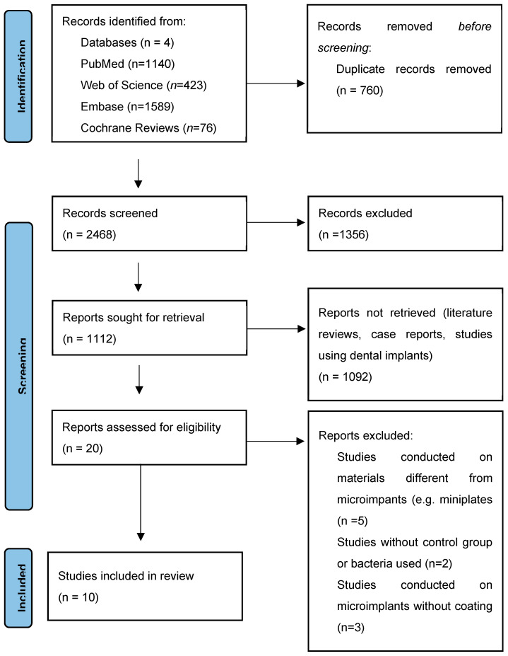 Figure 1
 Figure 2
 Figure 3
 Figure 4
Peer Reviews
No public reviews on file for this paper yet. If you reviewed it on a platform where reviews are public (OpenReview, ICLR, NeurIPS, ICML), you can paste yours below so the community can read it here.
Videos
No videos yet. Explain this paper in a talk, walkthrough, or lecture? Add one.
Taxonomy
TopicsOrthodontics and Dentofacial Orthopedics · Dental materials and restorations · Oral microbiology and periodontitis research
1. Introduction
The use of skeletal anchorage in the form of orthodontic microimplants has revolutionized orthodontic treatment. Temporary Intraoral Skeletal Anchorage Devices (TISADs) have enabled previously difficult dental rearrangements, such as alignment of the occlusal plane and intrusion of lateral sections of the dentition [1]. Unfortunately, their use in daily practice carries the risk of complications, which both doctor and patient need to know about before starting a treatment. These include peri-implantitis, defined as chronic progressive marginal inflammation occurring in tissues surrounding a microimplant [2]. The main symptoms of such a condition are hyperemia, hemorrhage, or proliferation of surrounding soft tissue, and even loosening and falling out of the implant because of the destruction of the implantation site [2,3]. Development of inflammation may also be influenced by the fact that the area of implantation is the most difficult for the patient to clean during brushing. According to the study published by de Freitas et al. [4], it is important to monitor microbial colonization in the implantation site, as conditions favorable for bacterial growth, including poor mini-implant hygiene, may lead to peri-implant infection.
While peri-miniscrew inflammation is a process influenced by many factors, bacterial infection appears to be one of the main causes responsible for this condition [5]. Attempts are underway to develop ways to reduce the risk of this complication, and to do so, it seems essential to understand the factors that affect it. Zhao et al. [5] tried to identify the specific bacteria responsible for peri-miniscrew inflammation. It was shown that around microimplants that failed, there was a stronger correlation with periodontal disease-associated bacteria, such as Fusobacterium nucleatum, Filifactor alocis, Porphyromonas gingivalis, and Prevotella nigrescens. The study by Huang et al. [6] demonstrated that in plaque found in the subgingival area around TISADs, the proportion of spirochetes was greater than that in the supragingival plaque, which may also contribute to the destructive effect on the surrounding soft tissues. The study by Garcez et al. [7] proved that a higher number of Porphyromonas gingivalis contamination was found around inflamed microimplants. According to the study published by Apel et al. [8], the absence of species that are markers of periodontal health, like Actinomyces viscosus and Cylindrotheca gracilis in failed microimplants, could be interpreted as a first symptom of a changing microflora, finally leading to peri-implantitis.
It seems no less important to understand the mechanism of inflammation around TISADs. According to the findings of He et al. [2], TLR-2, TLR-4, LOX-1, and BMP participate in the regulation of ILs (IL-1β, IL-6, IL-8, and IL-17), TNF-α, RANKL, MMP-2, and MMP-9 expression via JNK, Erk1/2, Wnt5a, NF-kBp65, OPN, and TAB/TAK signaling pathway, and among them IL-1 beta and IL-6 are critical inflammation mediators in the signaling pathways inducing inflammatory reactions surrounding implants. The discovery may, in the future, allow for better-targeted therapy focused on eliminating a specific factor.
Many attempts have been made to date to develop methods to reduce inflammation around the mini-implants and thus the frequency of complications in the form of reduced stability and loss of TISADs. These include gels and rinses containing antiseptics in their composition [9,10], possibly in combination with the use of irrigators, or the use of photobiomodulation therapy, whose effectiveness was proven in a systematic review published by Zhang et al. [11]. Another method to reduce inflammation around TISADs is to modify the surface of the microimplants by coating them with a material with antibacterial properties. This systematic review attempts to identify methods described to date for modifying the surface of microimplants to enhance their antibacterial properties.
2. Materials and Methods
The systematic review was written based on the principles detailed in Preferred Reporting Items for Systematic Reviews and Meta-Analyses (PRISMA Checklist in Supplementary Materials).
2.1. Questions
To pose the right question, facilitate the search for abstracts, and make sure that the relevant components of the question are well defined, the authors used the PICO format:
P (patient/population): in vivo studies involving humans or animals, in vitro studies, both concerning implants used for orthodontic treatment;
I (intervention): orthodontic implant modification causing antibacterial effect;
C (comparison): control group, without modification;
O (outcome): influence on the quantity of bacteria around orthodontic implants.
2.2. Study Identification and Search Method
An attempt was made to find a systematic review published to date on the studied topic. Such a study has not been found. The search began by identifying keyword combinations: orthodontic, implants, screw, inflammation, coating, antibacterial, antimicrobial, and bacteria.
The search was performed between 1 and 31 December 2024. The PubMed, Web of Science, Embase, and Cochrane Reviews databases were searched, and a literature review was conducted. Articles published in journals between 2003 (the appearance of the entry orthodontic microimplant in Angle Orthodontist for the first time) and 2023 were reviewed. This search yielded 3228 abstracts. Internal and external duplicates were removed, resulting in 2468 papers.
2.3. Eligibility Criteria
All the abstracts were analyzed for suitability for further review by two independent authors. Articles that were literature reviews and case reports were excluded. Studies using dental implants were also excluded because their use and mechanical properties differ from those desired for orthodontic microimplants. Studies in which the authors used plates or other materials instead of orthodontic microimplants were not eligible for the review. Studies without a control group were also excluded.
2.4. Study Selection
After final verification of the results, 10 studies were qualified for further analysis. They all turned out to be in vitro tests. All selected studies were published after 2017 (Figure 1).
2.5. Risk of Bias Assessment
An attempt was then made to find a suitable tool that could be used to assess the Risk of Bias (RoB). Unfortunately, despite the search, it was not possible to find a tool that fully matched the subject matter of the research. Such difficulties were also identified by Tran et al. [12]. Given the results of their work, after reviewing the methodology of the studies qualified for review, the authors decided to develop a proprietary analysis, which included questions collected from various studies and those developed by the authors.
2.6. Data Extraction and Data Synthesis
Each study was analyzed by all authors of the paper, independently. A table was created to include information obtained from the studies (Table 1). A meta-analysis of the obtained results was then attempted.
3. Results
The results obtained are summarized in Table 1. As the lack of a control group was an exclusion criterion, control groups were not included in the table.
Risk of Bias Assessment
In the next step, the authors conducted a Risk of Bias assessment of the results obtained to assess the quality of the studies. The analysis was performed according to the criterion developed from the subject analysis, as well as using questions from other analyses. This yielded 16 questions divided into three sections: Introduction, Methods, and Results (Table 2).
The authors divided the studies into three groups: Low, Moderate, and High Risk of Bias. The summary and description of the most important factors influencing the results are presented in Table 3.
Due to the excessive diversity of studies, especially in terms of methodology, it was not possible to conduct a meta-analysis.
4. Discussion
One of the most widely studied methods to modify the surface of microimplants has been the use of ZnO to impart antimicrobial properties. It was included in four studies by Bahrami [21], Abo-Elmahasen [19], Noorollahian [17], and Othman [22]. Efficacy against Porphyromonas gingivalis [17,21], Prevotella intermedia [21], Aggregatibacter actinomycetemcomitans [21], Enterobacter aeruginosa [19], Staphylococcus aureus [19], Streptococcus mutans [19], Enterococcus faecalis [19], Escherichia coli [19] bacteria, and Candida albicans [19] fungus has been demonstrated. The different methodologies and aims of the studies made it impossible to compare them. The antibacterial effect of ZnO was also proven in a study by Hammad et al. [23], which used NiTi arches in orthodontics coated with ZnO nanoparticles. Such surface modification has been proven to exert antibacterial activity against strains of Staphylococcus aureus, Streptococcus pyogens, and Escherichia coli.
Noorollahian et al. [17] did not evaluate whether the ZnO-containing coating itself exhibits antibacterial properties. However, the combination of this method with the use of the antibiotic doxycycline has been investigated. This combination proved effective in preventing the growth of Porphyromonas gingivalis for up to 30 days. Another study in which an antibiotic was used to enhance antibacterial properties is the study conducted by Anggani [16], rated High RoB. This study proved the greatest antibacterial potential against Porphyromonas gingivalis for the group using azithromycin alone, while the other methods (chitosan, chitosan with azithromycin) also showed antibacterial properties, but to a lesser extent.
In a study by Bahrami [21], ZnONPs-coated mini-screws impregnated with polymicrobial biofilm irrigated with PBS were studied, as well as the effect of their subsequent exposure to LED irradiation, ultrasound waves, and a combination of the latter two. Antibacterial activity against Porphyromonas gingivalis, Prevotella intermedia, and Aggregatibacter actinomycetemcomitans was obtained in each of the tested groups. The best result was obtained in the group where both methods were used, and also surprisingly in the group in which uncoated microimplants were immersed in 0.2% CHX solution for 5 min. This study points to the possibilities offered by ZnO coating, as well as exposure methods that improve antibacterial properties, such as LED irradiation and ultrasound waves.
The use of chlorhexidine and its antibacterial properties seems to be an interesting method. Often, gels or rinses containing this antiseptic are recommended as post-operative recommendations for patients in Poland. In a study by Chin et al. [24], the authors found that chlorhexidine gluconate and sodium fluoride exhibited significant antibacterial and plaque-forming activity on the surface of the microimplants, demonstrating their important role in oral hygiene in orthodontic patients.
Another substance that was often used in research was chitosan. Chitosan is considered to be a non-toxic, biocompatible, and biodegradable compound [25,26], and due to its versatile effects, including antibacterial, antifungal, analgesic, and anticancer, it is finding more and more applications in the cosmetic industry, medicine, or biomedical applications [27], such as wound healing, controlled drug delivery, and tissue regeneration [28]. It was used in the study by Alhazmi [18] (Moderate RoB), Anggani [16], and Sreenivasagan [14] (both High RoB), so the results obtained should be approached with caution. In the previously mentioned study by Anggani [16], an antibacterial effect was obtained with chitosan alone, chitosan with azithromycin, and azithromycin alone. In a study by Sreenivasagan [14], chitosan-based silver-impregnated nanoparticles were used, which also showed antibacterial properties but poor antifungal values. In a study by Alhazmi [18], the best antibacterial effect was obtained when chitosan was used in combination with hydroxyapatite. In these studies, efficacy against Streptococcus mutans [14,18], Streptococcus sanguis [18], Streptococcus salivarius [18], Enterococcus faecalis [18], Porphyromonas gingivalis [16], Staphylococcus aureus [14], Lactobacillus [14], and Candida albicans [14] was demonstrated. A study that also tested the antibacterial properties of chitosan was conducted by Ly et al. [29]. This paper was not qualified for review because the authors used titanium alloy discs for the study. However, the authors were able to prove that reduced biofilm formation of Streptococcus mutans and Streptococcus sobrinus was achieved with samples containing chitosan. The above studies show that chitosan is a promising option for modifying the surface of microimplants to impart antimicrobial properties, particularly when used in combination with another substance with similar effects.
In the study conducted by Alhazmi [18], hydroxyapatite was used. This substance was also used in the study [19], rated Low RoB. Both studies showed efficacy against Streptococcus mutans and Enterococcus faecalis bacteria. Both studies testify to the potential for HA to be used for antibacterial properties, particularly when used in combination with another substance. Similar results were obtained by Abdulkareem [30], who studied the antibacterial properties of three types of coatings (nano-ZnO; nZnO and nHA; and nHA) with which he coated titanium plates. All coatings have shown antibacterial activity. A study by Leelanarathiwat [31] tested the combination of antimicrobial activity for hydroxyapatite–tryptophan complex with Gray Titania coating and photocatalysis on titanium alloy substrate. The study was conducted on Porphyromonas gingivalis, Tannerella forsythia, and Aggregatibacter actinomycetemcomitans. The results showed that the photoactivated hydroxyapatite–tryptophan complex and Gray Titania as a photocatalytic coating have antibacterial effects; however, the coating itself did not show antibacterial effects against bacteria involved in the formation of peri-implantitis. The above studies indicate the possible use of HA, especially in combination with another method, to increase the antibacterial properties of the coating.
In the study conducted by Abo-Elmahasen [19], silver was used in addition to HA, yielding antibacterial properties. Many other studies confirm this result for various bacterial species, Streptococcus mutans and Porphyromonas gingivalis [32], or those less commonly present in the oral cavity—such as Staphylococcus aureus [33,34] or Escherichia coli [34]. The effect of this element on antibacterial properties was also studied by Venugopal [13] and Subramanian [15]. Venugopal et al. [13] evaluated a Moderate RoB, proving an antibacterial effect occurring only with AgNP-coated biopolymer. In contrast, no such properties were found for regular Ag-NPs. This study shows that it is not only the substance used that matters but also the form in which it is used. Both studies demonstrated the effectiveness of silver-containing coatings on Streptococcus mutans bacteria.
In the work of Subramanian [15], rated High RoB, it was proven that a coating containing AgNPs as well as SeNPs showed antibacterial properties, greater in the former case. In both cases, a biopolymer was used.
Studies show that SeNPs not only have antibacterial effects against Escherichia coli and Staphylococcus aureus [35] but also against Porphyromonas gingivalis, a bacterium commonly associated with peri-implantitis [36]. The antimicrobial effect of SeNPs was also described, in which SeNPs were shown to inhibit macrophage proliferation, thereby reducing the inflammatory response around implants [35].
Another way to impart antimicrobial properties to microimplants is to treat the microimplant surface with PEG polymerization plasma, as demonstrated in the study conducted by Rodriguez-Fernandez [20], rated as a high-quality study. All tested PEG samples showed decreased bacterial adhesion, either for Streptococcus sanguinis or Lactobacillus salivarius. This result confirms a study by Buxadera-Palomero et al. [37], who also proved reduced adhesion of Streptococcus sanguinis and Lactobacillus salivarius to PEG-like coating on the titanium surface. In addition, it has been shown that despite reducing bacterial adhesion to the surface, the coating does not affect fibroblast and osteoblast adhesion [37].
Limitations
All the studies analyzed in the review are in vitro studies. To date, there is no sufficiently large number of pertinent studies conducted on humans. Another problem is the diversity in testing methodology and the inability to compare all the methods described, which makes it difficult to assess the antibacterial properties of the coating.
5. Conclusions
All of the microimplant surface modifications analyzed showed antibacterial properties. The presented methods, such as the use of ZnO, antibiotics, chlorhexidine, silver compounds, selenium, hydroxyapatite, and PEG polymerization plasma, are an intriguing possibility for improving the properties of orthodontic microimplants, and thus reducing the risk of complications in the form of local inflammation. However, because of the still small number of studies on the subject and different methodologies, more studies are needed to assess the effectiveness of the given methods.In vitro studies are required to enable the implementation of new technology in the orthodontic treatment of patients.
The reference list from the paper itself. Each links out to its DOI / PubMed record.
- 1Antoszewska J. Raftowicz-Wójcik K. Kawala B. Matthews-Brzozowska T. Biological Factors Involved in Implant-Anchored Orthodontics and in Prosthetic-Implant Therapy: A Literature Review Arch. Immunol. Ther. Exp.20105837938310.1007/s 00005-010-0088-820676787 · doi ↗ · pubmed ↗
- 2He W. Zhuvand H. Liu C. Profiles of inflammation factors and inflammatory pathways around the peri-miniscrew implant Histol. Histopathol.20213689990610.14670/HH-18-33633834451 · doi ↗ · pubmed ↗
- 3Park H.S. Jeong S.H. Kwon O.W. Factors affecting the clinical success of screw implants used as orthodontic anchorage Am. J. Orthod. Dentofacial. Orthop.2006130182510.1016/j.ajodo.2004.11.03216849067 · doi ↗ · pubmed ↗
- 4de Freitas A.O. Alviano C.S. Alviano D.S. Siqueira J.F.Jr. Nojima L.I. Nojima M.d.C. Microbial colonization in orthodontic mini-implants Braz. Dent. J.20122342242710.1590/S 0103-6440201200040001923207860 · doi ↗ · pubmed ↗
- 5Zhao N. Zhang Q. Guo Y. Cui S. Tian Y. Zhang Y. Zhou Y. Wang X. Oral microbiome contributes to the failure of orthodontic temporary anchorage devices (TA Ds)BMC Oral Health 2023232210.1186/s 12903-023-02715-736650527 PMC 9844000 · doi ↗ · pubmed ↗
- 6Huang R. He Y.X. Jia X.T. Liu J.N. Fan X.C. Zeng N. Huang X.F. Investigation of periodontal status and bacterial composition around mini-implants Am. J. Orthod. Dentofac. Orthop.202316411612210.1016/j.ajodo.2022.11.01536858877 · doi ↗ · pubmed ↗
- 7Garcez A.S. Barros L.C. Fernandes M.R.U. Fujii D.N. Suzuki S.S. Nepomuceno R. Fluorescence image and microbiological analysis of biofilm retained around healthy and inflamed orthodontic miniscrews Photodiagn. Photodyn. Ther.20203010170710.1016/j.pdpdt.2020.10170732126307 · doi ↗ · pubmed ↗
- 8Apel S. Apel C. Morea C. Tortamano A. Dominguez G.C. Conrads G. Microflora associated with successful and failed orthodontic mini-implants Clin. Oral Implant. Res.2009201186119010.1111/j.1600-0501.2009.01756.x 19719743 · doi ↗ · pubmed ↗