Left ventricle strain and T1 mapping evaluation in a mouse model with myocardial infarction
Khaoula Bouazizi, Thulaciga Yoganathan, Frank Kober, Gwennhael Autret, Perrine Marsac, Clément Delacroix, Shannon Soulez, Mohamed Zarai, Vincent Nguyen, Adil Squalli, Paul Alayrac, Estelle Robidel, Bertrand Tavitian, Alban Redheuil, Jean-Sébastien Hulot, Nadjia Kachenoura

TL;DR
This study evaluates heart function and tissue changes in mice with heart attacks using MRI techniques, providing baseline data for future research.
Contribution
The study introduces a tailored MRI protocol to measure strain and T1 mapping in mice, offering normative values post-myocardial infarction.
Findings
Myocardial infarction caused LV dilation and reduced ejection fraction, but preserved stroke volume and cardiac output.
Circumferential strain decreased significantly in MI mice, especially at the apex.
Post-MI mice showed higher native T1 and lower post-contrast T1 values compared to wild-type mice.
Abstract
Myocardial strain and T1 mapping offer precise evaluation of cardiac function and tissue characteristics. The aim of this study is to provide normative myocardial strain and T1 values in mice model along with their changes after myocardial infarction (MI) using a tailored acquisition and analysis protocol. Healthy mice had MRI before and after MI. Imaging was performed to assess cardiac function and left ventricle (LV) strain. A Look-Locker Inversion-Recovery sequence was used for T1 maps reconstruction. Gadolinium doses and timing of image acquisitions were optimized. Radial and circumferential strain measurements were conducted using a custom software based on feature tracking. To address ECG signal interference, adjustments were made for accurate strain calculations. An LV dilation in MI compared to wild-type (WT) with a decrease in LV ejection fraction (p < 0.0001) were reported…
Genes, proteins, chemicals, diseases, species, mutations and cell lines named across the full text — each resolved to its canonical identifier and authoritative record.
Click any figure to enlarge with its caption.
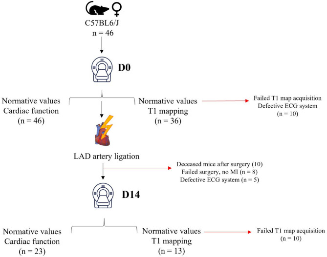 Figure 1
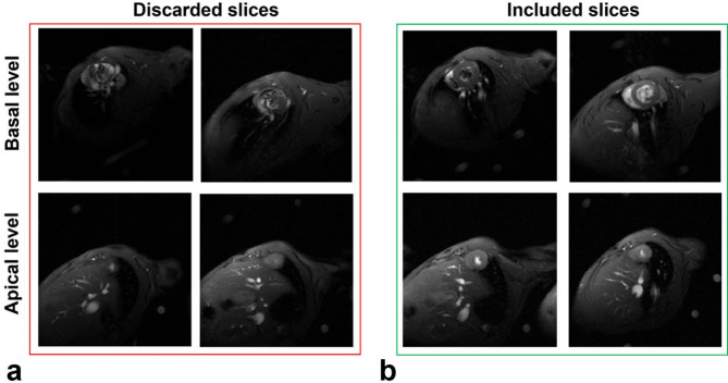 Figure 2
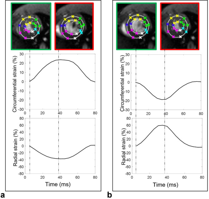 Figure 3
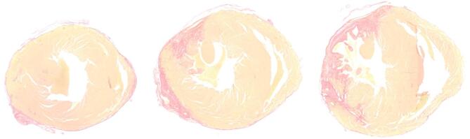 Figure 4
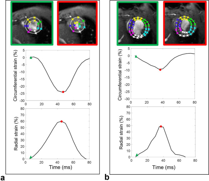 Figure 5
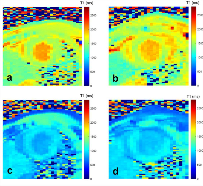 Figure 6
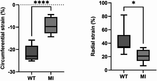 Figure 7
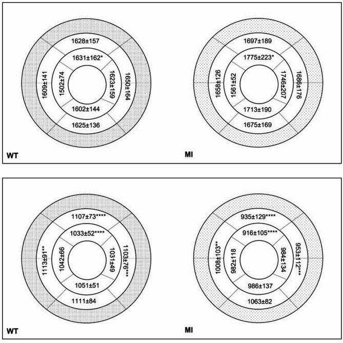 Figure 8
Peer Reviews
No public reviews on file for this paper yet. If you reviewed it on a platform where reviews are public (OpenReview, ICLR, NeurIPS, ICML), you can paste yours below so the community can read it here.
Videos
No videos yet. Explain this paper in a talk, walkthrough, or lecture? Add one.
Taxonomy
TopicsCardiovascular Function and Risk Factors · Cardiomyopathy and Myosin Studies · Cardiac Fibrosis and Remodeling