Comparison of the performance of digital variance angiography and digital subtraction angiography in children with arteriovenous malformations: a retrospective observational study
Balázs Bence Nyárády, Renáta Gubán, Ákos Pataki, András Bibok, Zsuzsanna Mihály, Dávid Korda, Dénes Horváthy, Anikó Ilona Nagy, János Pál Kiss, Edit Dósa

TL;DR
This study compares two imaging techniques in children with arteriovenous malformations, finding that one improves image quality but not visual assessment.
Contribution
The study provides the first evidence on the performance of DVA in pediatric endovascular procedures.
Findings
DVA significantly improved contrast-to-noise ratio compared to DSA across multiple anatomical regions.
Subjective image quality assessments showed DSA was superior in most comparisons, though differences were clinically negligible.
DVA's enhanced CNR suggests potential for reducing contrast agent and radiation exposure in pediatric patients.
Abstract
Reducing contrast agent and radiation exposure is paramount for pediatric patients. Digital variance angiography (DVA) might address this need by increasing the contrast-to-noise ratio (CNR). A total of 132 raw iodinated contrast angiograms of 10 children (mean age: 12 years) who had endovascular procedures for arteriovenous malformations were retrospectively processed for DVA analysis. The CNR of the DVA and digital subtraction angiography (DSA) images was calculated. The visual image quality was assessed using a four-point Likert scale. Statistical analyses were based on the Wilcoxon signed-rank test and one-sample t-test. The CNR was determined and compared for 3,318 regions of interest in 132 image pairs in four anatomical regions (upper limb (UL), lower limb (LL), head and neck (HN), and chest (CH)). DVA outperformed DSA, with a median overall CNRDVA/CNRDSA ratio of 2.00 (UL,…
Genes, proteins, chemicals, diseases, species, mutations and cell lines named across the full text — each resolved to its canonical identifier and authoritative record.
Click any figure to enlarge with its caption.
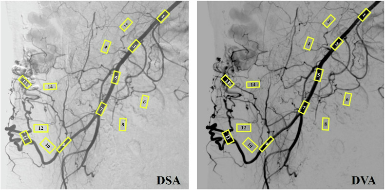 Figure 1
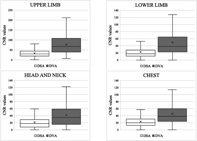 Figure 2
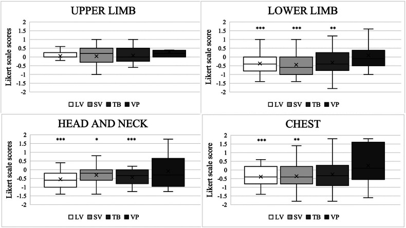 Figure 3
 Figure 4
Peer Reviews
No public reviews on file for this paper yet. If you reviewed it on a platform where reviews are public (OpenReview, ICLR, NeurIPS, ICML), you can paste yours below so the community can read it here.
Videos
No videos yet. Explain this paper in a talk, walkthrough, or lecture? Add one.
Taxonomy
TopicsVascular Malformations Diagnosis and Treatment · Intracranial Aneurysms: Treatment and Complications · Vascular anomalies and interventions