Point-of-care Ultrasound Diagnosis of Cardiac Myxoma
Joseph Brutico, Daniel Kreider

TL;DR
A case study shows how point-of-care ultrasound in the emergency department helped diagnose a rare heart tumor called a cardiac myxoma.
Contribution
Demonstrates the utility of point-of-care ultrasound in the emergency setting for diagnosing cardiac myxoma.
Findings
POCUS identified a left atrial mass in a patient with dyspnea and hematemesis.
Surgical excision confirmed the diagnosis of cardiac myxoma.
Early detection via POCUS enabled timely treatment.
Abstract
Cardiac myxomas are rare benign tumors of the heart that can become clinically relevant due to cardiovascular effects. Diagnosis can be challenging due to non-specific presenting symptoms. Point-of-care ultrasound (POCUS) provides a convenient first-line screening modality. A 65-year-old male with a history of tobacco use presented to the emergency department (ED) with a month of progressive dyspnea with exertion and hematemesis. Cardiac POCUS and pulmonary computed tomography with angiography revealed a left atrial mass consistent with a cardiac atrial myxoma. The patient underwent coronary artery bypass grafting with excision of the left atrial myxoma via right atriotomy and atrial septal defect repair. Presented is a case of a patient presenting with progressive dyspnea diagnosed with a cardiac myxoma using POCUS in the ED. Cardiac myxomas have a wide variety of clinical…
Click any figure to enlarge with its caption.
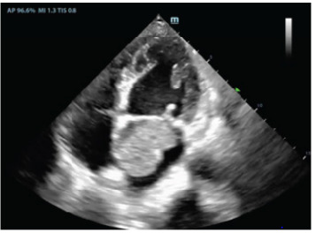 Figure 1
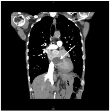 Figure 2
Peer Reviews
No public reviews on file for this paper yet. If you reviewed it on a platform where reviews are public (OpenReview, ICLR, NeurIPS, ICML), you can paste yours below so the community can read it here.
Videos
No videos yet. Explain this paper in a talk, walkthrough, or lecture? Add one.
Taxonomy
TopicsCardiac tumors and thrombi · Cardiovascular Effects of Exercise · Cardiac Structural Anomalies and Repair
CASE PRESENTATION
A 65-year-old male with a history of tobacco use presented to the ED with a month of progressive dyspnea with exertion and hematemesis. On review of systems, the patient reported unintentional weight loss and night sweats. Examination revealed a cachectic male appearing older than stated age with slight tachypnea. Cardiac point-of-care ultrasound (POCUS) (Video 1) and pulmonary computed tomography (CT) with angiography (Images 1 and 2) revealed a 3 x 3 x 5 centimeter (cm) left atrial mass consistent with a cardiac atrial myxoma, which was later confirmed by pathology.
After left heart catheterization, the patient underwent coronary artery bypass grafting with excision of the left atrial myxoma via right atriotomy and atrial septal defect repair.
DISCUSSION
Cardiac myxomas are the most common primary cardiac tumor. A vast majority of myxomas (80–90%) arise from the left atrium with fewer involving the right atrium (7–20%). Rarely, myxomas may be biatrial or arise from the ventricles.1 Symptoms vary and typically arise due to obstruction (heart failure), invasion of myocardial tissue (arrhythmias), or embolization (ischemia).2 Embolization occurs in up to 40% and is associated with villous tumors, size less than 4.5 cm, and valvular site of origin.3 Management begins with confirmation of diagnosis as common mimics include mural thrombi and valvular vegetations.2 While echocardiography is the diagnostic modality of choice, CT or cardiac magnetic resonance imaging may also be considered. Surgical resection and tumor histopathology remain the mainstay of treatment, with postoperative 30-day mortality < 5%.4 The most common complication is cardiac arrhythmia with reported rates of up to 20%.5 This clinical pathology requires a high index of suspicion and use of multiple diagnostic modalities including cardiac POCUS.
CPC-EM Capsule What do we already know about this clinical entity?Cardiac myxomas are rare benign tumors of the heart that can present with a wide range of clinical symptoms. Management includes confirmation of diagnosis and surgical resection.What is the major impact of the image(s)?This case highlights cardiac myxoma as an important diagnosis to consider in the evaluation of a patient with dyspnea and has unique and recognizable echocardiographic findings.How might this improve emergency medicine practice?With a high level of suspicion, emergency physicians can accurately recognize this disease state using point-of-care ultrasound, leading to appropriate management and good long-term survival.
The reference list from the paper itself. Each links out to its DOI / PubMed record.
- 1Lone R Ahanger A Singh S Atrial myxoma: trends in management Int J Health Sci 20082214151 PMC 306873421475496 · pubmed ↗
- 2Elbardissi A Dearani J Daly R Embolic potential of cardiac tumors and outcome after resection Stroke 2009401156621894860210.1161/STROKEAHA.108.525709 · doi ↗ · pubmed ↗
- 3Wang Z Chen S Zhu M Risk prediction for emboli and recurrence of primary cardiac myxomas after resection J Cardiothorac Surg 20161122182683280610.1186/s 13019-016-0420-4PMC 4736655 · doi ↗ · pubmed ↗
- 4Islam A Cardiac myxomas: a narrative review World J Cardiol 2022144206193558246610.4330/wjc.v 14.i 4.206PMC 9048271 · doi ↗ · pubmed ↗
- 5Okongwu C Olaofe O Cardiac myxoma: a comprehensive review J Cardiothorac Surg 20252011514008290310.1186/s 13019-024-03333-2PMC 11905437 · doi ↗ · pubmed ↗