Vascular Lesions on the Left Upper Gingiva in a Patient With Port‐Wine Stains
Tinglan Yang, Mengmeng Song, Qing Liu, Zhenlai Zhu

TL;DR
This paper discusses vascular lesions in the mouth of a patient with port-wine stains and emphasizes the importance of proper diagnosis and monitoring.
Contribution
Highlights the need for multidisciplinary management to distinguish oral vascular lesions from neoplasms in port-wine stain patients.
Findings
Asymptomatic intraoral capillary hemangiomas can occur in port-wine stain patients.
Regular oral monitoring helps detect vascular proliferation and dental abnormalities.
Multidisciplinary care is essential for managing bleeding risks and treatment optimization.
Abstract
Port‐wine stain patients may develop asymptomatic intraoral capillary hemangiomas ipsilateral to facial lesions. Multidisciplinary management is critical to differentiating these from neoplasms, mitigating bleeding risks, and optimizing therapies like photodynamic treatment. Regular oral monitoring is advised to detect vascular proliferation and dental abnormalities.
Genes, proteins, chemicals, diseases, species, mutations and cell lines named across the full text — each resolved to its canonical identifier and authoritative record.
Click any figure to enlarge with its caption.
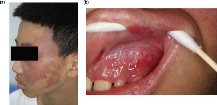 FIGURE 1
Peer Reviews
No public reviews on file for this paper yet. If you reviewed it on a platform where reviews are public (OpenReview, ICLR, NeurIPS, ICML), you can paste yours below so the community can read it here.
Videos
No videos yet. Explain this paper in a talk, walkthrough, or lecture? Add one.
Taxonomy
TopicsVascular Malformations and Hemangiomas · Oral and Maxillofacial Pathology · Oral and gingival health research
Case Presentation
1
A 13‐year‐old boy with a congenital port‐wine stain (PWS) on the left facial region (Figure 1a) had undergone multiple photodynamic therapy sessions over the past year, achieving significant erythema reduction. Two weeks prior to presentation, his parents noticed asymptomatic red lesions on his maxillary left gingiva (Figure 1b). The patient presented with no history of systemic diseases, seizures, or visual disturbances. Physical examination revealed an erythematous patch distributed along the left maxillary branch of the trigeminal nerve, with an irregular shape and well‐defined borders. Intraoral examination revealed bright fiery‐red areas on the maxillary left buccal gingiva, palatal gingiva, alveolar mucosa, and labial mucosa. The masticatory mucosa of the hard palate and soft palate mucosa were unaffected. Intraoral diascopy was performed, revealing blanching of the lesions. No systemic symptoms, bleeding tendency, or contralateral involvement were noted. The differential diagnoses considered included pyogenic granuloma and venous malformations. Pyogenic granuloma was deemed unlikely given the lack of bleeding tendency or trauma history, while venous malformations were ruled out based on the non‐purplish color and capillary morphology of the lesions. Following oral health education, the patient continued with photodynamic therapy for facial and oral lesions with the involvement of oral medicine specialists. The patient is currently under follow‐up with no adverse outcomes reported.
Clinical images. (a) Port‐wine stain along the distribution of the left maxillary nerve. (b) Lesions on the maxillary left gingiva, alveolar, and labial mucosa.
Discussion
2
PWSs are congenital vascular malformations that appear clinically as erythematous areas on the buccal mucosa, vermilion border of the lip, gingiva, or as pink to port‐wine‐colored patches on the skin. The intraoral capillary hemangiomas, observed in 40% of patients, are often overlooked due to their asymptomatic nature [1]. Regular oral examinations are critical for early detection of vascular proliferations and dental anomalies, such as malocclusion, associated with angiomatosis [2]. Our patient showed no features of syndromic PWS associations, specifically lacking both the neuro‐ophthalmic abnormalities characteristic of Sturge–Weber syndrome and the limb hypertrophy or lymphatic malformations of Klippel–Trenaunay–Weber syndrome. In some cases, there may be further proliferation on the basis of capillary hemangioma, leading to the formation of lobular capillary hemangioma [3]. Consequently, the involved mucosa becomes delicate and susceptible to even minor injuries and simple dental operations. It is noteworthy that most PWS patients do not have coagulation abnormalities but tend to bleed more easily during treatments. Additionally, recent studies have reported abnormal tooth maturation on the affected side in PWS patients; thus, attention should be paid to the tooth maturation on the affected side during oral examinations [2]. This case underscores the necessity of a multidisciplinary approach in managing PWS to ensure comprehensive care and achieve optimal outcomes.
Author Contributions
Tinglan Yang: writing – original draft, writing – review and editing. Mengmeng Song: data curation, investigation. Qing Liu: data curation, project administration, resources, supervision. Zhenlai Zhu: data curation, investigation, writing – original draft, writing – review and editing.
Consent
We have obtained the written informed consent from the patient's parents.
Conflicts of Interest
The authors declare no conflicts of interest.
The reference list from the paper itself. Each links out to its DOI / PubMed record.
- 1O. Pagin , N. N. Del , M. P. Battisti , A. L. Capelozza , and P. S. Santos , “Periodontal Manifestations and Ambulatorial Management in a Patient With Sturge–Weber Syndrome,” Journal of Craniofacial Surgery 23, no. 6 (2012): 1809–1811, 10.1097/SCS.0b 013e 318271016 c.23147344 · doi ↗ · pubmed ↗
- 2Z. Liu , H. Ying , X. Lin , et al., “Abnormal Tooth Maturation Associated With Port Wine Stains,” Orthodontics & Craniofacial Research 27, no. 3 (2024): 387–393, 10.1111/ocr.12734.38058228 · doi ↗ · pubmed ↗
- 3S. Priya , M. K. Rajaram , T. R. Pethagounder , S. M. Fenn , and K. Appusamy , “Concurrent Occurrence of Lobular Capillary Haemangioma and Port‐Wine Stain: A Case Report and Literature Review,” Cureus 15, no. 5 (2023): e 38642, 10.7759/cureus.38642.37284361 PMC 10241596 · doi ↗ · pubmed ↗