Benign Enchondroma in a 40-Year-Old Female: Emphasizing the Importance of Early 18F-FES PET/CT Utilization
Tamer M. Dawud, Abdullah T. Dawud

TL;DR
A 40-year-old woman with breast cancer had a benign bone lesion detected, and early use of 18F-FES PET/CT helped avoid unnecessary procedures.
Contribution
Highlights the clinical value of early 18F-FES PET/CT in breast cancer staging to reduce unnecessary interventions.
Findings
A bone scan showed abnormal femur uptake, but MRI revealed a benign enchondroma.
18F-FES PET/CT ruled out estrogen receptor expression and metastatic disease.
Biopsy confirmed the lesion was a benign enchondroma.
Abstract
A 40-year-old female with estrogen receptor–positive breast cancer underwent an initial staging using a technetium-99m methylene diphosphonate (Tc-99m MDP) bone scan, which revealed abnormal uptake in the femur without a patient history of prior trauma or associated symptoms. Subsequently, an MRI confirmed the presence of a well-defined lesion in the upper left femur. To rule out metastatic disease, an 18F-fluoroestradiol (FES) PET/CT was performed, demonstrating no ER expression. Following the PET/CT, a biopsy confirmed the presence of an enchondroma. This case underscores the importance of early utilization of 18F-FES PET/CT in breast cancer staging to minimize unnecessary additional procedures/imaging.
Genes, proteins, chemicals, diseases, species, mutations and cell lines named across the full text — each resolved to its canonical identifier and authoritative record.
Click any figure to enlarge with its caption.
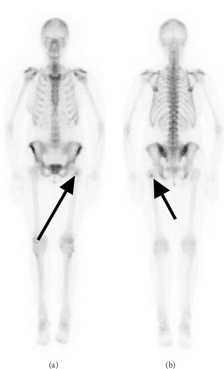 Figure 1
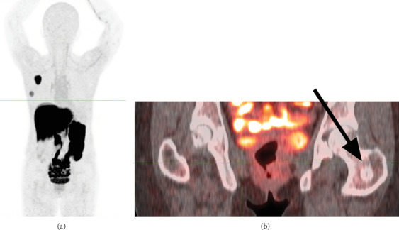 Figure 2
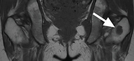 Figure 3
Peer Reviews
No public reviews on file for this paper yet. If you reviewed it on a platform where reviews are public (OpenReview, ICLR, NeurIPS, ICML), you can paste yours below so the community can read it here.
Videos
No videos yet. Explain this paper in a talk, walkthrough, or lecture? Add one.
Taxonomy
TopicsBone Tumor Diagnosis and Treatments · Sarcoma Diagnosis and Treatment · Bone health and treatments
Summary
- • Question: Should 18F-FES PET/CT replace bone scans as the initial imaging modality for evaluating suspected metastases in ER-positive breast cancer patients?
- • Pertinent findings: This diagnostic case study of a 40-year-old patient with ER-positive breast cancer demonstrated that negative 18F-FES PET/CT uptake (despite positive Tc-99m MDP bone scan) correctly predicted a benign enchondroma, as confirmed by MRI and biopsy.
- • Implications for patient care: Early integration of 18F-FES PET/CT in the diagnostic workflow may reduce unnecessary procedures and improve staging efficiency for ER-positive breast cancer patients with indeterminate skeletal findings.
1. Introduction
Breast cancer is the most commonly diagnosed malignancy in women in the United States [1], with bone being a frequent metastatic site [2]. Accurate distinction between benign and malignant bone lesions is critical for appropriate staging and treatment planning. Treatment often involves advanced imaging modalities that detect potential metastases and guide treatment decisions. Among these, technetium-99m methylene diphosphonate (Tc-99m MDP) bone scans are widely used to detect skeletal metastases due to their high sensitivity [3]. However, their limited specificity complicates the differentiation between malignant metastases and benign conditions. As noted by Cook, “Specificity for detecting metastatic lesions may also be limited without further morphological imaging as several benign processes can cause focal uptake in the skeleton” [4]. This diagnostic uncertainty often necessitates additional imaging—such as magnetic resonance imaging (MRI) and positron emission tomography/computed tomography (PET/CT)—or biopsy to confirm the nature of suspicious lesions.
18F-fluoroestradiol (FES) PET/CT has emerged as a valuable imaging modality for evaluating estrogen receptor (ER)–positive breast cancer, as it specifically targets ER expression in tumor cells [5]. Meta-analyses report 18F-FES PET/CT sensitivity of 71%–82% [6, 7], while a more recent 2022 study of 200 patients demonstrated improved sensitivity of 95% [8]. The radiotracer's specificity was recently reported at 98% in a recent meta-analysis [9]. However, 18F-FES PET/CT cannot reliably identify benign bone lesions such as enchondromas [6].
Enchondromas are benign cartilaginous tumors that typically arise in the medullary cavity of long bones, most frequently in the hands and feet [10]. Typically asymptomatic, they are often incidental findings on imaging performed for unrelated reasons. Their clinical and radiological features may mimic those of malignant bone lesions, complicating the diagnostic process [11].
2. Case Report
A 40-year-old female with a recent diagnosis of ER-positive breast cancer underwent an initial staging, including a Tc-99m MDP bone scan. The bone scan revealed an area of abnormal radiotracer uptake in the proximal left femur (Figure 1). The patient reported no history of trauma, pain, or other symptoms related to the femur. Subsequently, imaging with 18F-FES PET/CT revealed no radiotracer uptake in the femoral lesion (Figure 2), consistent with the absence of ER expression. Concurrently, MRI confirmed the presence of this low-signal-intensity T1 lesion (1.8 × 1.6 × 2.3 cm shown in Figure 3). Given the discordance between the abnormal bone scan and the absence of 18F-FES PET/CT uptake, a biopsy was performed to rule out metastasis definitively. The examination of the femoral lesion confirmed the diagnosis of an enchondroma. The patient's breast cancer was staged as T2N0M0 due to the absence of metastasis. No further intervention was required for the enchondroma, and she was referred to oncology for further management, including surgical resection of the primary tumor.
3. Discussion
18F-FES is approved by the US Food and Drug Administration as a diagnostic agent for “the detection of ER-positive lesions as an adjunct to biopsy in patients with recurrent or metastatic breast cancer” [12]. Following these guidelines, this case utilized 18F-FES PET/CT after a bone scan initially raised suspicion for metastasis. Although the FES PET/CT ultimately showed no uptake in the lesion, an MRI and biopsy were subsequently performed to confirm the lesion's benign nature. Had the FES PET/CT been incorporated earlier in the diagnostic workup, the absence of ER expression might have provided sufficient evidence against metastatic disease, potentially eliminating the need for additional imaging and an invasive biopsy. This case underscores the value of incorporating FES PET/CT earlier in the diagnostic process, which could reduce unnecessary procedures, optimize resource utilization, and ultimately improve diagnostic efficiency in similar scenarios.
The reference list from the paper itself. Each links out to its DOI / PubMed record.
- 1Siegel R. L. Kratzer T. B. Giaquinto A. N. Sung H. Jemal A. Cancer Statistics, 2025 CA: A Cancer Journal for Clinicians 2025751104510.3322/caac.2187139817679 PMC 11745215 · doi ↗ · pubmed ↗
- 2Coleman R. E. Clinical Features of Metastatic Bone Disease and Risk of Skeletal Morbidity Clinical Cancer Research 20061220 pt. 26243 s 6249 s 10.1158/1078-0432.CCR-06-09312-s 2.0-3375072607717062708 · doi ↗ · pubmed ↗
- 3Brenner A. I. Koshy J. Morey J. Lin C. Di Poce J. The Bone Scan Seminars in Nuclear Medicine 2012421112610.1053/j.semnuclmed.2011.07.0052-s 2.0-8185518369522117809 · doi ↗ · pubmed ↗
- 4Cook G. J. Imaging of Bone Metastases in Breast Cancer Seminars in Nuclear Medicine 202252553154110.1053/j.semnuclmed.2022.01.00535236615 PMC 7616189 · doi ↗ · pubmed ↗
- 5O’Brien S. R. Edmonds C. E. Lanzo S. M. Weeks J. K. Mankoff D. A. Pantel A. R. 18F-Fluoroestradiol: Current Applications and Future Directions Radiographics 2023433 e 22014310.1148/rg.22014336821506 · doi ↗ · pubmed ↗
- 6Chae S. Y. Son H. J. Lee D. Y. Comparison of Diagnostic Sensitivity of [18F]Fluoroestradiol and [18F]Fluorodeoxyglucose Positron Emission Tomography/Computed Tomography for Breast Cancer Recurrence in Patients With a History of Estrogen Receptor-Positive Primary Breast Cancer EJNMMI Research 2020101 p. 5410.1186/s 13550-020-00643-z 32448947 PMC 7246280 · doi ↗ · pubmed ↗
- 7Evangelista L. Vittoria Dieci M. Guarneri V. Franco Conte P. 18F-Fluoroestradiol Positron Emission Tomography in Breast Cancer Patients: Systematic Review of the Literature & Meta-Analysis Current Radiopharmaceuticals 20169324425710.2174/18744710096661610191449502-s 2.0-8500928817127774910 · doi ↗ · pubmed ↗
- 8van Geel J. J. Boers J. Elias S. G. Clinical Validity of 16α-[18F]Fluoro-17β-Estradiol Positron Emission Tomography/Computed Tomography to Assess Estrogen Receptor Status in Newly Diagnosed Metastatic Breast Cancer Journal of Clinical Oncology 202240313642365210.1200/JCO.22.0040035584346 · doi ↗ · pubmed ↗