Knee Pain: The Fine Line Between Malignant and Benign
Andrew Lew, Suleman Janjua, Tariq M Awan, Nicholas Moore, Gregory M Cibor, Paolo Balmaseda, David M Peck, Michael P Montico

TL;DR
FOPE is a normal knee finding in adolescents that can be mistaken for injury, but it often resolves with conservative care.
Contribution
The paper highlights FOPE as a normal variant to prevent misdiagnosis in adolescents with knee pain.
Findings
FOPE is commonly found in adolescent females aged 11-14 and is associated with physeal closure.
A case of a 14-year-old with chronic knee pain showed FOPE on MRI with no trauma history.
Conservative treatment led to symptom resolution, emphasizing the need to recognize FOPE as normal.
Abstract
Focal periphyseal edema (FOPE) is a normal physiological finding that is often incidentally discovered on MRI of the knee. FOPE zones are areas of periphyseal edema typically observed near the time of physeal closure. This common physiologic phenomenon is related to changes in the distribution of forces around the physis as it closes during adolescence, occurring more frequently in females between 11 and 14 years of age. The condition may be associated with pain or may be asymptomatic. It is often mistaken for pathological bone marrow edema or a Salter-Harris fracture. We present the case of a 14-year-old female patient with chronic knee pain and no history of trauma. MRI revealed two FOPE zones in the distal femur, with no other abnormalities. The patient was managed conservatively with observation and physical therapy, resulting in gradual symptom resolution. This case reinforces…
Genes, proteins, chemicals, diseases, species, mutations and cell lines named across the full text — each resolved to its canonical identifier and authoritative record.
Click any figure to enlarge with its caption.
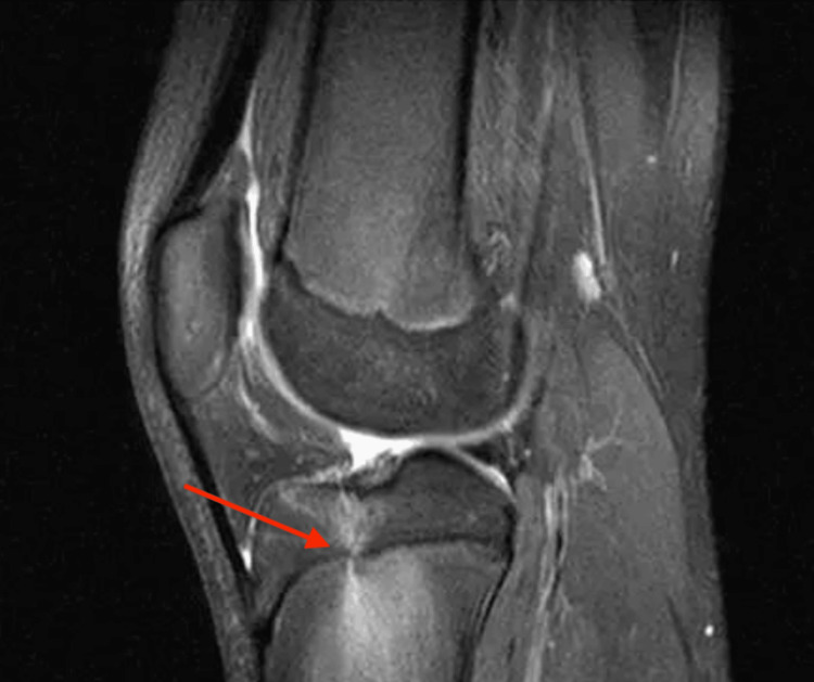 Figure 1
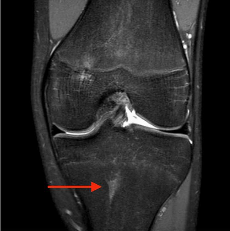 Figure 2
Peer Reviews
No public reviews on file for this paper yet. If you reviewed it on a platform where reviews are public (OpenReview, ICLR, NeurIPS, ICML), you can paste yours below so the community can read it here.
Videos
No videos yet. Explain this paper in a talk, walkthrough, or lecture? Add one.
Taxonomy
TopicsOrthopedic Infections and Treatments · Total Knee Arthroplasty Outcomes · Management of metastatic bone disease
Introduction
First described in 2011, focal periphyseal edema (FOPE) of the knee refers to transient bone marrow edema centered around the proximal tibial physis and distal femoral physis, most commonly observed during the process of physiologic epiphyseal fusion [1,2]. It typically presents in adolescents aged 11-14, often in active individuals, and may or may not be associated with pain or a history of trauma [1]. Patients can be further classified based on likely MRI findings and the presence or absence of symptoms, distinguishing between symptomatic and incidental FOPE. FOPE can present in unilateral or bilateral knees, as well as in the hips [1,3,4].
MRI is the most sensitive modality for detecting FOPE, revealing focal hyperintense signals on fluid-sensitive sequences [5]. Given the nature of FOPE, findings on X-ray and CT are often equivocal. On MRI, FOPE may be misinterpreted as other pathologies with similar imaging features, including stress fractures, chronic osteomyelitis, and contusional bone marrow edema [1,6,7]. Therefore, it is essential to correlate imaging findings with a detailed history, including the mechanism of injury, physical examination, and clinical context. One important differential diagnosis is a Salter-Harris fracture, which involves the physis of long bones and requires timely treatment [8].
Case presentation
Our case involves a 14-year-old female who presented to the hospital for evaluation of intermittent right anteromedial knee pain persisting for the past three months, first noticed while running during track practice. She has no significant medical history, recent illness, or relevant family medical history on file, and denies any injury or trauma. Her recent office visit revealed unremarkable clinical findings, and her X-rays were normal for her age.
On physical examination, her right knee showed no erythema or effusion, and there were no signs of infection or ligament involvement. She exhibited mild tenderness over the anteromedial knee. She had a full range of motion with bilateral hyperextension (-5° to 130° of flexion), and motor and sensory function were intact distally. Initially, her symptoms raised clinical suspicion for patellofemoral syndrome, prompting an MRI. The MRI revealed two focal zones of hyperintense signals around the knee in the distal femur, consistent with early stages of physiologic physeal closure (Figures 1-2). These may be a source of pain, especially in the absence of any other abnormality or injury on MRI. She also followed up with orthopedic surgery and opted for conservative management, including rest, cryotherapy, and activity modification. She was referred to physical therapy for gentle stretching and strengthening through low-impact exercises. Her symptoms gradually improved over three months without the need for surgical intervention. Given the symptomatic improvement, no additional imaging studies were pursued during subsequent follow-up visits.
T2-weighted, fat-suppressed sagittal view of the knee showing hyperintensities in the proximal tibial physis (arrow).
T2-weighted, fat-suppressed anteroposterior view of the knee showing hyperintense signals in the proximal tibial physis (arrow).
Discussion
FOPE is a transient bone marrow edema pattern centered around the closing physis, most commonly seen in adolescents during the early stages of physeal fusion. It is often an incidental MRI finding and may or may not be associated with pain. Despite its appearance, FOPE is considered a benign and self-limiting process that does not require further diagnostic testing or treatment [1].
Three key studies were identified in the literature review exploring FOPE in adolescent patients. In a 2018 study titled “Focal periphyseal edema: Are we overtreating physiologic adolescent knee pain?”, Giles E et al. reported on four patients (three females, one male; average age 13.7 years) who presented with MRI findings consistent with FOPE. The majority were active in high-impact sports such as soccer, basketball, and running. Notably, none reported recent trauma or mechanical symptoms, and their physical exams were unremarkable, showing no swelling, atrophy, or range of motion limitations. Conservative management led to complete symptom resolution. MRI findings were consistent with FOPE [9].
Similarly, Zbojniewicz AM and Laor T (2011) proposed that FOPE represents a physiological process related to early physeal closure and may explain knee pain in adolescents when no other abnormalities are detected on imaging. They emphasized that the MRI appearance of FOPE, a bone marrow edema pattern centered on the physis, should not be misinterpreted as pathology. In their view, FOPE requires neither invasive diagnostics nor follow-up imaging [1].
Ueyama H et al. (2018) presented three FOPE cases with long-term clinical and imaging follow-up. Their findings supported the benign nature of FOPE, as none of the patients developed deformity or leg length discrepancy. They concluded that observation alone was sufficient, though further longitudinal studies may help validate this approach [10].
Our case aligns with these findings. The patient’s clinical improvement over time without invasive intervention makes FOPE the likely cause of her symptoms. This supports the prevailing theory that FOPE is a physiological response during physeal closure in adolescents and should be recognized as a normal variant to prevent misdiagnosis and overtreatment.
Conclusions
As FOPE is a benign and self-limiting phenomenon that often coincides with normal physiologic physeal closure in adolescents, increased awareness among clinicians and radiologists can help distinguish it from more serious pathologies and guide appropriate care in the adolescent population.
As illustrated in our case, FOPE may present with nonspecific knee pain in the absence of trauma or other pathological findings. MRI is essential for accurate identification, and recognizing FOPE is crucial to avoid misdiagnosis and unnecessary interventions. Conservative management, including observation and physical therapy, is typically effective and remains the mainstay of treatment, with favorable outcomes. Despite its clinical relevance, FOPE remains a rare diagnosis, with only around 80 cases documented in the literature to date.
The reference list from the paper itself. Each links out to its DOI / PubMed record.
- 1Focal periphyseal edema (FOPE) zone on MRI of the adolescent knee: a potentially painful manifestation of physiologic physeal fusion?AJR Am J Roentgenol Zbojniewicz AM Laor T 998100419720112194059110.2214/AJR.10.6243 · doi ↗ · pubmed ↗
- 2Shoulder focal periphyseal edema: An unusual cause of shoulder pain in an adolescent J Rheumatol Larid G Duboe PO Gervais E 11925020233679211310.3899/jrheum.221291 · doi ↗ · pubmed ↗
- 3Focal periphyseal edema (FOPE): a newly reported cause of knee pain in adolescent athletes Orthop J Sports Med Wu M Bixby S Kramer DE Heyworth BE 232596711862018
- 4Unusual presentations of focal periphyseal edema zones: a report of bilateral symmetric presentation and partial physeal closure Case Rep Radiol Beckmann N Spence S 182015201510.1155/2015/465018 PMC 466001226640734 · doi ↗ · pubmed ↗
- 5Focal periphyseal edema of the knee in a junior youth tennis player: a case report Radiol Case Rep Yamaguchi J Funasaki H Kubota D Tanaka K Nagai S Saito M 358835912020254047568910.1016/j.radcr.2025.04.020PMC 12138506 · doi ↗ · pubmed ↗
- 6Growth plate of the normal knee: evaluation with MR imaging Radiology Harcke HT Synder M Caro PA Bowen JR 1191231831992154965710.1148/radiology.183.1.1549657 · doi ↗ · pubmed ↗
- 7The growing skeleton: MR imaging appearances of developing cartilage Magn Reson Imaging Clin N Am Khanna PC Thapa MM 4114211720091952419310.1016/j.mric.2009.03.012 · doi ↗ · pubmed ↗
- 8The high risk of infection with delayed treatment of open Seymour fractures: Salter-Harris I/II or juxta-epiphyseal fractures of the distal phalanx with associated nailbed laceration J Pediatr Orthop Reyes BA Ho CA 2472533720172632740110.1097/BPO.0000000000000638 · doi ↗ · pubmed ↗