Double aortic arch: a rare cause of adult-onset dysphagia
Xue-Mei Lin, Lin Xia, Xian-Fei Wang, Cong Yuan

Abstract
Genes, proteins, chemicals, diseases, species, mutations and cell lines named across the full text — each resolved to its canonical identifier and authoritative record.
Click any figure to enlarge with its caption.
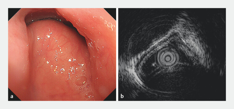 Fig. 1
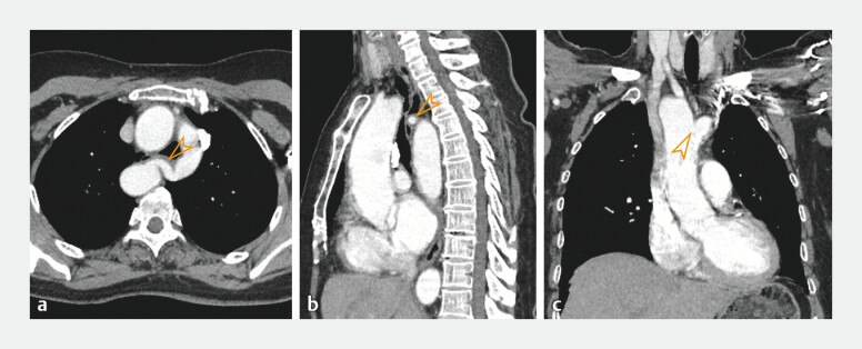 Fig. 2
Peer Reviews
No public reviews on file for this paper yet. If you reviewed it on a platform where reviews are public (OpenReview, ICLR, NeurIPS, ICML), you can paste yours below so the community can read it here.
Videos
No videos yet. Explain this paper in a talk, walkthrough, or lecture? Add one.
Taxonomy
TopicsTracheal and airway disorders · Congenital Heart Disease Studies · Aortic Disease and Treatment Approaches
A 61-year-old woman complained of intermittent dysphagia to solids for 4 months. There was no previous history of similar episodes, and dyspnea and stridor were also denied. A preliminary esophagogastroduodenoscopy indicated pulsatile subepithelial protrusions in the middle part of the esophagus (22 cm to 30 cm from the incisors), accompanied by significant deformation of the esophageal lumen ( Fig. 1 a , Video 1 ). Endoscopic ultrasonography showed that the subepithelial protrusions derived from extraluminal compression rather than intramural lesions ( Fig. 1 b ). Contrast-enhanced computed tomography scanning confirmed that the extraluminal compressions were being caused by aberrant vascular configuration, resulting from the double aortic arch (DAA) ( Fig. 2 ). Therefore, it became evident that the patient’s dysphagia was caused by a complete vascular ring due to DAA. The patient refused further surgical intervention.
Endoscopic and endosonographic findings of esophageal submucosal protrusions. a Endoscopic views showed an esophageal protrusion with normal overlying mucosa 22 cm from the incisors, suggestive of a subepithelial lesion. b Endoscopic ultrasonography revealed an intact esophageal wall covering the protrusion, indicating extraluminal compression.
Esophagogastroduodenoscopy indicated subepithelial protrusions and significant luminal deformation in the middle esophagus. Endoscopic ultrasonography and computed tomography confirmed that the changes arose from extraluminal compression by the double aortic arch.Video 1
Computed tomography (CT) findings of the chest. Contrast-enhanced CT scanning demonstrated a vascular ring (arrowhead) compressing the esophagus posteriorly on: a axial view; b sagittal view. The compression resulted in narrowing of the esophageal lumen, corresponding to the esophageal endoscopic views 22 cm away from the incisors. c Coronal images showed the ascending aorta bifurcating into the right and left aortic arches (arrowhead).
DAA is a rare congenital cardiovascular condition that results from the failure of the right fourth aortic arch to regress during embryonic development, constituting approximately 1% of cardiovascular congenital anomalies 1 . The vascular rings formed by the DAA may compress the encircled esophagus and trachea, causing dysphagia and wheezing. The clinical manifestations of DAA can differ depending on the degree of tightness of the ring and on subsequent tracheoesophageal compression. Some patients are completely asymptomatic, and others present late in life, as in this case. This patient developed dysphagia later in life, rather than earlier, which may be related to the altered elasticity of the arterial vessels in the elderly 2 .
Endoscopy_UCTN_Code_CCL_1AB_2AC_3AH
The reference list from the paper itself. Each links out to its DOI / PubMed record.
- 1Yaynishet YA Kibrom BT Abera MT Symptomatic vascular ring due to double aortic arch: a report of two cases Radiol Case Rep 2025209710010.1016/j.radcr.2024.09.13739444492 PMC 11497123 · doi ↗ · pubmed ↗
- 2Cavalcante JL Lima JA Redheuil A Aortic stiffness: current understanding and future directions J Am Coll Cardiol 2011571511152210.1016/j.jacc.2010.12.01721453829 · doi ↗ · pubmed ↗