Laser-guided rescue: endoscopic removal of complex esophageal foreign bodies
Priscilla Lopez, Mohan Ramchandani, Sundeep Lakhtakia, Krithi Krishna Koduri, Aniruddha Pratap Singh, Pramod Reddy, D. Nageshwar Reddy

Abstract
Genes, proteins, chemicals, diseases, species, mutations and cell lines named across the full text — each resolved to its canonical identifier and authoritative record.
Click any figure to enlarge with its caption.
 Fig. 1
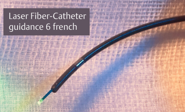 Fig. 2
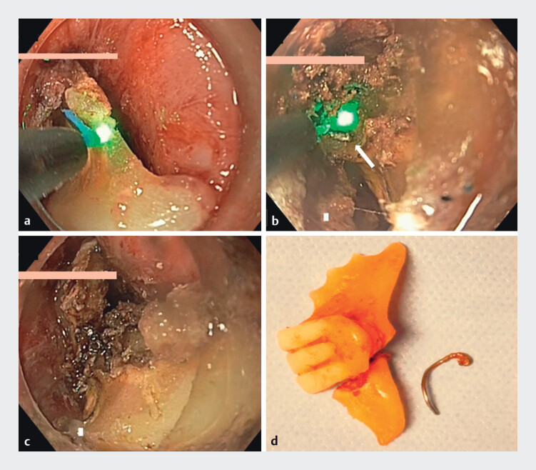 Fig. 3
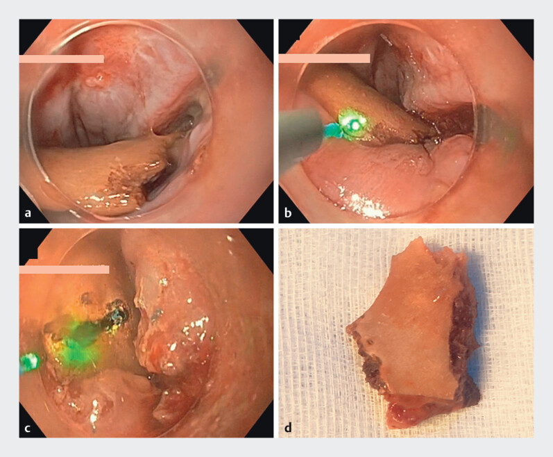 Fig. 4
Peer Reviews
No public reviews on file for this paper yet. If you reviewed it on a platform where reviews are public (OpenReview, ICLR, NeurIPS, ICML), you can paste yours below so the community can read it here.
Videos
No videos yet. Explain this paper in a talk, walkthrough, or lecture? Add one.
Taxonomy
TopicsForeign Body Medical Cases · Esophageal and GI Pathology · Airway Management and Intubation Techniques
About 10%–20% of esophageal foreign bodies require intervention 1 . Dentures are particularly difficult to remove due to their size, sharp edges, and metal parts 2 . While endoscopy is often effective, some cases need surgery 3 . We report two cases of successful laser-assisted removal of impacted foreign bodies.
Case 1 . A 35-year-old man with alcohol use disorder accidentally swallowed his denture. After failed removal attempts elsewhere, he was referred to our center. Computed tomography (CT) showed a radiopaque object at the D3 level ( Fig. 1 ). Endoscopy confirmed an impacted three-tooth denture with a sharp metallic edge, deeply embedded in the esophageal wall, with a contained perforation. Given the failure of conventional techniques, the patient was intubated and endoscopy-guided laser lithotripsy was used for fragmentation.
Computed tomography images. a–c A well-defined curvilinear radiopaque density measuring 3.2 × 6.2 cm was noted in the upper thoracic esophagus at the level of the D3 vertebral body with associated short segment circumferential wall thickening (9 mm).
Laser fiber (LightTrail Reusable 365 µm; Boston Scientific, Galway, Ireland) was preloaded onto a catheter (One Action Stent Introduction System [OASIS] internal catheter, 6 Fr; Cook Medical, Bloomington, Indiana, USA), compatible with the 2.8-mm channel ( Fig. 2 ). The laser source was a 360-nm Lumenis VersaPulse holmium laser (Boston Scientific, Marlborough, Massachusetts, USA), with settings of 9.6 W, Frequency 8 Hz, Energy 1200 mJ. This allowed precise disintegration. The narrowest section of the denture was cut ( Fig. 3 ), along with the metallic wire and acrylic resin. The entire procedure was completed in 15 minutes without collateral damage. The fragments were extracted with a snare ( Video 1 ). The decubitus ulcer-induced perforation was closed using the loop-and-clip technique.
Laser fiber (LightTrail Reusable 365 µm; Boston Scientific, Galway, Ireland) was preloaded onto a catheter (One Action Stent Introduction System [OASIS] internal catheter, 6 Fr, 203 cm; Cook Medical, Bloomington, Indiana, USA).
Case 1. a Laser-guided fragmentation of the denture. b Sectioning of the metallic wire. c Denture after complete fragmentation. d Retrieved denture fragments after removal.
Laser-assisted endoscopic fragmentation enabled successful removal of impacted esophageal foreign bodies and facilitated endoscopic closure, offering a safe alternative to surgery in complex cases.Video 1
Case 2 . A 67-year-old patient presented with dysphagia after eating chicken. CT revealed a foreign body in the upper esophagus. As in the previous case, retrieval failed. The bone was fragmented using the same laser technique without complications ( Fig. 4 ).
Case 2. a Impacted chicken bone. b, c Laser-guided fragmentation of the bone in the upper esophagus. d Retrieved bone fragment after removal.
Post-procedure assessments showed no leakage. Patients received prophylactic antibiotics, started oral intake the next day, and were discharged after 3 days, without complications.
Laser-assisted fragmentation is a safe and effective alternative for managing complex esophageal foreign bodies 4 5 , reducing surgery and enabling endoscopic closure in cases of perforation.
Endoscopy_UCTN_Code_TTT_1AO_2AL
The reference list from the paper itself. Each links out to its DOI / PubMed record.
- 1Birk M Bauerfeind P Deprez PH Removal of foreign bodies in the upper gastrointestinal tract in adults: European Society of Gastrointestinal Endoscopy (ESGE) Clinical Guideline Endoscopy 20164848949610.1055/s-0042-10045626862844 · doi ↗ · pubmed ↗
- 2Mughal Z Charlton AR Dwivedi R Impacted denture in the oesophagus: review of the literature and its management BMJ Case Rep 201912 e 22965510.1136/bcr-2019-229655 PMC 682777431653620 · doi ↗ · pubmed ↗
- 3Singh P Singh A Kant P An impacted denture in the oesophagus – an endoscopic or a surgical emergency – a case report J Clin Diagn Res 2013791992010.7860/JCDR/2013/5337.297623814744 PMC 3681071 · doi ↗ · pubmed ↗
- 4Yang Z Qin S Li X Esophageal foreign body removal under holmium laser-assisted gastroscope: a case report Front Surg 2023101.09416 E 610.3389/fsurg.2023.1094160 PMC 988686836733890 · doi ↗ · pubmed ↗
- 5Vishnu Swaroop Reddy N Shekhar S Sharma M Holmium:YAG laser-assisted removal of a lamb bone impacted in the upper oesophagus Indian J Otolaryngol Head Neck Surg 2023752338234110.1007/s 12070-023-03642-337636659 PMC 10447732 · doi ↗ · pubmed ↗