Neurosarcoidosis Limited to the Central Nervous System and Spleen, Presenting with Episodic Nonfluent Aphasia
Satoka Yano, Takashi Matsukawa, Kensho Sumi, Reo Yoshioka, Yusuke Baba, Hirotaka Maekawa, Masashi Hamada, Wataru Satake, Masako Ikemura, Tatsushi Toda

Abstract
Genes, proteins, chemicals, diseases, species, mutations and cell lines named across the full text — each resolved to its canonical identifier and authoritative record.
Click any figure to enlarge with its caption.
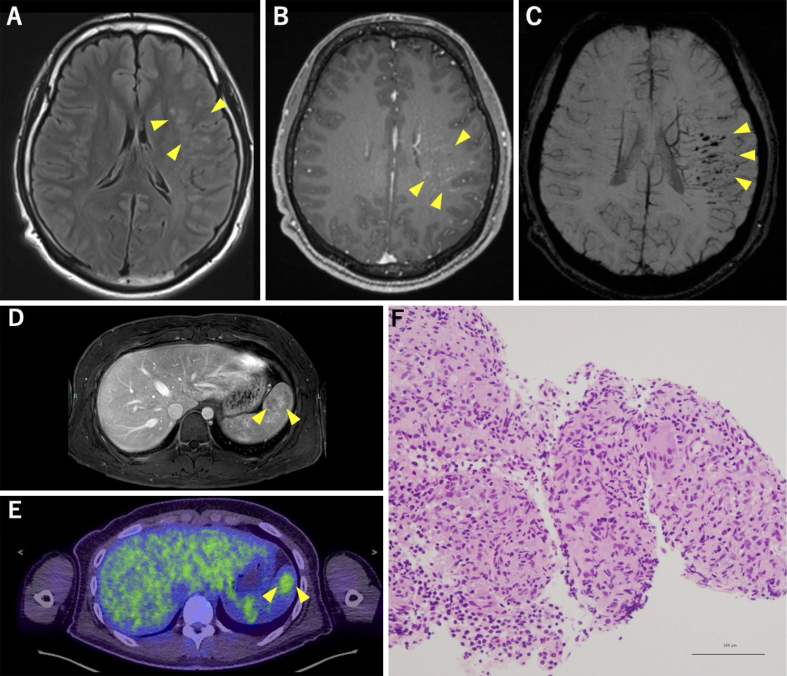 Figure 1
Peer Reviews
No public reviews on file for this paper yet. If you reviewed it on a platform where reviews are public (OpenReview, ICLR, NeurIPS, ICML), you can paste yours below so the community can read it here.
Videos
No videos yet. Explain this paper in a talk, walkthrough, or lecture? Add one.
Taxonomy
TopicsSarcoidosis and Beryllium Toxicity Research · S100 Proteins and Annexins · Radiopharmaceutical Chemistry and Applications
A 19-year-old right-handed previously healthy male presented with episodic, nonfluent aphasia lasting approximately 1 hour every few days for 4 months. Brain magnetic resonance imaging (MRI) revealed multiple hyperintense lesions in the superior temporal gyrus and supramarginal gyrus in fluid-attenuated inversion recovery images with linear contrast enhancement along the medullary veins (Figure 1A and B). Dilated medullary veins and multiple microbleeds were also noted in susceptibility-weighted imaging (Figure 1C). Abdominal MRI revealed mild splenomegaly and a heterogeneously enhanced lesion with variable contrast enhancement during the portal venous phase (Figure 1D). ^18^F-fluorodeoxyglucose positron emission tomography/computed tomography showed abnormal uptake corresponding to this splenic region, with a maximal standardized uptake value of 3.1 (Figure 1E). Ultrasound-guided splenic biopsy confirmed clustered epithelioid cell granulomas without caseous necrosis (Figure 1F), leading to the diagnosis of probable neurosarcoidosis. Comprehensive evaluations revealed no evidence of abnormalities in any other organs, including those in the pulmonary, cutaneous, cardiac, or ophthalmologic system.
Seizures in neurosarcoidosis ^(1)^ may result from brain parenchymal, meningeal lesions, or granuloma-associated small vessel vasculitis ^(2)^. In sarcoidosis, cases without pulmonary involvement are uncommon (8%) ^(3)^, and reports of lesions confined solely to the central nervous system (CNS) and spleen are exceedingly rare. However, considering the challenges associated with obtaining CNS biopsies, it is noteworthy that splenic biopsy aids in diagnosis.
Article Information
Conflicts of Interest
None
Author Contributions
Satoka Yano and Takashi Matsukawa acquired data and drafted the manuscript. Kensho Sumi, Reo Yoshioka, Yusuke Baba, Hirotaka Maekawa, Masashi Hamada, Wataru Satake, Masako Ikemura, and Tatsushi Toda edited and approved the final manuscript.
Informed Consent
Informed consent was obtained from the patient.
Approval by Institutional Review Board (IRB)
IRB approval was not required for this study.
The reference list from the paper itself. Each links out to its DOI / PubMed record.
- 1Fritz D, van de Beek D, Brouwer MC. Clinical features, treatment and outcome in neurosarcoidosis: systematic review and meta-analysis. BMC Neurol. 2016;16(1):220.27846819 10.1186/s 12883-016-0741-x PMC 5109654 · doi ↗ · pubmed ↗
- 2Lacomis D. Neurosarcoidosis. Curr Neuropharmacol. 2011;9(3):429-36.22379457 10.2174/157015911796557975 PMC 3151597 · doi ↗ · pubmed ↗
- 3James WE, Koutroumpakis E, Saha B, et al. Clinical features of extrapulmonary sarcoidosis without lung involvement. Chest.. 2018;154(2):349-56.29453944 10.1016/j.chest.2018.02.003 · doi ↗ · pubmed ↗