Evaluation of assumed tumour volume in multiple myeloma using dual-energy spectral CT and its correlation between haematological findings
Tetsuya Kosaka, Chisaki Masuda, Sachiho Tatebe, Risen Hirai, Akira Tanimura

TL;DR
This study uses dual-energy CT to measure tumor volume in multiple myeloma patients and finds a moderate correlation with β2-microglobulin levels.
Contribution
The novel use of dual-energy spectral CT to extract assumed tumor volume and its correlation with β2-microglobulin in multiple myeloma.
Findings
A moderate correlation was found between assumed tumor volume and β2-microglobulin levels (ρ = 0.69 for single threshold, ρ = 0.57 for double threshold).
Changes in assumed tumor volume and β2-microglobulin levels after treatment were similar in patients with multiple follow-ups.
Assumed tumor volume extracted via DESCT shows potential as a biomarker for multiple myeloma.
Abstract
To measure the assumed tumour volume in the humerus of patients with multiple myeloma using dual-energy spectral computed tomography (DESCT) and to evaluate the correlation with haematological indicators. We retrospectively analysed 82 DESCT examinations of 22 patients diagnosed with multiple myeloma. After extracting the bilateral humeri and removing the bone tissue, we measured the volume of the assumed tumour area using a single threshold based on Hounsfield unit values and double thresholds using material density images. We analysed the correlations between tumour volume and haematological indicators (β2-microglobulin, M-protein, free light chain, albumin, lactate dehydrogenase) and the trends after treatment intervention. A moderate correlation was identified between the assumed tumour volume in the initial scan and the β2-microglobulin level, with a correlation coefficient of ρ…
Genes, proteins, chemicals, diseases, species, mutations and cell lines named across the full text — each resolved to its canonical identifier and authoritative record.
Click any figure to enlarge with its caption.
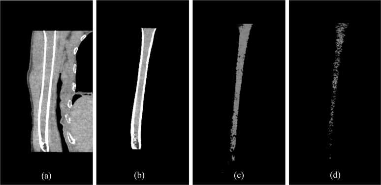 Figure 1
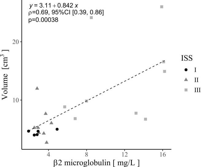 Figure 2
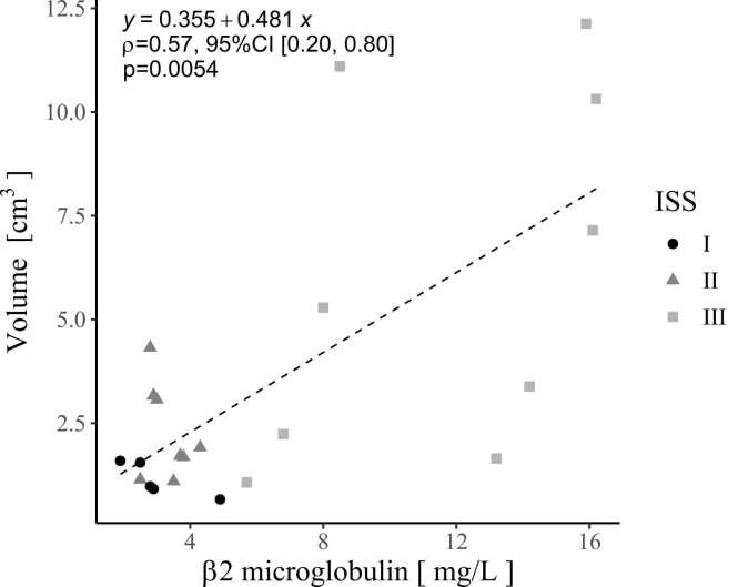 Figure 3
 Figure 4
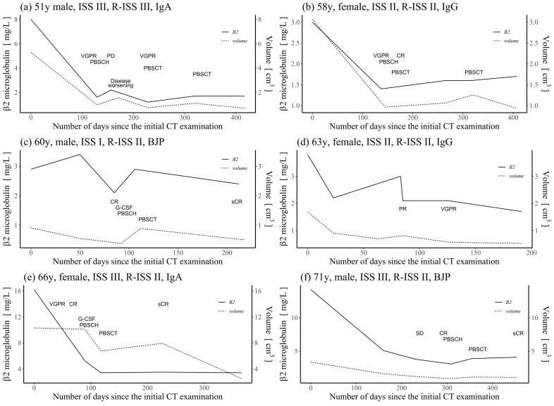 Figure 5
Peer Reviews
No public reviews on file for this paper yet. If you reviewed it on a platform where reviews are public (OpenReview, ICLR, NeurIPS, ICML), you can paste yours below so the community can read it here.
Videos
No videos yet. Explain this paper in a talk, walkthrough, or lecture? Add one.
Taxonomy
TopicsAdvanced X-ray and CT Imaging · Medical Imaging Techniques and Applications