Development of a deep learning based approach for multi-material decomposition in spectral CT: a proof of principle in silico study
Jayasai R. Rajagopal, Saikiran Rapaka, Faraz Farhadi, Ehsan Abadi, W. Paul Segars, Tristan Nowak, Puneet Sharma, William F. Pritchard, Ashkan Malayeri, Elizabeth C. Jones, Ehsan Samei, Pooyan Sahbaee

TL;DR
This study introduces a deep learning method for accurately identifying and measuring multiple materials in spectral CT scans using simulated data.
Contribution
A novel deep learning approach for multi-material decomposition in spectral CT is developed and validated using in silico datasets.
Findings
The model accurately classified and quantified iodine, gadolinium, and calcium in synthetic datasets.
Performance improved with increased inclusion of virtual patient phantoms in training.
The algorithm maintained strong performance under challenging imaging conditions like large patient size and reduced dose.
Abstract
Conventional approaches to material decomposition in spectral CT face challenges related to precise algorithm calibration across imaged conditions and low signal quality caused by variable object size and reduced dose. In this proof-of-principle study, a deep learning approach to multi-material decomposition was developed to quantify iodine, gadolinium, and calcium in spectral CT. A dual-phase network architecture was trained using synthetic datasets containing computational models of cylindrical and virtual patient phantoms. Classification and quantification performance was evaluated across a range of patient size and dose parameters. The model was found to accurately classify (accuracy: cylinders – 98%, virtual patients – 97%) and quantify materials (mean absolute percentage difference: cylinders – 8–10%, virtual patients – 10–15%) in both datasets. Performance in virtual patient…
Genes, proteins, chemicals, diseases, species, mutations and cell lines named across the full text — each resolved to its canonical identifier and authoritative record.
Click any figure to enlarge with its caption.
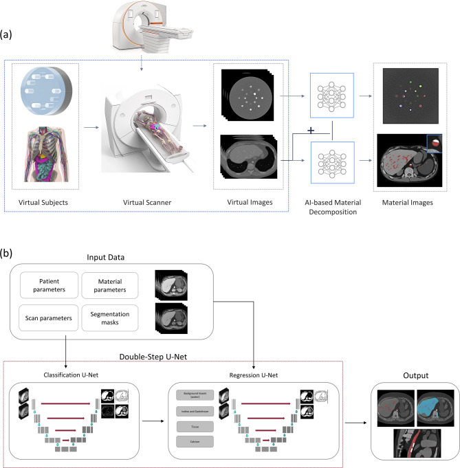 Figure 1
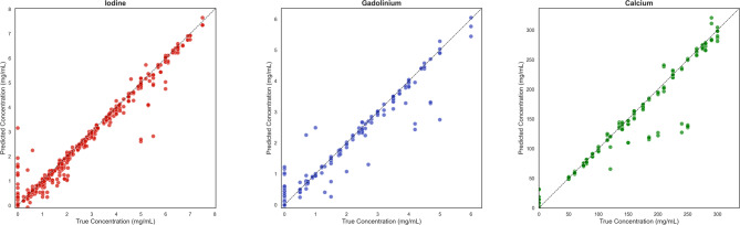 Figure 2
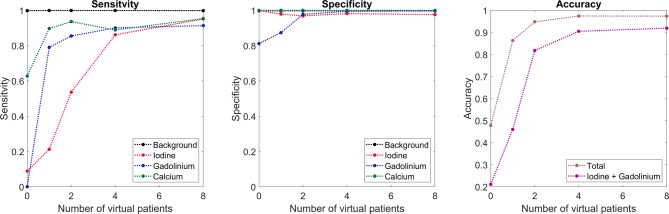 Figure 3
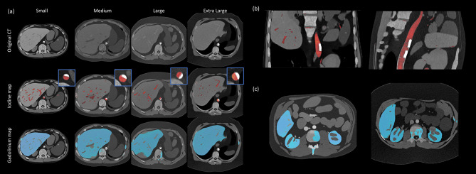 Figure 4
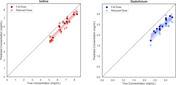 Figure 5
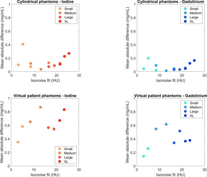 Figure 6
Peer Reviews
No public reviews on file for this paper yet. If you reviewed it on a platform where reviews are public (OpenReview, ICLR, NeurIPS, ICML), you can paste yours below so the community can read it here.
Videos
No videos yet. Explain this paper in a talk, walkthrough, or lecture? Add one.
Taxonomy
TopicsAdvanced X-ray and CT Imaging · Radiation Dose and Imaging · Medical Imaging Techniques and Applications