MRI-based multiregional radiomics for preoperative prediction of Ki-67 expression in meningiomas: a two-center study
Ming Luo, Guihan Lin, Duoning Chen, Weiyue Chen, Shuiwei Xia, Junguo Hui, Pengjun Chen, Minjiang Chen, Wangyang Ye, Jiansong Ji

TL;DR
This study uses MRI scans to predict Ki-67 levels in meningiomas, offering a non-invasive way to assess tumor aggressiveness before surgery.
Contribution
A novel clinical-radiomic model combining MRI features and clinical data to predict Ki-67 expression in meningiomas.
Findings
Peritumoral edema and heterogeneous enhancement were independent predictors of high Ki-67 expression.
The radiomics model achieved an AUC of 0.883 in training and 0.811 in validation.
A nomogram integrating clinical and radiomic features achieved an AUC of 0.904 for high Ki-67 prediction.
Abstract
High expression of Ki-67 in meningioma is significantly associated with higher histological grade and worse prognosis. The non-invasive and dynamic assessment of Ki-67 expression levels in meningiomas is of significant clinical importance and is urgently required. This study aimed to develop a predictive model for the Ki-67 index in meningioma based on preoperative magnetic resonance imaging (MRI). This study included 196 patients from one center (internal cohort) and 92 patients from another center (external validation cohort). Meningioma had to have been pathologically confirmed for inclusion. The Ki-67 index was classified as high (Ki-67 ≥ 5%) and low (Ki-67 < 5%). The internal cohort was randomly assigned to training and validation sets at a 7:3 ratio. Radiomics features were selected from contrast-enhanced T1-weighted MRI using the least-absolute shrinkage and selection operator…
Genes, proteins, chemicals, diseases, species, mutations and cell lines named across the full text — each resolved to its canonical identifier and authoritative record.
Click any figure to enlarge with its caption.
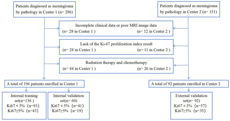 Figure 1
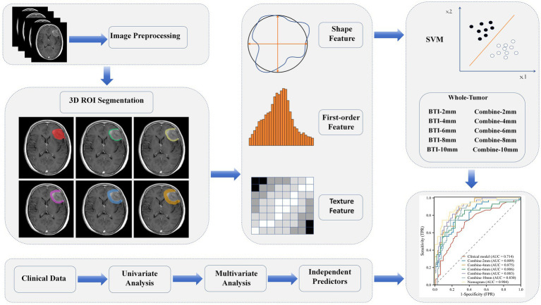 Figure 2
 Figure 3
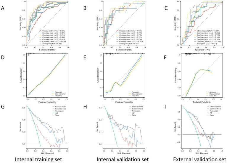 Figure 4
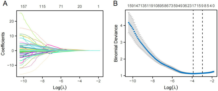 Figure 5
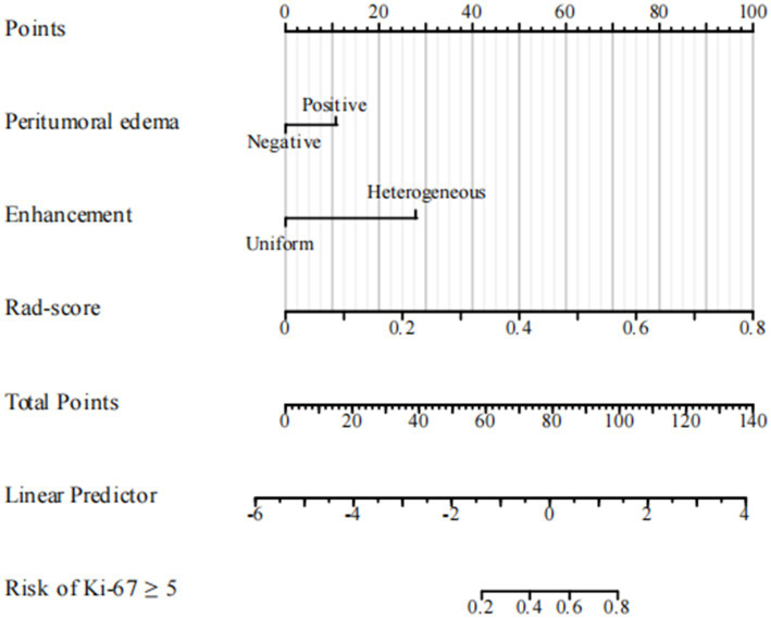 Figure 6
Peer Reviews
No public reviews on file for this paper yet. If you reviewed it on a platform where reviews are public (OpenReview, ICLR, NeurIPS, ICML), you can paste yours below so the community can read it here.
Videos
No videos yet. Explain this paper in a talk, walkthrough, or lecture? Add one.
Taxonomy
TopicsMeningioma and schwannoma management · Radiomics and Machine Learning in Medical Imaging · Neurofibromatosis and Schwannoma Cases