Effects of Immunostimulants on Physiological Performances, Immune Gene Expression, Liver, and Intestinal Protection Against Vibrio parahaemolyticus Infection in Juvenile Asian Seabass (Lates calcarifer)
Yun Liu, Qian-Qian Chen, Victor Charlie Andin, Wei-Kang Chor, Chou-Min Chong, Po-Tsang Lee, Crystale Siew-Ying Lim, Jiun-Yan Loh

TL;DR
This study explores how adding immunostimulants like ginger, garlic, and palm carotene to the diet of juvenile Asian seabass improves their immune response and protects against bacterial infection.
Contribution
The study introduces a combination of natural immunostimulants as a potential immune booster in aquaculture feed for Lates calcarifer.
Findings
Palm carotene supplementation reduced intestinal inflammation and liver damage in infected fish.
Combined treatment with ginger, garlic, and palm carotene improved intestinal and liver protection compared to individual supplements.
The combination increased goblet cell numbers and intestinal villus surface area in infected fish.
Abstract
The present study aims to develop a more valuable innovative aquaculture feed for the sustainable Lates calcarifer aquaculture industry. An experiment with a completely random design was developed with five groups containing 0% immunostimulant and 0.5% ginger, 1.0% garlic, 0.15% palm carotene alone or in combination. Each diet was randomly assigned to triplicate groups of 30 fish (10.06 ± 0.09 g) per tank. The fish were fed once daily to apparent satiation. Throughout the 56-day feeding trial, the results suggest that although no significant differences were observed among almost all experimental groups in growth performance, feed efficiency, fish composition and disease resistance, the palm carotene supplementations downregulated intestinal tumor necrosis factor alpha (TNF-α) and interleukin (IL)-6 expression, decreased sum scores of liver pathological changes, increased intestinal…
Genes, proteins, chemicals, diseases, species, mutations and cell lines named across the full text — each resolved to its canonical identifier and authoritative record.
Click any figure to enlarge with its caption.
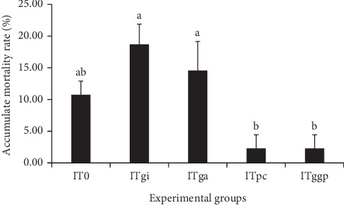 Figure 1
 Figure 2
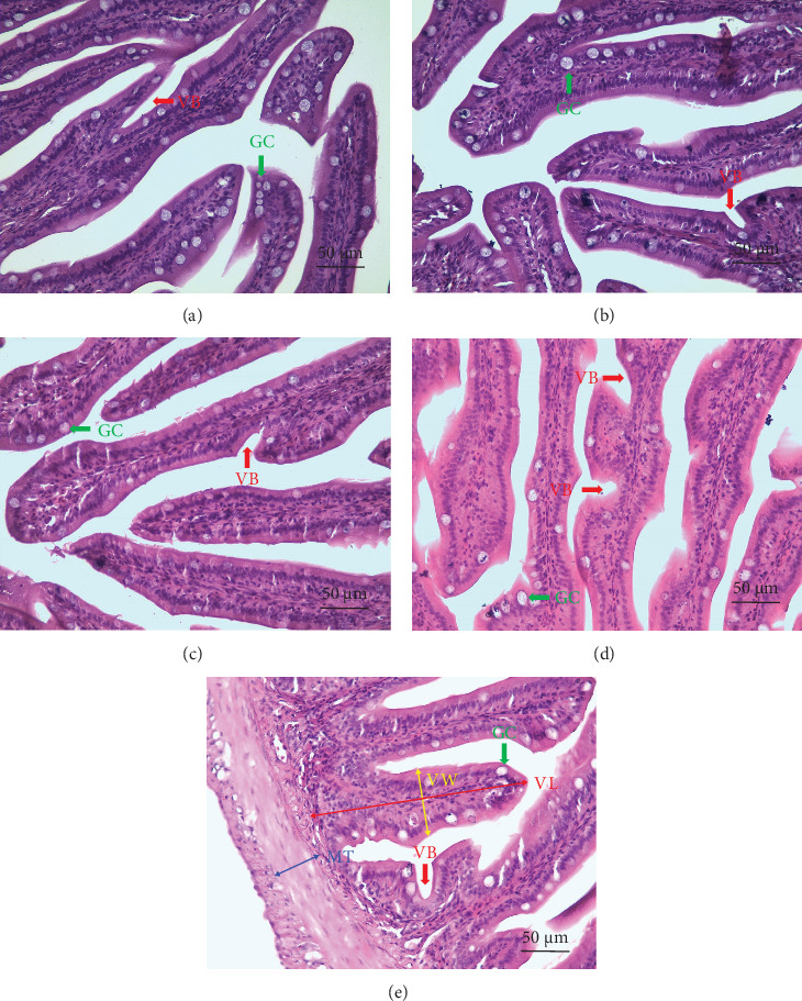 Figure 3
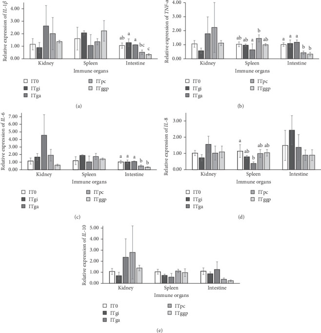 Figure 4
- —Worldwide Fund for Nature, Malaysia
- —Guangxi Youth Innovation Talent Research Program
- —Guangxi Young and Middle-Aged Teacher Scientific Research Ability Enhancement Program
- —Guangxi University of Science and Technology
- —UCSI University
Peer Reviews
No public reviews on file for this paper yet. If you reviewed it on a platform where reviews are public (OpenReview, ICLR, NeurIPS, ICML), you can paste yours below so the community can read it here.
Videos
No videos yet. Explain this paper in a talk, walkthrough, or lecture? Add one.
Taxonomy
TopicsAquaculture disease management and microbiota · Aquaculture Nutrition and Growth · Invertebrate Immune Response Mechanisms
1. Introduction
Asian seabass (Lates calcarifer), also commonly referred to as barramundi, is widely distributed across the Indo–Pacific and Australia. It is a highly profitable aquaculture fish because of its exceptional physiological tolerance, rapid growth, ease of maintenance, and productive qualities [1]. Based on the analysis by Future Market Insights (FMI), sales of L. calcarifer have experienced a 4.1% compound annual growth rate (CAGR) from 2016 to 2020. The FMI projections indicate that the worldwide market for L. calcarifer is projected to continue growing at a 5.5% CAGR through 2031 [2]. However, like any other cultured fish, L. calcarifer is affected by pathogens due to intensive culture [3]. This fish is prone to infectious diseases, such as Vibrio parahaemolyticus, which can cause tens of millions of dollars in annual economic losses [1].
Vibriosis, caused by pathogenic Vibrio species, has emerged as one of the most devastating bacterial diseases in global aquaculture. To combat this challenge, fish farmers have increasingly incorporated food additives or antibiotics into their practices over the past few decades. However, this approach constitutes a menace to the well-being of humans and the environment due to the occurrence of residues in fish bodies [4]. In addition, antimicrobial overuse and misuse are driving an increase in antimicrobial resistance (AMR), which has become a serious global health issue [5]. Instead of chemotherapeutic agents, increasing attention is being paid to the use of immunostimulants as a prophylactic measure for the prevention of diseases in aquaculture. Immunostimulants present an appealing and promising alternative to antibiotics, chemicals, and vaccines because they can activate the immune response by enhancing the activity of phagocytes and T and B cells [6]. Among others, plant-based immunostimulants have received more attention because they are cost-effective and eco-friendly. Ginger (Zingiber officinale, Roscoe) is one of the most widely used spices worldwide. This folk medicine is generally considered a safe herbal medicine for treating different diseases [7]. Studies have reported that the highest survival rate (SR) in L. calcarifer challenged with Vibrio harveyi [8] and in Nile tilapia (Oreochromis niloticus) infected with Aeromonas hydrophila [9] was achieved in supplemented diets containing 0.5% and 1% ginger, respectively. Another example, such as garlic (Allium sativum), is widely used around the world. Its functional properties have been extensively documented for their pivotal role in altering the principal risk factors associated with chronic diseases over an extended period [10]. Numerous studies and reports have consistently affirmed that garlic can efficiently eliminate pathogenic bacteria, including Aeromonas punctata, Edwardsiella tarda, Fibrobacter intestinalis, Myxococcus piscicola, Pseudomonas fluorescens, Vibrio anguillarum, and Yersinia ruckeri, in freshwater fish [11]. Most garlic research in aquaculture has involved garlic extracts [12], garlic powder [13], garlic peel [14], and fresh garlic [15, 16]. Among them, Irkin et al. [13] suggested that garlic powder supplementation is recommended in feeding regimens for European sea bass (Dicentrarchus labrax) juveniles; however, the dosage should not exceed 2%. On the other hand, Agbebi et al. [15] found that even 30% garlic inclusion in feed has no negative effect on the liver and gut of African catfish (Clarias gariepinus).
Like garlic, β-carotene is one of the main palm oil processing byproducts that can be used in the food, pharmaceutical, and cosmetics industries, apart from the oleochemical industries [17]. It is extracted from empty fruit bunches of oil palm, which are an abundant agricultural waste in Malaysia [18]. β-carotene, a carotenoid with no toxicity, exhibits immunomodulatory effects in both animals and humans [19]. It can be used as a natural immunostimulant to improve fish antioxidant capacity and immune status [20]. Several studies have indicated that the administration of palm carotene to mice at a dosage of 50 mg carotene per 100 g of diet can effectively prevent DNA impairment in bone marrow, reduce peripheral leukocyte counts, and enhance survival after X-ray irradiation [21]. Administration of 0.05% palm carotene through drinking water led to a significant decrease in the percentage of mice bearing tumors [22].
Our findings in a previous study [23] indicated that incorporating 10% black soldier fly larval (BSFL) meal into the diet of L. calcarifer can provide the best growth performance, feed efficiency, and economic returns. However, it does not significantly improve disease resistance. To optimize feed formula, the current study aimed to examine the effects of ginger, garlic, and palm carotene on physiological performances, immune gene expression, disease resistance, and morphological characteristics of juvenile L. calcarifer challenged with V. parahaemolyticus.
2. Materials and Methods
2.1. Experimental Diets and Proximate Analysis
Based on our findings in a previous study, a 10% BSFL meal in fishmeal substitution could enhance L. calcarifer growth performance, feed efficiency, and economic returns [23]. A completely random experimental design was developed with five groups based on 10% BSFL inclusion in the diet. Immune treatments including ginger, garlic, and palm carotene were supplemented in five isonitrogenous and isocaloric practical diets and were designated as IT0 (without any immunostimulant, serve as control), ITgi (containing 0.5% ginger), ITga (containing 1.0% garlic), ITpc (containing 0.15% palm carotene), and ITggp (containing 0.5% ginger + 1.0% garlic + 0.15% palm carotene). The BSFL meal was provided by Sentara Group (formerly known as Nutrition Technologies Sdn. Bhd.) (Johor, Malaysia), and palm carotene (the palm phytonutrient actives) manufactured by ExcelVite Sdn. Bhd. (Ipoh, Malaysia) for aquaculture. Ginger and garlic were obtained from the local market, chopped, and stirred for later use. All feed ingredients except for the above-mentioned were purchased from Nutri Vet Livestock Sdn. Bhd. (Negeri Sembilan, Malaysia). The feed raw materials were prepared in accordance with Table 1. The ingredients were well mixed, crushed, sieved, and pelleted into a feeding diet with a particle size of 1.5 mm through a feed pelletizer machine (Model 160, Guangzhou Juncheng Machinery Equipment Co. Ltd, China). The pellets were oven-dried at 55 °C for 12 h and subsequently sealed in vacuum-packed bags. Diets were stored in a dry and dark environment at room temperature until use.
The proximate composition of all diets was determined at Apical Scientific Sdn. Bhd. (Selangor, Malaysia), in accordance with the standard Association of Official Analytical Chemists (AOAC) methods. Ash, moisture, crude fat, and crude protein were analyzed using AOAC methods 942.05, 930.15, 920.39, and 988.05 (2016). Total carbohydrate was measured in accordance with MY/STP/378 based on US FDA 21 CFR101.9 Part 101 (2017). The proximate analysis results for all diets used in this study are shown in Table 1. The diets in all the treatments had a crude protein of between 33.10% and 34.65%, crude fat of between 6.70% and 7.20%, moisture of between 9.15% and 9.90%, ash of between 8.55% and 9.65%, and carbohydrate of between 39.90% and 41.60%. The above indices among all diets showed only slight variation.
2.2. Fish Feeding Trial
A total of 450 apparently healthy L. calcarifer juveniles (10.06 ± 0.09 g, 7.97 ± 0.04 cm) were purchased from F1 Aquaculture Sdn. Bhd. (Kuala Lumpur, Malaysia) and transported to the UCSI University Aquaculture Laboratory. The fish were divided into five groups at random, with each group consisting of three replicates. They were then placed into 15 round canvas tanks, each with a capacity of 300 L and accommodation for 30 fish. During the 7 days of acclimation, the fish were adequately fed with the IT0 diet. During the 56-day feeding trial, fish were manually fed to the point of visual satiation once daily (10:00) at approximately 4% of the fish's body weight (BW). The quantity of feed supplied to each experimental tank was recorded to calculate feed intake (FI) over the 56-day feeding trial. Feces were electrically siphoned out daily before morning feeding. Any remaining unconsumed feed was siphoned off after half an hour of each feeding. Every other day, one-third of the water in each tank was exchanged. Each week, water quality parameters, including salinity, dissolved oxygen (DO), water temperature, potential of hydrogen (pH), ammonia-N, and nitrite, were monitored. The salinity, water temperature, and pH were measured with a multifunctional water quality tester (C-600, Shenzhen Yieryi Technology Equipment Co. Ltd, China) and were 8.2–10.4 ppt, 28.7–30.1 °C, and 7.1–7.8,respectively. DO levels were measured using a DO analyzer (BLE-9100, Shenzhen Yieryi Technology Equipment Co. Ltd, China) and were maintained above 5.5 mg/L. Ammonia-N and nitrite were measured using a multiparameter portable colorimeter (DR900, Hach Company, America) and were consistently <0.1 and <0.01 mg/L, respectively.
2.3. Fish Physiological Performance and Body Composition
Following the feeding trial, the fish were subjected to a 24-h fasting period and subsequently anesthetized using tricaine methanesulfonate (MS-222) (100 mg/L) in small, aerated tanks. The number and weight of fish in each tank were recorded to calculate the SR, total weight gain (TWG), specific growth rate (SGR), and feed conversion ratio (FCR). Six fish per tank were randomly collected and killed. The fish's BW and body length (BL) were determined for condition factor (CF), viscera and liver were measured for viscerosomatic index (VSI) and hepatosomatic index (HSI), and the body was preserved at −20 °C for subsequent proximate composition determination. The following parameters were calculated to evaluate growth performance and feed efficiency [24]:
The proximate composition of the fish body was determined as described above for the proximate analysis of diets.
2.4. RNA Extraction, Complementary DNA (cDNA) Synthesis, and Quantitative Real-Time Polymerase Chain Reaction (qPCR) Analysis
Head kidney, spleen, and mid-intestine from fish in each treatment (n = 3) were collected at the end of the feeding trial and preserved in NucleoProtect RNA (Macherey-Nagel, Germany), incubated at 4 °C for 24 h, and stored at −20 °C until RNA extraction. Total RNA was extracted from the tissue samples homogenized via liquid nitrogen grinding using the Monarch Total RNA Miniprep Kit (New England Biolabs, USA) in accordance with the manufacturer's protocol, with a final elution at 30 µL. The concentration of RNA was determined using a Biophotometer (D30, Eppendorf, Germany), and purity was evaluated by measuring the OD260/280 and OD260/230 absorption ratios. RNA integrity was verified by running 5 µL RNA on an ethidium bromide-stained 1% agarose gel submerged in 1× Tris Acetate EDTA (TAE) buffer, and the results were visualized using a gel documenter (Gel Doc XR+, Bio-Rad, USA). RNA was preserved at −80 °C for later use. cDNA was synthesized from 9 µL of RNA using a RevertAid RT Reverse Transcription Kit (Thermo Scientific, USA) according to the manufacturer's instructions. Mastercycler nexus gradient (Eppendorf, Germany) was used to run the cDNA synthesis with the following parameters of 25 °C for 5 min, followed by 42 °C for 60 min, and 70 °C for 5 min. The synthesized cDNA was preserved at −20 °C until it was used for qPCR analysis.
TaqMan-based qPCR was performed to determine the target gene expression between interleukin (IL)-1 beta (IL-1β), tumor necrosis factor alpha (TNF-α), IL-6, IL-8, and IL-10. The StepOne Real-Time PCR system (Applied Biosystems, USA) used in this experiment could run duplex probe reactions; hence, this experiment was designed using two probes, FAM and HEX, for all reactions. The primers and probes were designed by Apical Scientific Sdn. Bhd. (Selangor, Malaysia), and the relative information is listed in Table 2. The cDNA was diluted to a concentration of 13.5 ng/µL with a final volume of 10 µL, and then the reaction master mix was prepared. Each reaction comprised of 10 µL of SensiFAST Probe MasterMix, Hi-ROX (Bioline, UK), 7 µL of ddH_2_O, 1 µL of primer/probe mix 1, 1 µL of primer/probe mix 2, and 1 µL of cDNA samples. The thermal profile for all reactions was 2 min at 95 °C, followed by 40 cycles of 10 s at 95 °C, and 35 s at 60 °C. The expression levels of the target genes were calculated using the 2^−ΔΔCt^ method, and the data were normalized with β-actin as the housekeeping gene. Each sample assay was performed in triplicate.
2.5. In Vivo Bacterial Challenge
The bacteria employed in this study, Vibrio parahaemolyticus, was isolated from diseased Pacific white shrimp (Litopenaeus vannamei) cephalothorax collected from aquaculture farms and provided by the Laboratory of Marine Biotechnology, Institute of Bioscience, Universiti Putra Malaysia. The pathogenic strain was further confirmed using polymerase chain reaction (PCR) (with universal primers 27F and 1492R, 27F: 5′-AGAGTTTGATCMTGGCTCAG-3′, 1492R: 5′-TACGGYTACCTTGTTACGACTT-3′). Following overnight incubation in 3% marine Luria broth at 30 °C, bacteria were harvested by centrifugation (×10,000 rpm) and resuspended in 0.8% physiological saline solution (PSS) in a 50-mL centrifuge tube. Before the bacterial challenge test, a preliminary experiment was conducted to determine the dose of the pathogenic bacteria (LD_50_ was 0.03 × 10^7^ CFU/g). For the vibrio-challenge test, 20 fish were selected randomly and retained in each tank at the end of the 56-day feeding trial. Subsequently, each fish was anesthetized using MS-222 and then subjected to an intraperitoneal injection of 0.3 mL of a pathogenic strain at a concentration of 1 × 10^7^ CFU/mL. On the third day, each tank was poured with 30 mL of the same concentration of bacteria. Fish were subjected to a 24-h fasting period before the bacterial challenge test and re-fed with the respective experimental diets after 12 h. A range of vibriosis symptoms, including the presence of a thick layer of mucus on the body surface, fin congestion, and skin and muscle tissue hemorrhage and ulceration, were observed and recorded daily for 7 days. The cumulative mortality rate (CMR) of the fish was recorded and calculated as follows [25]:
2.6. Histological Analysis of Liver and Intestinal Samples
Moribund fish in the challenge experiments were instantly collected for histological analysis. The liver and mid-intestine from each fish of the tanks (n = 3) were dissected, removed, and stored in 10% formalin solution for histological analysis. The tissues were dehydrated using a range of graded (80%, 90%, 95% and 100%) ethanol baths, equilibrated with xylene, and subsequently embedded in paraffin wax, sections with a thickness of 5 µm were prepared, whereby two slides were cut for each sample at different depths in the wax block using a rotary microtome (RM2235, Leica, Germany), and subsequently stained with hematoxylin eosin (HE) using conventional histological methods. Slides were examined and representative pictures were taken under a light microscope (E200MV, Nikon, Japan) at 400-fold magnification, which was equipped with a photomicrograph connected to a computer with SmartV digital imaging system software (Jiangsu Jieda Technology Development Co. Ltd, China).
Morphometric evaluation of the liver was performed following the methodology outlined by Caimi et al. [26] with minor adjustments. In brief, six images per liver section were used to assess the following histological alterations: vascular congestion (VC), cytoplasmic vacuolization (CV), necrotic tissue (NT), inflammatory cell infiltration (II), and nuclear displacement (ND). The assessment of observed findings in liver sections employed a 5-point semiquantitative scoring system, where 0 denotes no alterations, 1 indicates minimal histopathology present in less than 25% of the observed areas, 2 represents minor histopathology present in less than 50% of the areas, 3 signifies moderate histopathology present in less than 75% of the areas, and 4 indicates severe histopathology observed in more than 75% of the areas.
To measure intestinal mucosal morphology, 10 villi per intestinal section were randomly selected from a cross-section. Villus length (VL), villus width (VW), muscular thickness (MT), and the number and density of goblet cells (GCs) per villus were measured following the protocols outlined by Hisano et al. [27] and Wassef et al. [28]. The villus surface (VS) was calculated using the formula [29] as follows:
2.7. Statistical Analysis
All data were analyzed using SPSS version 23.0. Before initiating statistical analysis, normality and homogeneity of variance for all dependent variables were assessed using Shapiro–Wilk's and Levene's tests. The histopathological scores were analyzed by Kruskal–Wallis ANOVA, and the results are expressed as the mean. The other indices underwent one-way ANOVA, and the subsequent Tukey's test was employed for post hoc analysis. The results are shown as means ± SE. Significant differences were declared at p < 0.05.
3. Results
3.1. Growth Performance and Feed Efficiency
The growth performance and feed efficiency of L. calcarifer under different dietary treatments are detailed in Table 3. At the start of the feeding trial, initial BW, BL, and CF values exhibited no statistically significant differences (p > 0.05). During the feeding trial, the SR was 88.65%–96.25% and was unaffected by any dietary treatment. At the end of the feeding trial, the VSI was significantly (p < 0.05) decreased due to palm carotene supplementation (ITpc and ITggp groups) compared with the control group. No statistically significant differences (p > 0.05) were observed among all experimental groups in terms of final BW, BL, HSI, TWG, SGR, FI, and FCR. The final CF of the fish fed with the immunostimulant supplement was comparable (p > 0.05) to that of the control group.
3.2. Proximate Composition of the Fish Body
Proximate analysis of the fish body after the feeding trial is presented in Table 4. In comparison with the control group, the crude protein in the ITgi group and the moisture in the ITggp group showed a significant (p < 0.05) increase. No significant difference (p > 0.05) was found in crude fat, ash, and carbohydrate in all immunostimulant supplementation groups compared with the control group.
3.3. Postchallenge Cumulative Mortality
The CMR of L. calcarifer after infection with V. parahaemolyticus is shown in Figure 1. The CMRs of the ITpc and ITggp groups were low at 2.22% compared with 10.69% in the control group. However, no statistical differences (p > 0.05) were found among the three aforementioned groups. In contrast, the CMRs of the ITgi (18.65%) and ITga (14.52%) groups were high and similar to those of control group (p > 0.05).
3.4. Histology of the Liver
The histomorphological changes in the liver morphological examination of L. calcarifer challenged with V. parahaemolyticus are shown in Figure 2. Clear VC and CV with rare NT, II, and ND were observed. The histopathological assessment of the liver is shown in Table 5. In comparison to the control group, congestion scores exhibited a significant (p < 0.05) decrease with immunostimulant supplementation; the vacuolization score exhibited a significant (p < 0.05) increase in the ITpc group; necrosis scores and sum scores of pathological changes exhibited a significant (p < 0.05) decrease in the ITga, ITpc, and ITggp groups; and inflammation scores were significantly (p < 0.05) decreased in the ITpc and ITggp groups. However, the ND scores did not show significant differences (p > 0.05) across all experimental groups. Apart from the inflammation score, the poorest values of the other indices were recorded in the ITggp group.
3.5. Histology of the Intestine
The histomorphological changes in the intestinal morphological examination of L. calcarifer challenged with V. parahaemolyticus are shown in Figure 3. Occasional instances of villus breakage and the presence of GC were observed in all experimental groups. Morphometric analysis of the intestinal mucosa is shown in Table 6. The ITggp group showed the highest values with statistical significance (p < 0.05) in VW, VS, and GC numbers per villus. Meanwhile, VS exhibited a significant (p < 0.05) increase in the ITpc group compared with the control group. However, the GC density exhibited a significant (p < 0.05) reduction in the ITgi group among treatments. No significant differences (p > 0.05) were noted in VL and MT.
3.6. Immune Gene Expression
The relative expression of IL-1β, TNF-α, IL-6, IL-8, and IL-10 in the kidney, spleen, and intestine of L. calcarifer is shown in Figure 4. IL-1β expression in the intestine of the ITggp group, TNF-α and IL-6 expression in the intestine of the ITpc and ITggp groups, and IL-8 expression in the spleen of the ITga group were significantly (p < 0.05) downregulated compared with the control group. No significant difference (p > 0.05) was observed in the above-mentioned genes among other experimental groups in other organs. Furthermore, the relative expression of IL-10 in all experimental groups was significantly unaffected (p > 0.05) by immunostimulant supplementation.
4. Discussion
Incorporation of immunostimulants in aquaculture has paved the way for advancements in fish health protection [30]. Many studies have proven that there are many types of immunostimulants that can activate the fish immune system. According to their sources, they can be roughly divided into saccharides and their complexes from microbes [31], animals [32], plants [33], organisms [34], and their extracts [35], chemical compounds [28], nutritional factors [36], hormones [37], and other cytokines [38]. Among them, plant immunostimulants have the advantages of abundant resources, low cost, drug residue-free, drug resistance-free, and convenient use, making them an important strategy for promoting the healthy and sustainable development of the aquaculture industry. Ginger, garlic, and palm carotene were chosen in the present study because of their availability, wide cultivation, and high production in Malaysia [39], especially palm oil, as the country holds a strategic position in the global production and export of palm oil. Biochemical conversion of palm oil solid waste into the extraction of carotene can promote faster growth in the sustainability of the industry [40]. There have been reports of supplementing the diet with ginger in L. calcarifer [8] and O. niloticus [9], garlic in D. labrax [13], and C. gariepinus [15, 16], β-carotene in O. niloticus [41] and Piaractus mesopotamicus [42]. Immunostimulants are crucial for enhancing nonspecific immune responses. For example, they can promote the synthesis of complement, lysozyme, protease inhibitor, C-reactive protein, natural hemolysin, agglutinin, α-macroglobulin, macrophage activating factor, and interferon [43]. They can activate the phagocytic and bactericidal functions of macrophages, neutrophils, and specific cytotoxic cells [44]. In addition, immunostimulants can improve the level of fish IgM antibodies and enhance the level of fish-specific immune responses [45]. Among the immunostimulants used in the present study, ginger and garlic are used worldwide for different chronic diseases, β-carotene is generally treated as an antioxidant in human and animal food/feed additives. The results indicated that the supplement of ginger, garlic, and palm carotene had almost no effect on the proximate composition of the feed, body composition, growth performance, and feed utilization of L. calcarifer. However, the VSI of the ITpc and ITggp groups decreased, indicating that palm carotene may have a positive effect on improving the health and reducing the inflammatory status of fish visceral organs.
Vibrio parahaemolyticus is a gram-negative pathogen that widely exists in aquaculture. These bacteria infect various aquatic animals, such as fish, shrimp, and shellfish. It is one of the main pathogens of aquatic animals [46]. The bacterial challenge test can directly and effectively evaluate the disease resistance of fish [47]. Our results indicate that palm carotene supplementation could enhance the V. parahaemolyticus resistance of L. calcarifer. Palm oil is renowned as one of the most abundant natural sources of plant-derived β-carotene, a vital nutrient essential for maintaining health [48]. β-carotene possesses potent antioxidant, anticancer, and anti-inflammatory properties [49]. The application of this substance in aquaculture feed has been reported in numerous papers and patents. For instance, dietary β-carotene supplement increased immune gene expression and immune-oxidative stress biomarkers of O. niloticus [50], improved antioxidant status, reduced harmful effects induced by cold in pacu (Piaractus mesopotamicus) [42, 51], and enhanced the mucosal immune responses of platy fish (Xiphophorus maculatus) [52]. Thus, β-carotene as a feed additive can improve the overall health of fish [53]. However, ginger and garlic alone supplements may not have exhibited positive effects on the disease resistance of L. calcarifer in the current study. This finding is incongruent with the observed results of L. calcarifer in the same percentage of ginger diet infected with V. harveyi [8] and olive flounder (Paralichthys olivaceus) in the same percentage of garlic extract diet challenged with V. anguillarum [54]. The disease protection could be attributed to the major pharmacologically active component of ginger, gingerol [55], and a complex of various allyl-containing sulfides, allicin, in garlic [56]. This study is an improvement based on our previous research on substituting fishmeal with BSFL meal, and 10% BSFL is included in each group (data unpublished). Antimicrobial peptides (AMPs), chitin, lauric acid, and other antibacterial substances from BSFL are effective immunostimulants for fish and shellfish. However, dietary BSFL and ginger or garlic did not show a synergistic effect on the disease resistance of L. calcarifer challenged with V. parahaemolyticus in the present study. Further research is needed to evaluate the long-term effects of BSFL and immunostimulants combination on the disease resistance of L. calcarifer. It has been reported that the antibacterial effect of ginger extract decreases in acidic environments or at temperatures above 80 °C [57], and garlic extract is prone to oxidation and has poor antibacterial ability when the temperature is above 50 °C [58]. Hence, exploring other feed production and preservation regimes should be considered.
Histology stands out as the gold standard for morphological analysis because it enables the study of substantial biological sections, allowing for the examination of the internal architecture of diverse tissues and cells. Limited information is currently available regarding the effects of immunostimulants on the histomorphology of the liver and intestine in bacteria-infected fish. The present study evaluated the liver and intestinal protection effects of ginger, garlic, and palm carotene by observing the histomorphological changes in L. calcarifer juveniles challenged with V. parahaemolyticus. The liver is an important target organ when infected with V. parahaemolyticus [59]. In this study, typical symptoms of bacterial septicemia gradually occurred in L. calcarifer after 12 h of infection with V. parahaemolyticus, such as organ bleeding and edema, hydropic degeneration, and VC. Inflammation was observed in liver HE-stained sections. This aligns with the pathological alterations observed in zebrafish (Danio rerio) challenged with V. parahaemolyticus [60]. It is noteworthy that ginger, garlic, and palm carotene all exhibit a certain degree of liver protection. Some studies have found that plant essential oils and many Chinese medicinal herbs have shown strong antimicrobial effects against V. parahaemolyticus by disrupting cell walls and membranes, interfering with cellular energy metabolism, and causing DNA loss or denaturation [61–63]. This may also be the antibacterial mechanism of ginger, garlic, and palm carotene in the present study. The NT and II in the ITpc and ITggp groups decreased, further supporting the view based on the VSI that palm carotene has an anti-inflammatory effect. However, the increased CV in the ITpc group may be a noteworthy side effect. At the same time, the NT in the ITga group also decreased, where the CMR increased. This suggests that the reduction of necrotic cells may not be the only factor determining mortality, and other factors, such as the expression of proinflammatory cytokines, may also play an important role.
The intestine is essential for the digestion and absorption of food and is considered a primary portal of entry for pathogens, given its direct exposure to the external environment [64]. In the current study, histological analyses did not reveal any indications of severe intestinal inflammation in any of the experimental groups. The lack of intestinal inflammation observed could be attributed to the fatty acid composition of BSFL (there was 10% BSFL inclusion in each treatment). BSFL is rich in saturated fatty acids (SFAs) with a notable concentration of lauric acid (C12). Many reports have shown its potential in improving intestinal health, attributed to its anti-inflammatory, antibacterial, and antiviral activities within the intestine [65]. However, high levels of inclusion of BSFL in fish feed may be indicative of liver lipid accumulation, potentially leading to the induction of intestinal inflammation. This agrees with previous reports of L. calcarifer [66], largemouth seabass (Micropterus salmoides) [67], Thai climbing perch (Anabas testudineus) [68], grass carp (Ctenopharyngodon idellus) [69], and zebrafish (Danio rerio) [70] fed BSFL meal. Hence, the high level of SFA in BSFL poses a limiting factor for their inclusion in feed. Among all experimental groups in this study, the combination of ginger, garlic, and palm carotene exhibited significant protective effects on the intestine. This evidence was shown in an increase in VW, VS, and the number of GCs in this treatment group. The intestinal villus correlates with an improvement in the digestion and absorption of nutrients. An increase in the number of GC in the intestine is typically linked to an immune response during the inflammation process [28]. In contrast, the decrease in GC density in the ITgi group resulted in a weakened intestinal barrier function, which may be related to an increased CMR.
BSFL has high fat content, leading to potential inflammation. To comprehensively evaluate the effect of immunostimulants on this potential inflammation, pro and anti-inflammatory genes were selected in this study. The cytokine IL-1β is a proinflammatory cytokine that plays a vital role in host immunological reactions against microbial infections, leading to proliferation of leucocytes and mediating the secretion of other cytokines. TNF-α exerts not only a cytotoxic effect on tumor cells and engages in a variety of pathophysiological processes, including viral and bacterial resistance, coagulation disorders, fever, inflammation, shock, multiorgan dysfunction, and formation of malignant fluid [71]. IL-6 is recognized to be important to hematopoiesis and to have both pro and anti-inflammatory properties. It fosters the advancement of disease or aids in sustaining immunological responses [72]. Palm carotene supplementation led to a significant downregulation in the intestinal expression of pro-inflammatory cytokines IL-1β, TNF-α, and IL-6 in this study. This supported the histological results in the intestinal morphological examination of L. calcarifer challenged with V. parahaemolyticus. These findings indicate that palm carotene may alleviate inflammation by regulating the intestinal immune response. IL-8 serves as a chemokine that mobilizes neutrophils and is vital in orchestrating inflammatory reactions [73]. The relationship between the decrease in the IL-8 expression in the spleen of the ITga group and the increase in the CMR is still unclear, and further research is needed to reveal the mechanism behind it. However, the relative expression of IL-10 in all experimental groups was significantly unaffected by immunostimulant supplementation in this study. IL-10 is an anti-inflammatory cytokine. It is the primary inhibitor of immune reactions and other factors [71]. This indicates that the anti-inflammatory effects of these immunostimulants on L. calcarifer are not dependent on the regulation of the IL-10 pathway. These findings are inconsistent with the immune responses of L. calcarifer to dietary licorice (Glycyrrhiza uralensis) and probiotics (probiotic yeast Saccharomyces cerevisiae coupled with lactic acid bacteria Lactobacillus casei). In Yang et al.'s study [74], 1% dietary licorice upregulated the expression of TNF, IL-8, and IL-10 in the kidneys of L. calcarifer. However, IL-8 expression was significantly downregulated as the proportion of licorice increased to 3%, and IL-10 expression was only downregulated when the licorice concentration reached 5%. TNF expression was always upregulated with an increase in licorice content. In Siddik et al. [75], significant upregulated expression of TNF-α and IL-10 was observed in the intestine of probiotic-supplemented L. calcarifer. However, IL-6 expression was unaffected by dietary probiotic supplementation. Little information is available about the effects of BSFL incorporated with immunostimulants on the gene expression of cytokines.
The downregulation of proinflammatory cytokine gene expression in this study may be due to immunostimulant supplementation effectively reducing the potential intestinal inflammation and liver damage caused by high levels of BSFL inclusion. To provide stronger support for the healthy development of the aquaculture industry, further research is required to explore the mechanisms, dose–response, and impacts on other physiological and immune indicators of BSFL incorporated with immunostimulants; further investigations are required to observe the long-term effects of BSFL incorporated with immunostimulants on the overall health of L. calcarifer.
5. Conclusion
The palm carotene supplements downregulated intestinal TNF-α and IL-6 expression, decreased the sum scores of liver pathological changes and increased the intestinal VS compared with the control group. Moreover, the ginger, garlic, and palm carotene combined supplement show more promising immune gene expression (intestinal IL-1β was also downregulated compared to the control), liver and intestinal protection (lower sum scores of liver pathological changes and larger intestinal VS than palm carotene alone supplementation, GC number per intestinal villus was also increased compared to the control) effects on L. calcarifer infected with Vibrio parahaemolyticus. Therefore, it is recommended that the L. calcarifer diet could be supplemented with a combination of 0.5% ginger, 1% garlic, and 0.15% palm carotene in the feed formulation for sustainable L. calcarifer aquaculture industry.
The reference list from the paper itself. Each links out to its DOI / PubMed record.
- 1Nor N. M. Yazid S. H. M. Daud H. M. Azmai M. N. A. Mohamad N. Costs of Management Practices of Asian Seabass (Lates calcarifer Bloch, 1790) Cage Culture in Malaysia Using Stochastic Model That Includes Uncertainty in Mortality Aquaculture 201951034735210.1016/j.aquaculture.2019.04.0422-s 2.0-85067600797 · doi ↗
- 2Future Market Insights (FMI), Asian Sea Bass Market Accessed on September 12, 2023 https://www.futuremarketinsights.com/reports/sea-bass-market
- 3Shen X. Niu Y. C. Uichanco J. A. V. Mapping of a Major QTL for Increased Robustness and Detection of Genome Assembly Errors in Asian Seabass (Lates Calcarifer) BMC Genomics 202324110.1186/s 12864-023-09513-z PMC 1041368537558985 · doi ↗ · pubmed ↗
- 4Yamaguchi T. Quillet E. Boudinot P. Fischer U. What Could Be the Mechanisms of Immunological Memory in Fish Fish and Shellfish Immunology 2019853810.1016/j.fsi.2018.01.0352-s 2.0-8504464097029410093 · doi ↗ · pubmed ↗
- 5Zhao A. Xiao S. Kandelaki K. Knowledge, Perception, and Educational Status of Antimicrobial Resistance Among Chinese Medical Students Microbial Drug Resistance 201925(10)1458-14643136933910.1089/mdr.2018.0411 · doi ↗ · pubmed ↗
- 6Vakaloloma U. Ho T. H. Loh J. Y. Modulation of Immune Genes in the Mucosal-Associated Lymphoid Tissues of Cobia by Sarcodia Suae Extract Veterinary Research Communications 20234741973199010.1007/s 11259-023-10152-837349590 · doi ↗ · pubmed ↗
- 7Annunziato G. Falavigna C. Pieroni M. Faccini A. Costantino G. In Vitro Digestion of Zingiber officinale Extract and Evaluation of Stability as a First Step to Determine Its Bioaccessibility Natural Product Communications 201813911431146
- 8Talpur A. D. Ikhwanuddin M. Bolong A. A. Nutritional Effects of Ginger (Zingiber officinale Roscoe) on Immune Response of Asian Sea Bass, Lates calcarifer (Bloch) and Disease Resistance Against Vibrio Harveyi Aquaculture 2013400-4011465210.1016/j.aquaculture.2013.02.0432-s 2.0-84875357635 · doi ↗