Correction to: Transplantation of R-GSIK scaffold with mesenchymal stem cells improves neuroinflammation in a traumatic brain injury model
Sajad Sahab Negah, Mohammad Moein Shirzad, Ghazale Biglari, Farzin Naseri, Hassan Hosseini Ravandi, Ali Hassani Dooghabadi, Ali Gorji

Abstract
Genes, proteins, chemicals, diseases, species, mutations and cell lines named across the full text — each resolved to its canonical identifier and authoritative record.
Click any figure to enlarge with its caption.
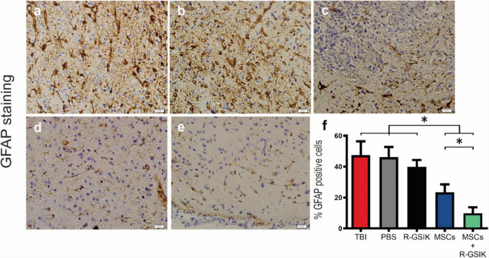 Figure 1
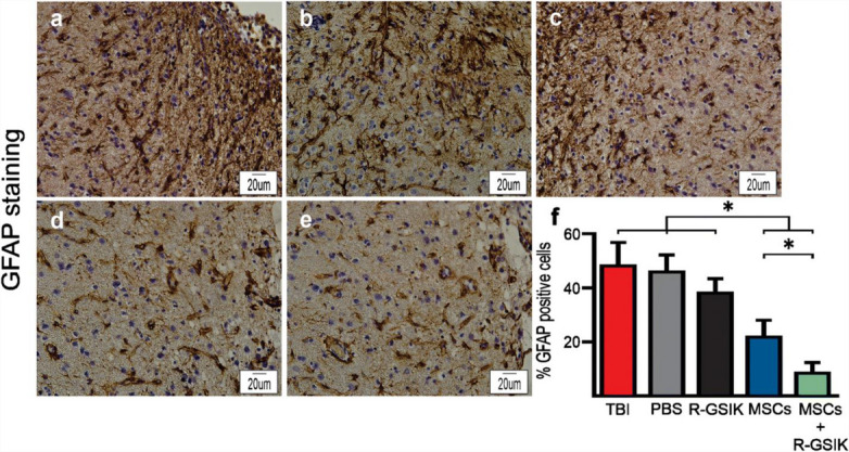 Figure 2
Peer Reviews
No public reviews on file for this paper yet. If you reviewed it on a platform where reviews are public (OpenReview, ICLR, NeurIPS, ICML), you can paste yours below so the community can read it here.
Videos
No videos yet. Explain this paper in a talk, walkthrough, or lecture? Add one.
Taxonomy
TopicsMesenchymal stem cell research · Neonatal Respiratory Health Research
Correction to: Cell and Tissue Research (2020) 382:575–583
10.1007/s00441-020-03247-0
The authors regret that the version of Figure 3 that appeared in the original published article is incorrect.
An error occurred during the assembly of the upper portion of Figure 3, specifically in the panels displaying GFAP staining. Images originating from the staining setup process were inadvertently included in the panels representing experimental groups. As a result, two sets of images in the GFAP panels partially overlapped, leading to incorrect representation of different experimental groups. Figure 3 has now been corrected to display the appropriate representative images corresponding to each experimental group in the GFAP staining section. All associated quantitative analyses have been carefully reviewed and confirmed to be based on the correct underlying data. The main results and conclusions remain unaffected. Both the original and corrected versions of Figure 3 are provided below. We sincerely apologize for this oversight and any confusion it may have caused.
Correct Figure 3 (upper panel). Representative immunohistochemistry (IHC) images show the expression of GFAP (brown cells) within the injury site. Bar graphs show the mean percentage of GFAP-positive cells in the lesion site 30 days after TBI in different animal groups. Administration of MSCs+R-GSIK and MSCs decreased the number of GFAP- positive cells within the injury site compared with the control groups. Data are expressed as mean ± SD. *P < 0.05
Incorrect Figure 3 (upper panel). Representative immunohistochemistry (IHC) images show the expression of GFAP (brown cells) within the injury site. Bar graphs show the mean number of GFAP-positive cells in the lesion site 30 days after TBI in different animal groups. Administration of MSCs+R-GSIK and MSCs decreased the number of GFAP-positive cells within the injury site compared with the control groups. Data are expressed as mean ± SD. *P < 0.05
The original article has been corrected.
The original article can be found at https://doi.org/10.1007/s00441-020-03247-0.