Anaemia and Other Haemogram Parameters Associated With Benign Maxillomandibular Odontogenic Lesions
Mamadou Diatta, Macoura Gadji, Marie Joseph Diémé, Abdoulaye Keita, Abdou Ba, Bintou Catherine Gassama, Mouhammad Kane, Khadim Seck, Babacar Tamba, Soukeye Dia Tine

TL;DR
This study found that benign dental lesions are linked to types of anemia and low neutrophil counts, suggesting a possible connection between oral health and blood parameters.
Contribution
The study is the first to explore the association between benign odontogenic lesions and specific anemia types and neutropenia in a clinical setting.
Findings
21 out of 50 patients with benign odontogenic lesions showed signs of anemia.
Normocytic normochromic and microcytic hypochromic anemia were the most common types observed.
Neutropenia was present in 23 patients, indicating a potential inflammatory or immune response.
Abstract
Introduction: Dental alterations associated with benign odontogenic lesions can alter patients' diet, possibly leading to anaemia. Indeed, cytological studies of fluid contained in benign odontogenic lesions report the presence of blood cells. The aim of this study was, therefore, to investigate anaemia and haemogram parameters in relation to benign odontogenic lesions. Material and Method: We conducted a descriptive cross-sectional study over 24 months in the Odontostomatology Department of the Idrissa Pouye General Hospital in Dakar, Senegal. The selection criteria included all patients who had received treatment for benign odontogenic lesions with an available cell blood count. The collected variables were demographic, clinical and paraclinical, with calculation of inflammation marker ratios. The data were analysed using SPSS 20.0 software, and the Kruskal–Wallis and Fisher tests…
Genes, proteins, chemicals, diseases, species, mutations and cell lines named across the full text — each resolved to its canonical identifier and authoritative record.
Click any figure to enlarge with its caption.
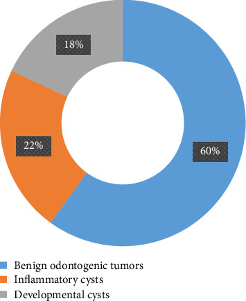 Figure 1
Peer Reviews
No public reviews on file for this paper yet. If you reviewed it on a platform where reviews are public (OpenReview, ICLR, NeurIPS, ICML), you can paste yours below so the community can read it here.
Videos
No videos yet. Explain this paper in a talk, walkthrough, or lecture? Add one.
Taxonomy
TopicsOral and Maxillofacial Pathology · Salivary Gland Tumors Diagnosis and Treatment · Vascular Malformations and Hemangiomas
1. Introduction
Maxillomandibular odontogenic lesions develop from dental bud cells [1, 2]. Benign lesions, which are more common, include pseudo tumours (cysts) and odontogenic tumours [3, 4]. They account for more than 20% of pathologies affecting the maxillomandibular bone bases [5]. These lesions develop slowly, progressively and sometimes silently, requiring consultation only when pain, functional and/or aesthetic discomfort appear [4, 6]. Some of these lesions lead to bone lysis, sometimes containing a liquid whose cytological study reveals the presence of red blood cells [7, 8]. In addition, the neovascularisation that is essential for the proliferation of cancerous cells, combined with bone and tooth lysis, leads to displacement, mobility and/or loss of teeth. This later will affect mastication, thus disrupting patients' diet, with possible repercussions on their general condition [9, 10]. According to some authors [11–13], there is also a relationship between anaemia and dentoparodontal lesions; they have reported that the inflammatory phenomena encountered during chronic generalised periodontitis lead to anaemia and an increase in leucocyte and lymphocyte counts, MCV values and RDW [11–13]. Odontogenic lesions have also been reported in patients with general pathologies leading to anaemia, such as sickle cell disease [14].
This relationship between benign odontogenic lesions and anaemia prompted the initiation of this study, aiming to investigate anaemia and other biological variables of the haemogram during the course of benign maxillomandibular odontogenic lesions.
2. Materials and Methods
We conducted a descriptive cross-sectional study of patients with benign odontogenic lesions managed at the Odontostomatology Department of the Idrissa Pouye General Hospital in Dakar, Senegal. Sampling was exhaustive, with the selection of all patients who presented with a benign maxillomandibular odontogenic lesion after free and informed consent. This study was approved by the Ethics Committee of Cheikh Anta Diop University in Dakar (026612017/ASS/CER/UCAD). The selection criteria were all patients who had their benign odontogenic maxillomandibular lesion treated in the department, under local or general anaesthesia, with a cell blood count and regular follow-up. The collected variables include epidemiological aspects (age and sex), clinical aspects (duration of evolution, location of the lesion, etc.) and paraclinical aspects (haemogram, nosological group and histological type). These variables were recorded in a data collection form over a 24-month period. A total of 7 other variables were calculated, representing indices and good markers of inflammation [11, 15, 16]. These were platelet to red blood cell ratio (PGGR) ∗ 1000, red blood cell to platelet ratio (GRPR) ∗ 1000, neutrophil to lymphocyte ratio (NLR) ∗ 1000, neutrophil to platelet ratio (NPR) ∗ 1000, neutrophil to monocyte ratio (NMR) ∗ 1000, monocyte to lymphocyte ratio (MLR) ∗ 1000 and platelet to lymphocyte ratio (PLR) ∗ 1000 [11, 15, 16].
The data were entered into a Microsoft Excel 13 spreadsheet and analysed using SPSS 20 on Windows. Data were expressed as frequencies and means with standard deviations after verification of the Gaussian distribution for quantitative variables. As the variables did not follow the normal distribution, the Kruskal–Wallis test was used to determine their distribution according to histological type or nosological group. Finally, the Fisher test was used to assess the association between certain variables.
3. Results
3.1. Sociodemographic Aspects
Women represented 70% (n = 35) of the studied population with a sex ratio of 0.43 (Table 1).
The mean age was 32.6 ± 17.8 years, with a median of 27.5 years and extremes of 7 and 76 years. The 16–30 age group represented 34% (n = 17) of the studied population, the 31–45 age group 22% (n = 11) and the 1–15 age group 20% (Table 1).
3.2. Clinical Aspects
The duration mean of the disease was 41.5 ± 25.5 months, with a median of 36 months and extremes of 18 and 120 months. Progression time between 24 and 59 months was noted in 56% (n = 28) of the patients (Table 1).
Mandibular location was the most represented with 76% (n = 38) of the patients (Table 1).
3.3. Paraclinical Aspects
3.3.1. Histological Types
Ameloblastomas and cemento-osseous dysplasias, both represented each 24% (n = 12) of the patients, inflammatory cysts 22% (n = 11) and dentigerous cysts 18% (n = 9) (Table 2).
3.3.2. Nosological Groups
Benign odontogenic tumours were found in 60% (n = 30) of the patients, inflammatory cysts in 22% (n = 11) and developmental cysts in 18% (n = 9) (Figure 1).
3.3.3. Haemogram
Analysis of the red blood cell line (erythrocyte count, haemoglobin level and erythrocytic indices) revealed anaemia in 21 patients (42%), including 11 cases of normocytic normochromic anaemia, 8 cases of microcytic hypochromic anaemia and 2 cases of normocytic hypochromic anaemia. In addition, a decrease in mean corpuscular haemoglobin content (MCHC) was noted in 14 patients (28%), a decrease in RBC count in 7 patients (14%), a decrease in MCHC in 5 patients (10%) and anisocytosis in 4 patients (8%).
Analysis of the platelet lineage revealed thrombocytosis (hyperplaquettosis) in 4 patients (8%) and thrombocytopaenia in 1 patient (2%).
Analysis of the leucocyte counts and repartition described neutropenia in 23 patients (46%) and lymphopenia in 7 patients (14%). It was also noted an increase in the count of eosinophilic polymorphonuclear cells in 7 patients (14%) (Table 3).
3.4. Age Groups According to MCHC
Of the 5 patients with a decreased MCHC value, 4 (80%) had a benign odontogenic tumour with a significant difference (p value = 0.040) (Table 4).
Furthermore, the type of anaemia was not associated with the nosological groups of benign odontogenic lesions.
3.5. Nosological Groups According to Haematocrit (HCT)
Of the 12 patients with a reduced HCT value, 50% were found in developmental cysts with a p value < 0.05 (Table 5).
3.6. Platelets/Red Blood Cells Ratio ((PRBCR) ∗ 1000) According to Histological Types of Lesions
The means for odontogenic keratocysts, dentigerous cysts and complex odontoma are higher with a significant difference (p=0.002) (Table 6).
3.7. Platelets/Red Blood Cells Ratio ((PRBCR) ∗ 1000) According to Nosological Groups of Lesions
The means for developmental cysts and benign odontogenic tumours are higher with a significant difference (p=0.047) (Table 7).
The associations sought between certain ratios and histological types and nosological groups of benign odontogenic lesions were not significant (Appendices: Tables A1, A2, A3, A4, A5 and A6).
4. Discussion
Benign odontogenic lesions are highly polymorphic lesions, in which several are easy to diagnose but others are difficult to diagnose because of their similar clinical and/or radiological features. A positive diagnosis is often confirmed after pathological examinations. That is why some biological parameters, such as haemograms, could make a major contribution to the diagnosis and management of these lesions.
4.1. Sociodemographic Aspects
In this present study, a predominance of females (70%) was noted, with a sex ratio of 0.43. These results are similar to those reported by certain authors with a female predominance in benign maxillomandibular odontogenic lesions [5, 10]. However, other studies have described a male predominance [1, 17]. This difference could be explained by the small sample size in this study. These lesions can occur at any age [10]. In addition, the affected population was young, with an average age of 32.6 years and a predominance of the 16–30 age group (34%). These results are similar to those reported by Kaur et al. 2021 [1], with an average age of 31.9 years. However, higher mean ages have been reported by other authors [10, 18] on a larger sample size. Besides the sociodemographic aspects, clinical symptoms were also studied.
4.2. Clinical Symptoms
The duration of odontogenic lesions varies from a few months to several years [10, 17]. These lesions are sometimes discovered by chance during a routine radiological examination [19]. The mean duration of evolution was 41.5 months, with more than 50% of cases having a duration of evolution in between 24 and 59 months. These results differ from those reported by Sayela et al. in 2020, with 52.8% of the patients having a duration of less than 24 months [10]. In addition, shorter durations of progression were also reported [10, 20]. This difference could be explained by poverty, ignorance, societal beliefs, reliance on traditional practitioners and lack of infrastructure [20].
Benign odontogenic lesions can be located in the maxilla as well as the mandible. The mandible was the most frequently reported site in the literature, accounting for more than 60% of the cases [1, 5, 10]. This is in perfect agreement with the results of this present study, in which the mandibular location was found in 76% of the patients.
4.3. Paraclinical Aspects
According to the literature, ameloblastoma, with its various histological forms, is the most frequently encountered benign odontogenic maxillomandibular tumour [1, 18]. In fact, the high number of cases of cemento-osseous dysplasia (24%) found in this study could be explained by the frequency of this lesion, which is most often found in the elderly, particularly women, in Senegal [21]. Ameloblastomas are locally aggressive lesions prompted to recur. They are clinically manifested by deformation of the bone tables, displacement and mobility of the teeth, thus impairing the patient's quality of life. On radiography, the image may be unigeodic radiolucent or multigeodic (classic forms) with rhizalysis of the dental roots.
Other lesions such as inflammatory cysts have also been reported by some authors [5, 22]. Inflammatory cysts are chronic lesions of the periodontal tissue resulting from bacterial infection of the endodontium or periodontium, which, as they develop, lead to deformation of the bony bases and may even break out after rupture of the bony cortex.
Cemento-osseous dysplasias are fibro-osseous lesions associated with dental apices, which develop slowly and silently. They may be discovered by chance during a radiographic examination. In florid forms, the radiograph shows mixed images (osteolytic and osteocondensing) poorly limited around the apices of several teeth. The lesion may be painful if superinfected. In focal and florid clinical forms of cemento-osseous dysplasia, the lesions are often surrounded by granulation tissue, which is highly haemorrhagic during surgical removal.
These classical clinical manifestations might impair the patient nutrition and lead to nutritional anaemia. Thus, besides the clinical and paraclinical aspects, study of benign maxillomandibular odontogenic lesions requires the use of complementary examinations such as haemogram.
4.4. Cell Blood Counts (Haemograms)
Benign maxillomandibular odontogenic lesions, as they progress, will lead to bone deformations associated with displacements, retentions, mobility or loss of teeth, which may impair the patient's mastication, with a possible impact on the general state of health and thus on certain cell blood count parameters, including the haemoglobin level. In this present study, anaemia was found in 21 patients (42%), including 11 cases of normocytic normochromic anaemia, 8 cases of microcytic hypochromic anaemia and 2 cases of normocytic hypochromic anaemia. According to the literature, microcytic anaemia is most often hypochromic with variable aetiologies that may be related to nutritional deficiency in addition to inflammatory phenomena [23, 24]. According to Lanier et al. 2021, anaemia in adults may be asymptomatic and discovered fortuitously [25]. The most commonly reported causes are nutritional deficiencies and/or insufficiencies, which may be encountered in patients with benign odontogenic lesions due to impaired mastication [9, 10]. In addition, as odontogenic lesions progress, they create neovascularisation to satisfy the need of blood nutriments, which is essential for the proliferation and development of cancerous cells [26]. According to Fatemeh et al., 2017 [27], the cytological study of puncture fluids of benign odontogenic lesions reports the presence of red blood cells.
Moreover, the 2 cases of normocytic hypochromic anaemia encountered could be an intermediate phase due either to an iron deficiency in the body or to a defect in iron utilisation due to the presence of inflammatory phenomena [28].
A decrease in HCT values and mean corpuscular haemoglobin concentration was associated with developmental cysts, which could be explained by the blood cell requirements of these lesions, particularly neoangiogenesis, for their growth and development [26].
Thrombocytosis was associated with the 1–15 year age group and with a duration of evolution of 1–23 months. In contrast, thrombocytopaenia was associated with the age range of 60 years and later as well as with a duration of evolution of 60 months and above. According to Sharma et al., 2013 [6], besides their involvement in haemostasis, platelets are thought to mediate the growth, dissemination and angiogenesis of tumour lesion cells [6]. The association of thrombocytopaenia with advanced age could be explained by a decrease in platelet levels reported beyond the age of 60 [29].
The anisocytosis associated with cemento-osseous dysplasia could be explained by the fact that these lesions are often surrounded by highly haemorrhagic granulation tissue due to chronic infiltration of inflammatory cells into the lesion site [30]. These chronic inflammatory phenomena promote the supply of blood cells to the site, which is essential for the cellular repair reaction. The histological appearance of cemento-osseous dysplasia shows the presence of small blood-filled vessels in the stroma, which may explain the influx of blood cells into the bone tissue. In the initial stage, these lesions consist of unencapsulated fibrous connective tissue with numerous small blood vessels [31]. According to Urs et al. [32], histological sections of cemento-osseous dysplasia sometimes show haemorrhagic areas.
Furthermore, the statistically significant associations observed between high mean PRBCR ratios in developmental cysts (dental cysts) and benign odontogenic tumours (odontogenic keratocysts and complex odontoma) might be explained by the fact that platelets are essential for the development and progression of tumour cells in certain types of lesion [6].
5. Conclusion
Benign odontogenic lesions are diverse and varied, with different clinical and radiological manifestations. They may affect the patient's mastication, with possible repercussions on patient nutrition and his general condition. Neutropenia and anaemia were the most common abnormalities of haemogram parameters. Anaemia as normocytic normochromic anaemia was mostly recorded, followed by microcytic hypochromic anaemia and finally by fewer normocytic hypochromic anaemia. Thus, haemogram appears to be very useful for stratification and follow-up of benign odontogenic lesions. However, a more inclusive study should be undertaken to gain a better understanding of the variations in haemogram parameters and especially anaemia in benign maxillomandibular odontogenic lesions.
The reference list from the paper itself. Each links out to its DOI / PubMed record.
- 1Kaur H. Gosavi S. Hazarey V. K. Gupta V. Bhadauria U. S. Kherde P. Impact of Changing Classification Systems on Prevalence and Frequency Distribution of Odontogenic Tumors in Tertiary Care Center of Nagpur Brazilian Journal of Otorhinolaryngology 202288 Suppl 1S 3S 1310.1016/j.bjorl.2021.02.00633757753 PMC 9734271 · doi ↗ · pubmed ↗
- 2Guimarães L. M. Coura B. P. Gomez R. S. Gomes C. C. The Molecular Pathology of Odontogenic Tumors: Expanding the Spectrum of MAPK Pathway Driven Tumors Frontiers in Oral Health 2021211310.3389/froh.2021.740788 PMC 875781435048058 · doi ↗ · pubmed ↗
- 3Chacham M. Almoznino G. Zlotogorski-Hurvitz A. Buchner A. Vered M. Expression of Stem Cell Markers in Stroma of Odontogenic Cysts and Tumors Journal of Oral Pathology and Medicine 2020491068107710.1111/jop.1310232840915 · doi ↗ · pubmed ↗
- 4Dwivedi D. Prabhakar N. Kasetty S. Ahuja R. Peripheral Adenomatoid Odontogenic Tumor in a Cloak of an Epulis: Report of a Rare Case BMC Oral Health 2019191811010.1186/s 12903-019-0759-82-s 2.0-8506555209431077195 PMC 6511215 · doi ↗ · pubmed ↗
- 5Ali A. Asif M. Ali I. Analysis of CD 10 Expression in the Epithelial Lining of Odontogenic Cysts Pakistan Armed Forces Medical Journal 2020701 S 10S 14
- 6Sharma D. Brummel-Ziedins K. E. Bouchard B. A. Holmes C. E. Platelets in Tumor Progression: A Host Factor That Offers Multiple Potential Targets in the Treatment of Cancer Journal of Cellular Physiology 201422981005101510.1002/jcp.245392-s 2.0-8489907265624374897 · doi ↗ · pubmed ↗
- 7Shalley S. Chand N. Aggarwal A. Garg L. N. Yadav V. Yadav A. Diagnostic Accuracy of Fine Needle Aspiration Cytology in Lesions of Oral Cavity and Salivary Glands: A Clinico-Pathological Study The Open Dentistry Journal 201812178279010.2174/17450179018140107822-s 2.0-8505575607430369988 PMC 6182874 · doi ↗ · pubmed ↗
- 8Sethi S. Kumar M. Aggarwal P. Indra Kumar H. S. Sugandhi C. D. Singh S. A Case Report and Short Review on Changing Trends in the Site of Occurrence of Adenomatoid Odontogenic Tumor: Unravelling the Past 15 Years Dental Research Journal 201613546247110.4103/1735-3327.1923122-s 2.0-8499211110627857774 PMC 5091007 · doi ↗ · pubmed ↗