Compressive Fourier-Domain Intensity Coupling (C-FOCUS) enables near-millimeter deep imaging in the intact mouse brain in vivo
Renzhi He, Yucheng Li, Brianna Urbina, Jiandi Wan, Yi Xue

TL;DR
A new technique called C-FOCUS improves two-photon microscopy to image deep into the mouse brain with high resolution and clarity.
Contribution
C-FOCUS combines Fourier-domain intensity modulation and compressive sensing to enable deep-tissue scattering correction in two-photon microscopy.
Findings
C-FOCUS achieves high-resolution imaging of neurons and blood vessels beyond 900 μm in the intact mouse brain.
Transcranial imaging of dendritic structures is possible through the adult mouse skull using C-FOCUS.
Fluorescence intensity is enhanced over 20-fold compared to uncorrected imaging at deep tissue depths.
Abstract
Two-photon microscopy is a powerful tool for in vivo imaging, but its imaging depth is typically limited to a few hundred microns due to tissue scattering, even with existing scattering correction techniques. Moreover, most active scattering correction methods are restricted to small regions by the optical memory effect. Here, we introduce compressive Fourier-domain intensity coupling for scattering correction (C-FOCUS), an active scattering correction approach that integrates Fourier-domain intensity modulation with compressive sensing for two-photon microscopy. Using C-FOCUS, we demonstrate high-resolution imaging of YFP-labeled neurons and FITC-labeled blood vessels at depths exceeding 900 μm in the intact mouse brain in vivo. Furthermore, we achieve transcranial imaging of YFP-labeled dendritic structures through the intact adult mouse skull. C-FOCUS enables high-contrast…
Genes, proteins, chemicals, diseases, species, mutations and cell lines named across the full text — each resolved to its canonical identifier and authoritative record.
Click any figure to enlarge with its caption.
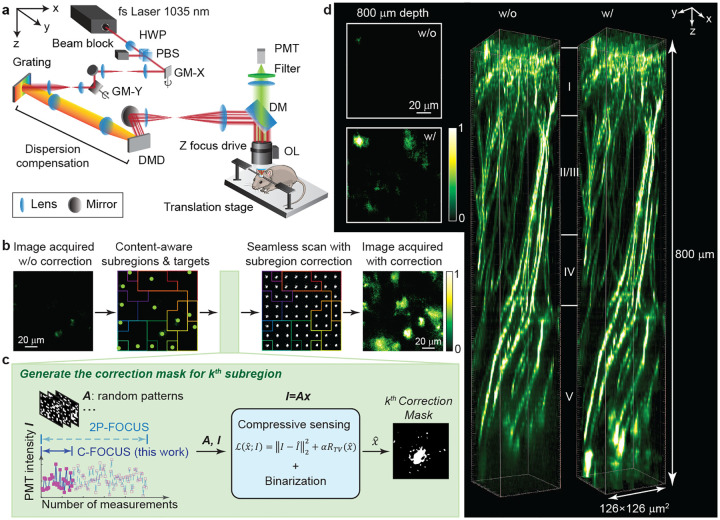 Figure 1
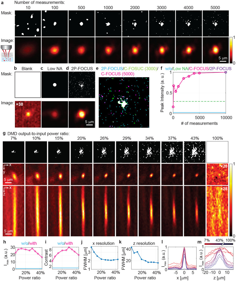 Figure 2
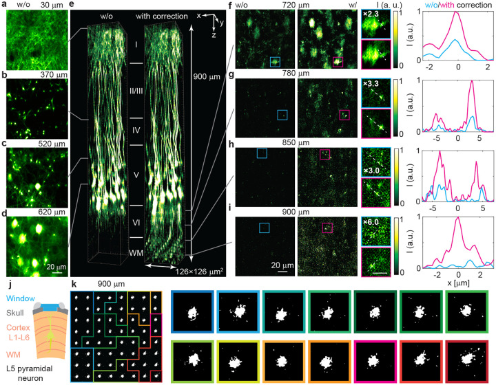 Figure 3
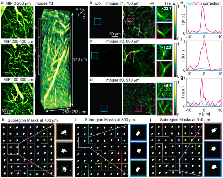 Figure 4
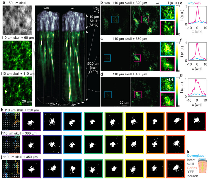 Figure 5
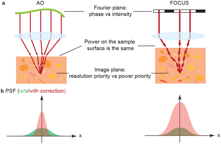 Figure 6
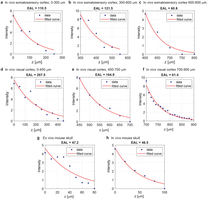 Figure 7
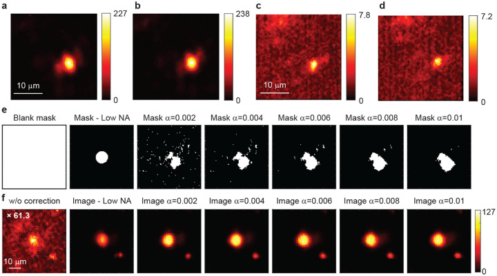 Figure 8
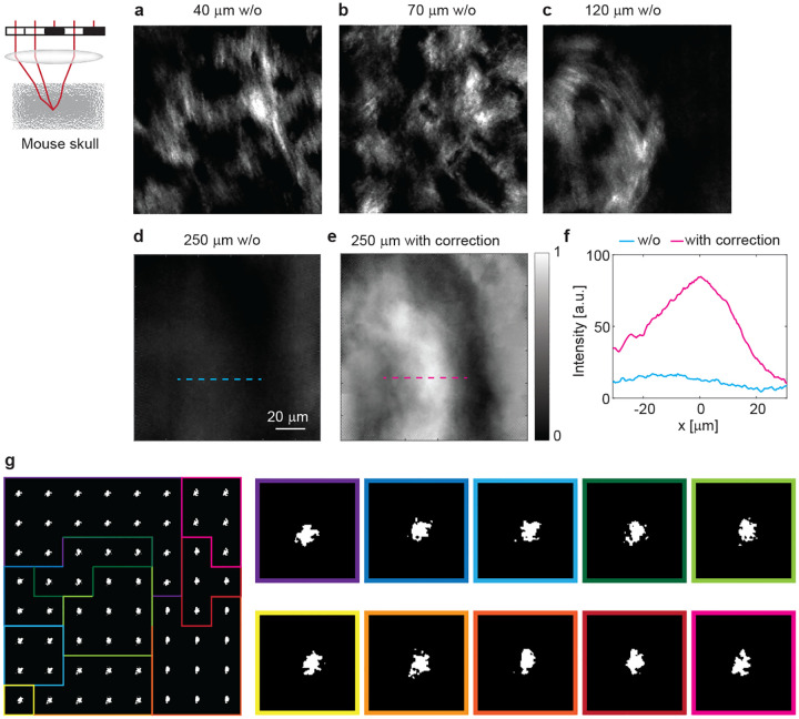 Figure 9
Peer Reviews
No public reviews on file for this paper yet. If you reviewed it on a platform where reviews are public (OpenReview, ICLR, NeurIPS, ICML), you can paste yours below so the community can read it here.
Videos
No videos yet. Explain this paper in a talk, walkthrough, or lecture? Add one.
Taxonomy
TopicsAdvanced Fluorescence Microscopy Techniques · Photoacoustic and Ultrasonic Imaging · Optical Imaging and Spectroscopy Techniques