Photon counting detector CT-derived virtual non-contrast images of the liver: comparison of conventional and liver-specific algorithms across arterial and portal venous phase scans
Anna-Katharina Gerstner, Franka Risch, Luca Canalini, Gerlig Widmann, Elke R. Gizewski, Stefanie Bette, Simon Hellbrueck, Thomas Kroencke, Josua A. Decker

TL;DR
This study compares virtual non-contrast liver CT images generated using different algorithms and scan phases, finding they closely match true non-contrast images regardless of patient BMI.
Contribution
The study introduces a comparison of liver-specific and conventional algorithms for generating virtual non-contrast CT images and evaluates their performance across BMI groups.
Findings
Virtual non-contrast images from both conventional and liver-specific algorithms showed strong correlations with true non-contrast images.
No significant differences in liver attenuation were found between BMI groups using either algorithm.
Bland-Altman plots and Passing-Bablok regression confirmed good agreement and absence of systematic differences.
Abstract
The aim of this retrospective study is to compare photon-counting detector computed tomography (PCD-CT) derived virtual non-contrast (VNC) images of the liver reconstructed from both arterial and portal venous phase using conventional and liver-specific VNC algorithm to true non-contrast images, in context of the body mass index (BMI). VNC images reconstructed from multiphase (non-contrast, arterial and portal venous phase) PCD-CT scans performed between April 2021 and February 2023 were analysed retrospectively. For each patient, four VNC series were generated: two series (arterial and portal venous) using a conventional VNC algorithm (VNCconvart; VNCconvpv) and two using a liver-specific “Liver VNC” algorithm (VNCLiverart; VNCLiverpv). Regions of interest were placed in the left and right liver lobes and in the spleen, avoiding large vessels and focal lesions. The VNC CT-values were…
Genes, proteins, chemicals, diseases, species, mutations and cell lines named across the full text — each resolved to its canonical identifier and authoritative record.
Click any figure to enlarge with its caption.
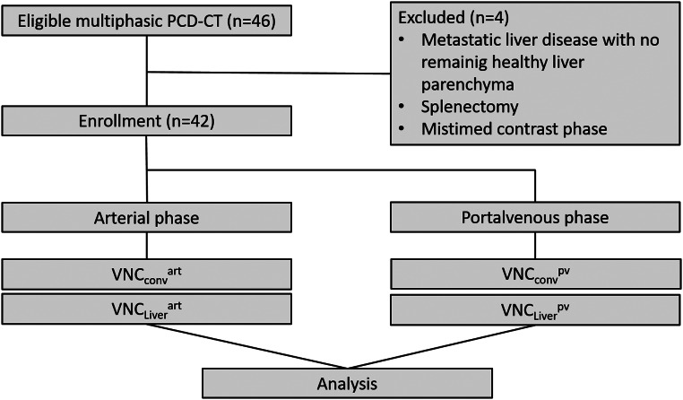 Figure 1
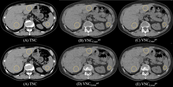 Figure 2
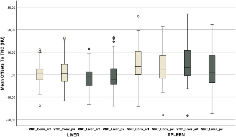 Figure 3
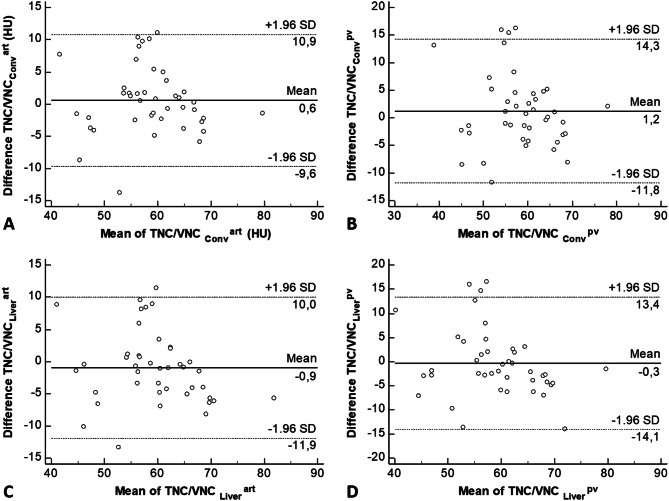 Figure 4
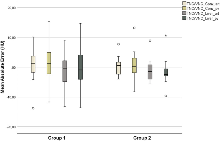 Figure 5
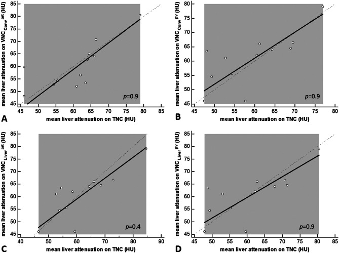 Figure 6
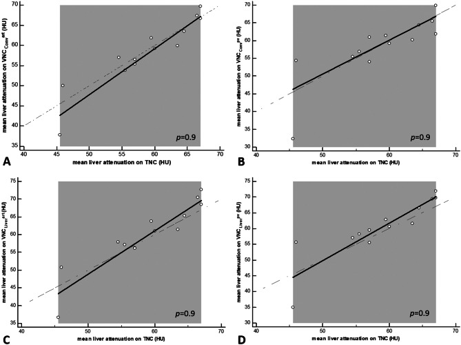 Figure 7
Peer Reviews
No public reviews on file for this paper yet. If you reviewed it on a platform where reviews are public (OpenReview, ICLR, NeurIPS, ICML), you can paste yours below so the community can read it here.
Videos
No videos yet. Explain this paper in a talk, walkthrough, or lecture? Add one.
Taxonomy
TopicsAdvanced X-ray and CT Imaging · Radiation Dose and Imaging · Medical Imaging Techniques and Applications