Krabbe disease: a differential cause of the hyperdense boomerang sign
Luis Alcides Quevedo Canete, Sérgio Ferreira Alves Júnior, Ângelo Dante de Carvalho Côrrea, Nina Ventura

Abstract
Genes, proteins, chemicals, diseases, species, mutations and cell lines named across the full text — each resolved to its canonical identifier and authoritative record.
Click any figure to enlarge with its caption.
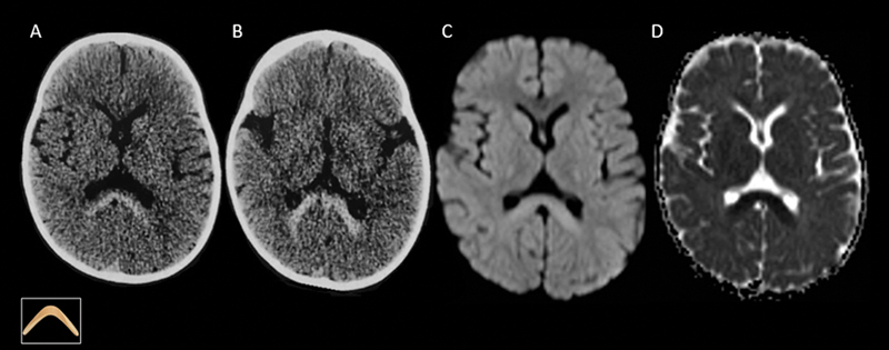 Figure 1
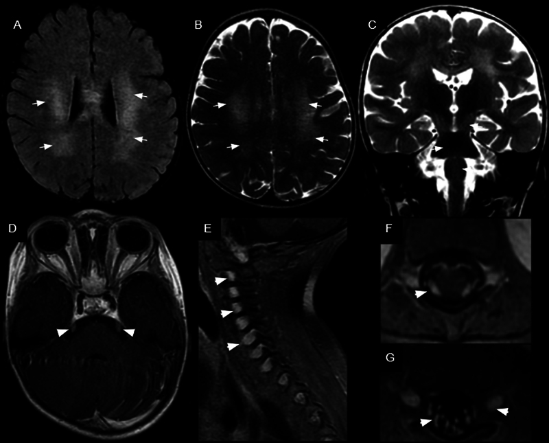 Figure 2
Peer Reviews
No public reviews on file for this paper yet. If you reviewed it on a platform where reviews are public (OpenReview, ICLR, NeurIPS, ICML), you can paste yours below so the community can read it here.
Videos
No videos yet. Explain this paper in a talk, walkthrough, or lecture? Add one.
Taxonomy
TopicsLysosomal Storage Disorders Research · Hereditary Neurological Disorders · Parasitic Diseases Research and Treatment
A 1.5-year-old female child presented with regression of developmental milestones, spastic tetraparesis, and fever. Computed tomography (CT) scans showed hyperdensity, and magnetic resonance imaging (MRI) scans revealed restricted diffusion in the splenium of the corpus callosum, characterizing the boomerang sign ( Figure 1 ). On the follow-up examination, bilateral and symmetrical T2 and fluid-attenuated inversion recovery (FLAIR) hyperintense lesions were observed in the cerebral white matter, predominantly in the parieto-occipital regions, presenting a tiger- or leopard-skin pattern, as well as involvement of the brainstem, corticospinal tracts, and dentate nuclei ( Figure 2 A–C ). Additionally, bilateral thickening and enhancement of the cranial nerves were noted, most prominently in the cisternal portions of the III, V, and VI pairs, and in the intracanalicular portions of the VII and VIII pairs. Diffuse thickening and enhancement of the spinal roots were also observed ( Figure 2 D–G ). Krabbe disease was confirmed through genetic testing, which identified the c.884A>T variant in heterozygosity in the GALC gene. In clinical presentations featuring hyperdense lesions on CT and restricted diffusion on MRI in the corpus callosum (splenium), Krabbe disease should be considered. 1 2 3 4
Brain computed tomography (CT) and magnetic resonance imaging (MRI) scans revealing hyperdensity ( A,B ) and restricted diffusion ( C,D ) in the splenium of the corpus callosum.
Brain MRI scans revealing bilateral and symmetrical T2 and fluid-attenuated inversion recovery (FLAIR) hyperintense lesions in the white matter of the cerebral hemispheres, predominantly in the parieto-occipital regions, displaying a tiger-like or leopard-skin pattern (white arrows in A and B ). Involvement of the brainstem and corticospinal tracts (white arrows in C ) was observed, along with thickening and enhancement of the trigeminal nerves (white arrowhead in D ) and spinal roots (white arrowheads in E , F , and G ).
The reference list from the paper itself. Each links out to its DOI / PubMed record.
- 1Muthusamy K Sudhakar S V Thomas M Yoganathan S Christudass C S Chandran M Revisiting magnetic resonance imaging pattern of Krabbe disease - Lessons from an Indian cohort J Clin Imaging Sci 201992510.25259/JCIS-18-201931448176 PMC 6702867 · doi ↗ · pubmed ↗
- 2Abdelhalim A N Alberico R A Barczykowski A L Duffner P K Patterns of magnetic resonance imaging abnormalities in symptomatic patients with Krabbe disease correspond to phenotype Pediatr Neurol 2014500212713410.1016/j.pediatrneurol.2013.10.00124262341 · doi ↗ · pubmed ↗
- 3Loonen M C Van Diggelen O P Janse H C Kleijer W J Arts W F Late-onset globoid cell leucodystrophy (Krabbe's disease). Clinical and genetic delineation of two forms and their relation to the early-infantile form Neuropediatrics 1985160313714210.1055/s-2008-10525584047347 · doi ↗ · pubmed ↗
- 4Bernal O G Lenn N Multiple cranial nerve enhancement in early infantile Krabbe's disease Neurology 200054122348234910.1212/wnl.54.12.2348 PMID 10881274 · doi ↗ · pubmed ↗