Identification of IGF2BP3 for the Expression and Prognosis in Gastrointestinal Cancers
Fengfeng Han, Chen He, Chen Qian, Yujie Wang, Mingge Zhou, Ruirui Yang, Zhou Zhang

TL;DR
This study explores how IGF2BP3 is expressed in gastrointestinal cancers and how it could help in diagnosis and predicting patient outcomes.
Contribution
The novel contribution is identifying IGF2BP3 as a potential diagnostic and prognostic biomarker in gastrointestinal cancers.
Findings
IGF2BP3 is highly expressed in four types of gastrointestinal cancers.
High IGF2BP3 levels correlate with poor survival in liver cancer patients.
IGF2BP3 shows the highest diagnostic accuracy for esophageal cancer.
Abstract
Purpose: This study investigates the expression, diagnostic, and prognostic significance of IGF2BP3 in gastrointestinal cancers, providing new insights for clinical management and patient care. Methods: An integrated approach was used to analyze the expression and mutation of IGF2BP3 across various cancers, utilizing multiple online analytical tools. Data from the TCGA database were examined to identify differentially expressed genes (DEGs) in four types of gastrointestinal cancers and to assess the specific expression of IGF2BP3. Further validation was carried out through western blotting (WB), quantitative real-time PCR (qRT-PCR), and immunohistochemistry (IHC). Protein-protein interaction (PPI) network was also employed to investigate the relationships between IGF2BP3 and its associated genes. Additionally, bioinformatics databases were used to investigate the impact of IGF2BP3 on…
Genes, proteins, chemicals, diseases, species, mutations and cell lines named across the full text — each resolved to its canonical identifier and authoritative record.
Click any figure to enlarge with its caption.
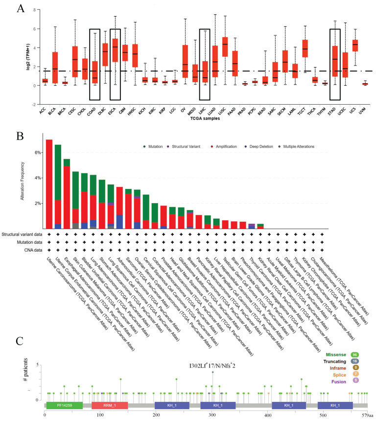 Figure 1
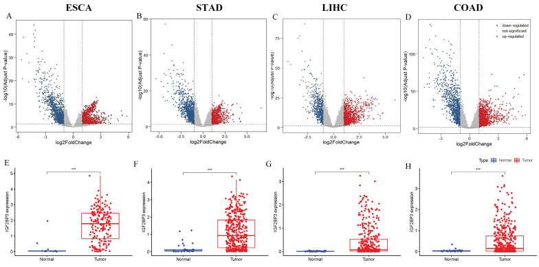 Figure 2
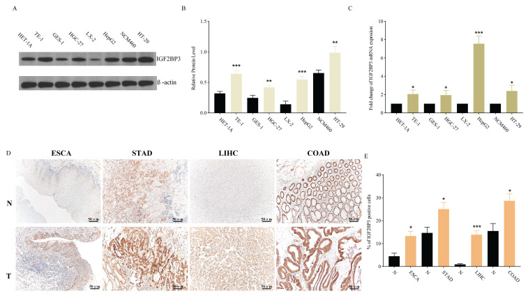 Figure 3
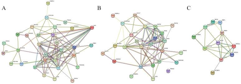 Figure 4
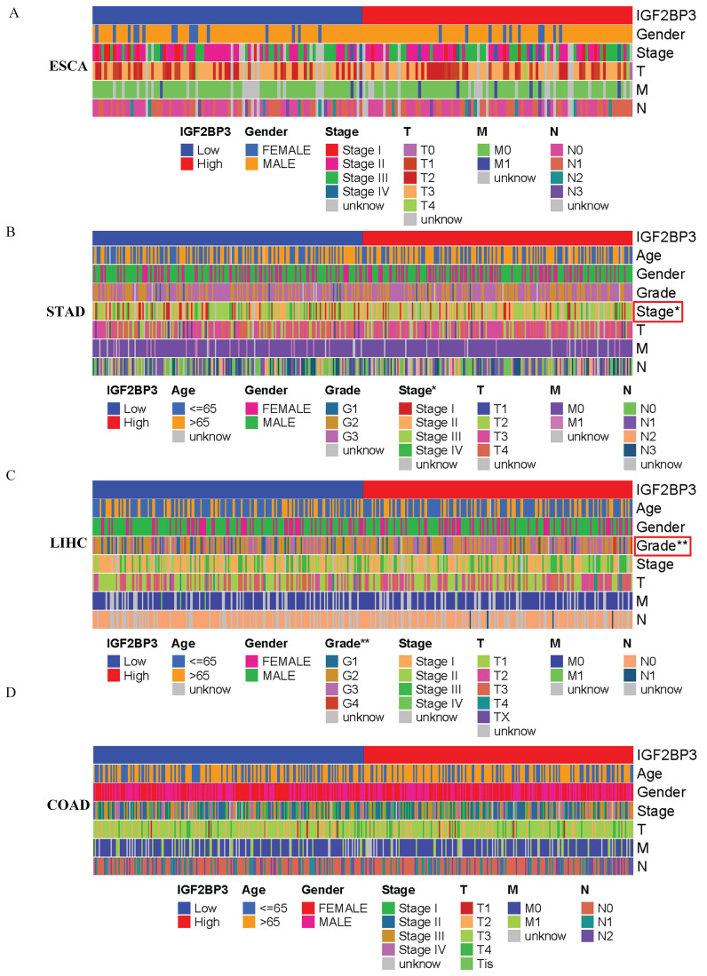 Figure 5
 Figure 6
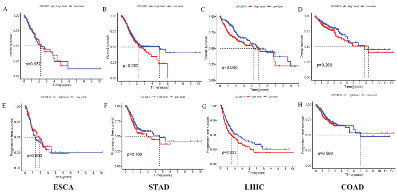 Figure 7
Peer Reviews
No public reviews on file for this paper yet. If you reviewed it on a platform where reviews are public (OpenReview, ICLR, NeurIPS, ICML), you can paste yours below so the community can read it here.
Videos
No videos yet. Explain this paper in a talk, walkthrough, or lecture? Add one.
Taxonomy
TopicsGrowth Hormone and Insulin-like Growth Factors · Gastric Cancer Management and Outcomes · Genetic factors in colorectal cancer
1. Introduction
Gastrointestinal cancers are the most prevalent type of malignancy, constituting roughly 25% of all cancer cases and accounting for about one-third of cancer-related fatalities globally 1. The rising occurrence of these cancers is intricately associated with demographic shifts, such as population expansion, aging populations, and alterations in lifestyle 2. These malignancies primarily encompass esophageal, gastric, liver, and colorectal cancer 3. The early identification of conditions such as esophageal, gastric, and liver cancer is currently inadequate, leading to generally unfavorable patient outcomes 4. Consequently, it is essential to pinpoint innovative diagnostic and therapeutic targets to enhance early detection and treatment, thereby establishing a scientific basis for future treatment protocols.
Some literatures reveal that IGF2BP3 (Insulin-like growth factor 2 mRNA-binding protein 3) is considered an oncogene in multiple tumors, including leukemia 5, glioma 6, oral cancer 7, among others 8. However, the role of IGF2BP3 in gastrointestinal tumors and its potential as a biomarker for gastrointestinal tumors are worth more exploring and summarizing. IGF2BPs form a conserved group of RNA-binding proteins (RBPs) 9, 10. RBPs 11-14 are proteins that bind to RNA, forming ribonucleoprotein complexes. These proteins are predominantly functional in a methylated state, notably in N6-methyladenosine (m6A) 15-17. The research of Oh et al points to the fact that the upregulation of m6A-reading proteins, particularly IGF2BP3, is connected to adverse survival, increased proliferation, and epithelial-mesenchymal transition (EMT) activation across diverse cancers, including gastric and colorectal carcinomas 18. Studies have shown that IGF2BPs are critical in various cellular functions, including cell polarization, migration, morphology, metabolism, proliferation, and differentiation 19. Among them, IGF2BP3, notably abundant in pancreatic cancer tissues and initially named KOC, is particularly overexpressed in various human cancers. Beyond influencing growth and chemoresistance, IGF2BP3 also enhances the invasive capacity of tumor cells in vitro 20-22. Upregulation of this protein in cancers is known to promote tumor growth by increasing IGF2 expression. Additionally, IGF2BP3 aids in tumor cell proliferation by interacting with HNRNPM in the nucleus 23, thereby boosting cyclin protein expression.
The field of bioinformatics, which integrates genomics, information science, mathematics, and biology, is crucial in managing, processing, and interpreting large-scale biological data. It helps uncover the biological significance of such data, facilitating the identification of key tumor biomarkers and laying a foundation for enhanced cancer diagnosis and prognosis 24-26.
In this study, we applied bioinformatics techniques to explore the relationship between IGF2BP3 expression and gastrointestinal cancers, focusing on gene mutations, pathological features, diagnostic potential, and prognostic value. Our findings offer insights that could guide future research into the role of IGF2BP3 in these cancers.
2. Materials and methods
2.1 Expression and mutation analysis of IGF2BP3 in pan-cancer
The expression patterns of IGF2BP3 across pan-cancer tissues were assessed using the University of Alabama at Birmingham Cancer Data Analysis Portal (UALCAN). Afterwards, extensive genomic data, representing various cancer types, was analyzed using the cBioPortal platform. This data included mutation frequency, variant categorization, copy number modifications, and mutation localization.
2.2 Gene mutations and related genes analysis of IGF2BP3 in gastrointestinal cancers
Initially, the cBioPortal platform was used to examine the origins and patterns of IGF2BP3 mutations in gastrointestinal cancers. Further analysis through the UALCAN database identified significant correlations between IGF2BP3 and its core regulatory genes across prevalent gastrointestinal cancers types, including the quantification of their respective correlation coefficients.
2.3 Identification of DEGs in gastrointestinal cancers
To show the gene differential expression landscape among four gastrointestinal cancers types, volcano plots were made using datasets from The Cancer Genome Atlas (TCGA) 4. Statistical analysis was then performed to identify significant differences, which were shown in box plots, after the IGF2BP3 expression levels were quantified using TCGA data.
2.4 Protein-protein interaction (PPI) networks in gastrointestinal cancers
The online database STRING was used to analyze the functional interactions between IGF2BP3 and its correlated genes including positive and negative correlated genes in gastrointestinal cancers.
2.5 Survival analysis and diagnostic efficacy of IGF2BP3 in gastrointestinal cancers
Using TCGA cohort data, this study evaluated the prognostic significance of IGF2BP3. Subsequent ROC curve analysis quantified diagnostic performance through systematic comparison of AUC values and threshold optimization, identifying the statistically optimal discrimination cutoff for clinical application.
2.6 Cell lines and cell culture
The Cell Bank of the Chinese Academy of Sciences (China) was used to procure the following human cancer cell lines: TE-1 (esophageal cancer), HGC-27 (gastric cancer), HepG2 (liver cancer), and HT-29 (colorectal cancer). The following human cell lines were obtained from IMMOCELL: HET-1A (esophageal epithelium), GES-1 (gastric epithelium), LX-2 (hepatocyte), and NCM460 (colorectal epithelium). All cells, except for HET-1A cells, were cultured in RPMI-1640 with 10% FBS, while HET-1A cells were grown in a distinct medium. All cells were kept at 37°C in 5% CO_2_.
2.7 Quantitative real-time PCR (qRT-PCR)
The Trizol reagent (Ambion) was employed for the extraction of total RNA. The application of Vazyme's HiScript® II Q Select RT SuperMix facilitated the reverse transcription process for qRT-PCR. To quantify mRNA levels, qRT-PCR was conducted utilizing the SYBR Green Master Mix. The primer sequences of IGF2BP3 were as follows: Forward, 5'-GGCAAAACGGTGAATGAACT-3' and Reverse, 5'-GTCCACTTTGCAGAGCCTTC-3'. Using β-actin as an internal reference gene and the primer sequence were as follows: Forward, 5'-CCCTGGAGAAGAGCTACGAG -3'and Reverse, 5'-CGTACAGGTCTTTGCGGATG -3'. The 2^-ΔΔCt^ method was employed to assess the levels of IGF2BP3. There were three iterations of the experiments.
2.8 Western blotting (WB)
The whole cell protein was isolated for Western blotting by transferring it to PVDF membranes (Millipore, Billerica, MA, USA) after being separated by 12% SDS-PAGE in RIPA lysis buffer (Invitrogen, Carlsbad, CA, USA). Priming antibodies: rabbit anti-IGF2BP3 (diluted 1:5000; Proteintech, China) and mouse anti-β-actin (dilution 1:20000, Affinity, China) were applied to the membranes after blocking them with 5% non-fat milk and incubating them overnight at 4°C. Proteintech of China diluted goat anti-rabbit/mouse secondary antibodies 1:2000 into the membranes and left them to incubate at 37°C for 60 minutes. Millipore, Germany's ECL reagents were used to identify signals after incubation. As an internal control, β-actin was utilized. There was at least one triplicate of each experiment conducted separately.
2.9 Immunohistochemistry (IHC)
The IHC was carried out using a kit from Boster Biological Technology, China, in accordance with the instructions provided by the manufacturer. Thin slices measuring 5 μm were cut from tumor tissues that had been fixed in paraffin. Dewaxing and rehydration were followed by the application of 3% H_2_O_2_ to inactivate endogenous peroxidase. Primary antibodies against IGF2BP3 (diluted 1:200; Proteintech, China) were incubated overnight at 4°C after blocking the slices with 5% BSA for 1 hour. Slices were stained with 3,3-diaminobenzidine substrate (DAB) after incubation with the secondary antibody such that IGF2BP3 expression could be observed under a microscope.
2.10 Statistics analysis
Data analysis was conducted using R software (version 4.02) and presented as mean ± SD. Statistical differences were assessed using GraphPad Prism software 8.0.2 (USA). Differential gene expression analysis (|Log2FC| > 1, adjusted P-value (FDR) < 0.05) was performed by comparing tumor tissues with normal controls using the limma R package. Gene expression data were log2-transformed, and correlations were calculated using Spearman's rank correlation coefficient. The diagnostic potential of IGF2BP3 in gastrointestinal cancers was evaluated through receiver operating characteristic (ROC) curves. Survival analysis was conducted using Kaplan-Meier survival curves to assess the prognostic value of IGF2BP3 expression in these cancers. Statistical significance was determined using either an independent t-test for two groups or one-way analysis of variance (ANOVA), with a P-value of <0.05 considered significant. *P<0.05, **P<0.01, ***P<0.001.
3. Results
3.1 Clinical parameters of patients
Clinical datasets for four cancer types were systematically retrieved from TCGA. The cohort included 184 validated esophageal cancer cases, 403 qualified gastric cancer samples, 349 confirmed liver cancer specimens, and 404 validated colorectal cancer records that met the inclusion criteria (Table 1).
3.2 Expression and mutation of IGF2BP3 in pan-cancer
Initially, the expression profile of IGF2BP3 across various cancers was examined. Figure 1A demonstrates that IGF2BP3 expression is higher in esophageal cancer and gastric cancer compared to the median expression across pan-cancer. Additionally, the mutation frequency, types of mutations, and copy number variations of IGF2BP3 were analyzed. Figure 1B displays the frequency and types of alterations in IGF2BP3 across pan-cancer. The main alteration in esophageal cancer is amplification, with a frequency of 4.95%. In liver cancer, the main alteration is mutation, with a frequency of 0.54%. In gastric cancer, amplification is the primary alteration, with a frequency of 2.05%. In colorectal cancer, the primary alteration is mutation, with a frequency of 1.52%. Figure 1C shows multiple mutation sites of IGF2BP3 across pan-cancer.
3.3 Expression and gene mutations of IGF2BP3 in gastrointestinal cancers
Next, the expression profile of IGF2BP3 in gastrointestinal cancers was explored. DEGs in gastrointestinal cancers were identified using R programming. Volcano plots based on RNA-seq differential analysis were generated to visualize DEG distribution in four pairs of gastrointestinal cancers and adjacent noncancerous tissues, as shown in Figure 2A-D. Figure 2E showed that the expression levels of IGF2BP3 was significantly higher in esophageal cancer compared to adjacent noncancerous tissues(P<0.001). In addition, gastric cancer (Figure 2F), liver cancer (Figure 2G), and colorectal cancers (Figure 2H), have a similar expression profile. Compared to paired normal tissues, IGF2BP3 exhibited significant upregulation across all four types of gastrointestinal cancers (P<0.001), suggesting its potential involvement in tumorigenesis and progression. The mutational landscape of IGF2BP3 in gastrointestinal cancers was also explored, revealing that somatic alterations predominantly occur through amino acid substitutions, with missense mutations and single nucleotide polymorphisms (SNPs) being the most common types (Table 2).
3.4 Validation of IGF2BP3 expression in gastrointestinal cancers
In addition to bioinformatics analysis of IGF2BP3 expression in gastrointestinal cancers, experimental validations were conducted at both cellular and tissue levels. WB and QRT-PCR results showed that IGF2BP3 exhibited significantly higher expression at both protein and mRNA levels in all four tumor types compared to their corresponding normal tissues. The expression of IGF2BP3 was observed to be elevated in multiple cancer cell lines when compared to their normal counterparts: specifically, in TE-1 cells relative to HET-1A cells, in HGC-27 cells compared to GES-1 cells, in HepG2 cells versus LX-2 cells, and in HT-29 cells against NCM460 cells, with statistically significant differences noted (Figure 3A, B). Figure 3C demonstrated that the expression of IGF2BP3 mRNA was consistently elevated in gastrointestinal cancers cell lines. The upregulation of IGF2BP3 expression in tumor tissues across all four cancer types, as well as in their adjacent normal tissues, was further substantiated by IHC analysis (Figure 3D, E).
3.5 Related gene analysis of IGF2BP3 in gastrointestinal cancers
We further investigated genes related to IGF2BP3 in four types of gastrointestinal cancers. In esophageal cancer, four genes were positively correlated with IGF2BP3: KLHL7, C7orf30, NUPL2, and RAD51L3. In colorectal cancer, six genes were positively correlated: ANLN, ACOT7, OSBPL3, SEC61G, DDX56, and TRIM10. In liver cancer, ten genes were positively correlated and five genes were negatively correlated with IGF2BP3: WDR77, RAN, CCT6A, HDAC1, FAM136A, RBM19, TMEM48, E2F6, RNF34, HOMER1, RBP4, APOC1, ACSM2A, HSD17B6, and SLC27A5. However, no related genes were found in gastric cancer (Table 3). Subsequently, PPI network analysis was performed on all relevant genes associated with IGF2BP3 (Figure 4A), all positively correlated genes (Figure 4B), and all negatively correlated genes (Figure 4C).
3.6 Correlation Between IGF2BP3 and Clinicopathological Characteristics of gastrointestinal cancers
Based on the median IGF2BP3 expression level from the TCGA database, patients were categorized into high-expression and low-expression groups. The correlation between IGF2BP3 expression and clinical features of patients with gastrointestinal tumors was then analyzed. Figure 5 shows that IGF2BP3 expression was significantly associated with cancer stage in gastric cancer and tumor grade in liver cancer (all p<0.05). However, there was no significant correlation between the level of IGF2BP3 expression and the sex, age, TMN grade of patients.
3.7 Diagnostic Efficacy of IGF2BP3 in gastrointestinal cancers
The diagnostic utility of IGF2BP3 in gastrointestinal cancers was assessed using ROC curve analysis. The confidence intervals for the impact of IGF2BP3 expression on diagnostic efficacy in gastrointestinal cancers were as follows: 0.834-1.000 for ESCA (Figure 6A), 0.803-0.911 for STAD (Figure 6B), 0.774-0.858 for LIHC (Figure 6C), and 0.656-0.763 for COAD (Figure 6D). The calculated AUC values were 0.919 (ESCA, Figure 6A), 0.857 (STAD, Figure 6B), 0.816 (LIHC, Figure 6C), and 0.710 (COAD, Figure 6D). According to established clinical criteria (AUC > 0.7: moderate diagnostic value; AUC>0.9: high diagnostic utility), IGF2BP3 demonstrated high diagnostic capacity for esophageal cancer, suggesting strong diagnostic value for its detection. It also showed moderate diagnostic capacity for the other three cancers.
3.8 Survival Analysis of IGF2BP3 in gastrointestinal cancers
The correlation between IGF2BP3 expression and survival outcomes, including PFS, was assessed in four types of gastrointestinal cancers (Figure 7). Notably, high expression of IGF2BP3 significantly reduced survival time only in liver cancer, with this difference being statistically significant. In contrast, no significant survival differences were observed in the other three gastrointestinal cancers. These results suggest that IGF2BP3 may have particular relevance in liver cancer prognosis.
4. Discussion
Gastrointestinal cancers, encompassing those of the esophagus, stomach, liver, and colorectal regions, pose a considerable challenge to global health due to their widespread occurrence and high mortality rates. A comprehensive understanding of the molecular pathways that underpin the initiation and advancement of these malignancies is essential for enhancing early detection methods and treatment efficacy. IGF2BP3, belonging to the RNA-binding protein family, has emerged as a pivotal factor in regulating cellular growth and differentiation throughout development. Its potential as a diagnostic and prognostic marker for gastrointestinal tumors merits further investigation. Current research indicates that IGF2BP3 is implicated in the processes of proliferation, invasion, and resistance to therapeutic agents across a spectrum of tumor types 27. Furthermore, IGF2BP3 has been shown to interact with various signaling pathways, including the PI3K and MAPK pathways, which are critical for cancer cell survival and proliferation 28.
In this study, we primarily focused on the expression and mechanisms of IGF2BP3 in gastrointestinal cancers using bioinformatics methods. Initial analysis revealed that IGF2BP3 expression in gastrointestinal cancers exceeded the median expression levels observed across a broad range of cancers, laying the groundwork for further investigation into its diagnostic and prognostic significance in gastrointestinal cancers. This finding aligns with previous studies 29-32.
Using the TCGA database, volcano plots were generated to identify DEGs in gastrointestinal cancers. To further explore IGF2BP3 expression, box plots were created to compare its levels across four types of gastrointestinal cancers and their corresponding adjacent tissues. The analysis revealed significantly elevated expression of IGF2BP3 in all four cancer types. To experimentally validate these findings, WB, QRT-PCR, and IHC experiments were performed to assess IGF2BP3 expression at both the protein and RNA levels. These results were consistent with the findings from the box plots. Additionally, a literature review provided supporting data on the expression level of IGF2BP3 in gastrointestinal tumors 15, 16, 33-37.
To explore the molecular mechanisms associated with IGF2BP3 in gastrointestinal cancers, a thorough examination of gene mutations and genes related to IGF2BP3 was conducted. The most common type of mutation identified was the missense mutation, with SNPs representing the most frequently occurring variant. The analysis revealed the presence of IGF2BP3-related genes across three types of gastrointestinal cancers, excluding gastric cancer, indicating that mutations in IGF2BP3 could be crucial in the initiation and progression of these cancers. The results establish a basis for subsequent investigations into the molecular roles of IGF2BP3 within gastrointestinal cancers.
Additionally, the relationship between IGF2BP3 expression and clinical-pathological characteristics was examined. Elevated IGF2BP3 levels were significantly associated with advanced tumor stages in gastric cancer and higher tumor grades in colorectal cancer, indicating its involvement in tumor progression and differentiation. The diagnostic efficacy of IGF2BP3 was evaluated through ROC curves, which revealed that IGF2BP3 displayed the highest diagnostic accuracy for esophageal cancer and the lowest for colorectal cancer. Survival analysis using Kaplan-Meier curves derived from the TCGA database indicated that high IGF2BP3 expression was associated with reduced survival times only in liver cancer patients, while no significant differences were noted for the other three types of gastrointestinal cancers. These findings corroborate those of Sun et al. 38, which also found that high IGF2BP3 expression correlates with more aggressive tumor features and poorer outcomes, particularly in liver cancer, where elevated IGF2BP3 levels were linked to shortened OS and PFS. These results highlight the potential role of IGF2BP3 in the pathogenesis of liver cancer.
Despite the significant insights provided by this study, certain limitations exist. The role of IGF2BP3 in gastrointestinal cancers was primarily explored through bioinformatics and existing literature. Although this method holds merit, it is deficient in comprehensive molecular and cellular investigations to validate the functional role of IGF2BP3 in gastrointestinal cancers. Moreover, the lack of actual patient data from gastrointestinal cancers in this study constrains our capacity to reinforce and authenticate our conclusions. Future investigations will seek to overcome these constraints by integrating these experimental methodologies.
In summary, this study demonstrates that IGF2BP3 is highly expressed in gastrointestinal cancers and is associated with both diagnostic and prognostic factors in these cancers. These findings could provide valuable insights for clinical practice and offer potential avenues for future research.
The reference list from the paper itself. Each links out to its DOI / PubMed record.
- 1Wang S Zheng R Li J Zeng H Li L Chen R Global, regional, and national lifetime risks of developing and dying from gastrointestinal cancers in 185 countries: a population-based systematic analysis of GLOBOCAN Lancet Gastroenterol Hepatol 2024922937 doi: 10.1016/s 2468-1253(23)00366-73818512910.1016/S 2468-1253(23)00366-7PMC 10849975 · doi ↗ · pubmed ↗
- 2Davila RE Davila ML Recent Advancements in the Diagnosis and Treatment of Gastrointestinal Cancers Gastroenterol Clin North Am 202251 xiiixivdoi: 10.1016/j.gtc.2022.07.00910.1016/j.gtc.2022.07.00936153117 · doi ↗ · pubmed ↗
- 3Huang J Lucero-Prisno DE 3rd Zhang L Xu W Wong SH Ng SC Updated epidemiology of gastrointestinal cancers in East Asia Nat Rev Gastroenterol Hepatol 20232027187 doi: 10.1038/s 41575-022-00726-33663171610.1038/s 41575-022-00726-3 · doi ↗ · pubmed ↗
- 4Zhang Z Zhou P Liu M Pei B Expression And Prognostic Role of PRDX 1 In Gastrointestinal Cancers J Cancer 2023142895907 doi: 10.7150/jca.865683778107210.7150/jca.86568 PMC 10539570 · doi ↗ · pubmed ↗
- 5Wen D Xiao H Gao Y Zeng H Deng JN 6-methyladenosine-modified SENP 1, identified by IGF 2BP 3, is a novel molecular marker in acute myeloid leukemia and aggravates progression by activating AKT signal via de-SUM Oylating HDAC 2Mol Cancer 202423116 doi: 10.1186/s 12943-024-02013-y 3882235110.1186/s 12943-024-02013-y PMC 11141000 · doi ↗ · pubmed ↗
- 6Zheng X Li S Yu J Dai C Yan S Chen GN(6)-methyladenosine reader IGF 2BP 3 as a prognostic Biomarker contribute to malignant progression of glioma Transl Cancer Res 2023129921005 doi: 10.21037/tcr-23-4493718066710.21037/tcr-23-449PMC 10174994 · doi ↗ · pubmed ↗
- 7Qin Z Bai J He H Li B Expression and significance of m 6A-RNA-methylation in oral cancer and precancerous lesion Front Oncol 2023131013054 doi: 10.3389/fonc.2023.10130543679359310.3389/fonc.2023.1013054 PMC 9923020 · doi ↗ · pubmed ↗
- 8Wan W Ao X Chen Q Yu Y Ao L Xing WMETTL 3/IGF 2BP 3 axis inhibits tumor immune surveillance by upregulating N(6)-methyladenosine modification of PD-L 1 m RNA in breast cancer Mol Cancer 20222160 doi: 10.1186/s 12943-021-01447-y 3519705810.1186/s 12943-021-01447-y PMC 8864846 · doi ↗ · pubmed ↗