Primary bladder diffuse large B-cell lymphoma: a rare case report and literature review
Ya Li, Xianwen Hu, Jiong Cai

TL;DR
A rare case of bladder lymphoma is reported, highlighting how a diuretic-enhanced PET/CT scan improved diagnosis and treatment monitoring.
Contribution
Demonstrates the clinical utility of dual-phase 18F-FDG PET/CT with diuretics in diagnosing and staging bladder lymphoma.
Findings
Diuretic-enhanced delayed PET/CT imaging revealed a focal bladder lesion obscured in initial scans.
The approach enabled accurate staging and confirmed complete remission after chemotherapy.
This method improves lesion visualization and has significant clinical value for PB-DLBCL.
Abstract
Primary bladder diffuse large B-cell lymphoma (PB-DLBCL) is an exceedingly rare form of non-Hodgkin lymphoma, accounting for only 0.2% of all extranodal lymphomas. Here we report a case of PB-DLBCL in a woman presenting with hematuria. To determine the disease stage, an 18F-FDG PET/CT scan was performed after surgery. However, intense physiological 18F-FDG activity within the bladder lumen obscured potential lesions and concurrent CT imaging revealed suboptimal bladder distension limiting anatomical delineation. To address these limitations, 20 mg of intravenous furosemide was administered. Delayed imaging was performed 2 h after 18F-FDG injection (1 h after diuretic administration), revealing a focal hypermetabolic lesion in the left bladder wall. This metabolic pattern confirmed the diagnosis of PB-DLBCL, staged as IE according to the Ann Arbor classification. Following 4 cycles of…
Genes, proteins, chemicals, diseases, species, mutations and cell lines named across the full text — each resolved to its canonical identifier and authoritative record.
Click any figure to enlarge with its caption.
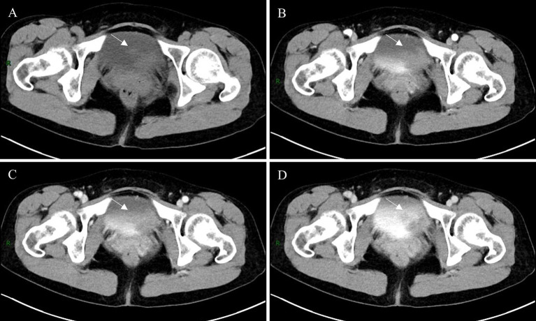 Figure 1
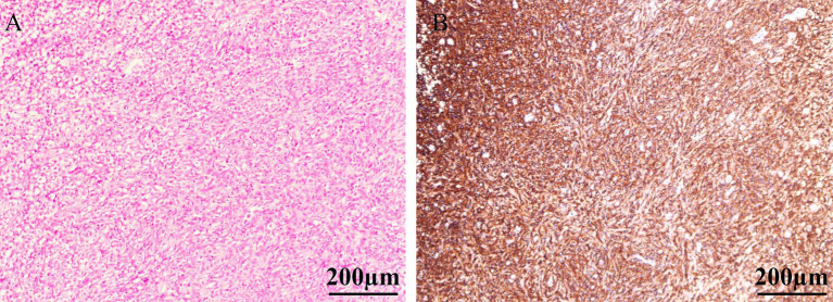 Figure 2
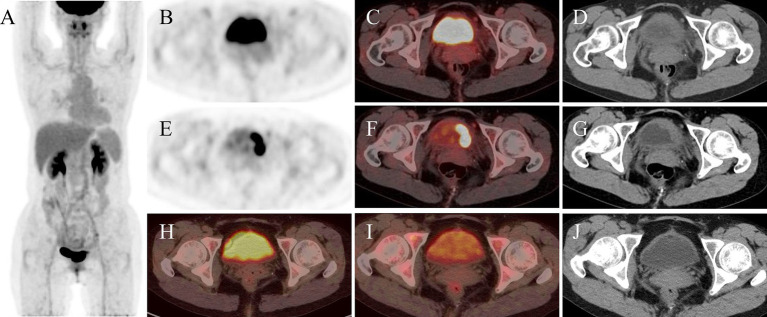 Figure 3
| Case no. | First author, year | Age (y)/sex | Symptoms | Location/bladder | US | CT/MRI | Cystoscopy | Bone marrow aspiration | Treatment | Recurrence |
|---|---|---|---|---|---|---|---|---|---|---|
| 1 ( | Jian-Hui Xin, 2024 | 83/F | Frequency urination | Posterior wall | Uneven hypoechoic lesion | CE: irregular enhancing nodule | Mild trigonal congestion, no neoplasia (post-treatment) | (−) | R-CHOP | (−) |
| 2 ( | Hideshige Seki, 2024 | 77/M | Fever, anorexia, general malaise, pyuria, bacteremia | NA | NA | Bladder wall thickening with perivesical stranding | NA | (−) | R-CHOP | (+) |
| 3 ( | Po-Sung Liang, 2024 | 79/F | Urinary frequency and urgency, suprapu-bic pain | Anterior wall | A suspicious 32 mm bladder tumor | CE: 39 mm polypoid enhancing lesion | Multiple bladder tumors | (−) | Radiation therapy | (−) |
| 4 ( | Xi Tu, 2023 | 81/F | Frequent urination and painful urination | Right side and posterior wall | A hypoechoic mass | Long T1 signal/short T2 signal, homogeneous CE enhancement, DWI restriction | Two nodular lesions | NA | TURBT | (−) |
| 5 ( | Atsuto Katano, 2022 | 65/F | Macroscopic hematuria, chronic cystitis | Bladder trigone | NA | Intermediate T2 signal intensity | A submucosal tumor | NA | TURBT, Radiation therapy | (−) |
| 6 ( | Zhen-Zhen Jiang, 2021 | 48/F | Macroscopic hematuria | NA | A large hypoechoic mass | Bladder wall thickening; ill-defined low-density nodules | Multiple bulging bladder masses | (−) | R-CHOP | (−) |
| 7 ( | Naoya Ishibashi, 2021 | 77/F | Hematuria and severe perineal pain | Bladder trigone | NA | Enlargement of an irregular elevated lesion | Non-papillary nodular tumor | NA | Radiation therapy | (−) |
| 8 ( | Rodolfo Montironi MD, 2015 | 81/M | Macroscopic hematuria, pain on micturition | Anterior wall to the right lateral bladder wall | NA | A solitary mass lesion | A wide-based mass | (−) | TURBT | (−) |
| 9 ( | Nischith D’Souza, 2017 | 56/M | Clot retention | Right lateral wall | A mass | NA | Polypoidal mass | NA | TURBT | (−) |
| 10 ( | Hewei Xue, 2020 | 77/F | Frequent urination, urinary urgency, dysuria | NA | Roughness in the inner walls of the bladder | Multiple nodules | Bladder occupying tumor | (−) | TURBT | (−) |
Peer Reviews
No public reviews on file for this paper yet. If you reviewed it on a platform where reviews are public (OpenReview, ICLR, NeurIPS, ICML), you can paste yours below so the community can read it here.
Videos
No videos yet. Explain this paper in a talk, walkthrough, or lecture? Add one.
Taxonomy
TopicsLymphoma Diagnosis and Treatment · Ovarian cancer diagnosis and treatment · Viral-associated cancers and disorders
Introduction
As an exceptionally rare extranodal malignancy, primary bladder lymphoma comprises just 0.2% of all non-Hodgkin lymphoma cases originating outside lymph nodes (1). Extranodal lymphoma diagnosis relies on comprehensive histopathological examination and immunohistochemistry (2). ^18^F-FDG PET/CT combining functional and anatomical imaging, forms the cornerstone of lymphoma patient management (3). PET/CT demonstrates superior accuracy and sensitivity to CT in identifying involved nodal sites (4). However, diagnostic challenges persist in regions with physiological ^18^F-FDG uptake or excretion, such as the brain and urinary tract. We present a rare case of primary bladder DLBCL. Initial ^18^F-FDG PET/CT imaging at 45 min post-intravenous tracer administration revealed that physiological tracer excretion limited lesion visualization. Accurate identification of bladder lesion sites presented significant challenges. To address this limitation, intravenous diuretic administration was implemented to mitigate physiological uptake within the bladder. Following intravenous diuretic administration, delayed imaging performed at 2-h post-radiotracer injection clearly delineated the bladder wall lesion. Compared with post-chemotherapy PET/CT images without diuretics, the diuretic-assisted protocol showed reduced urinary activity and clearer lesion visualization.
Case presentation
A 50-year-old woman presented to the Urology Department in November 2024 with a 1-week history of gross hematuria and nocturia. She had no associated symptoms such as urinary frequency, dysuria, voiding difficulty, flank pain, or fever. The patient had a 6-year history of chronic hepatitis B, with no documented family history of lymphoma or hematologic malignancies. The patient’s vital signs were stable and superficial lymph nodes were not palpably enlarged. Urinalysis revealed nitrite positivity, leukocyturia (2 cells/μL), and occult blood (3 cells/μL). Serological testing for hepatitis B demonstrated: Hepatitis B surface antigen (HBsAg): Positive (>250,000 IU/mL), Anti-hepatitis Be antibody (Anti-HBe): Negative (0.02 S/CO), Anti-hepatitis B core antibody (Anti-HBc): Positive (7.88 S/CO), HBV DNA: Negative (<500 IU/mL). Routine urological color ultrasound examination suggested that a low and weak echogenic mass measuring about 33 mm × 20 mm was seen in the left wall of the bladder. Uro-pelvic CT scan and enhancement showed a mass on the left posterior wall of the bladder, which was considered to be a high possibility of bladder cancer (Figure 1). Cystoscopy revealed a mucosal bulge extending from the 12 to 5 o’clock position, involving the anterior bladder wall and left wall near the bladder neck, measuring approximately 40 mm × 30 mm. After exclusion of contraindications to surgery, “transurethral cystectomy” was performed. Postoperative pathology confirmed DLBCL of the bladder, Hans classification: anaplastic B-cell subset. Immunohistochemistry showed positivity for CD20, PAX-5, BcL-6, MUM1, Ki-67 (90%), and wild-type P53; negative for CD10, BCL-2, CD21, CD3, CD5, CK, c-Myc, CyclinD1, and GATA3 (Figure 2). To further evaluate disease staging, a PET/CT scan was performed after the transurethral cystectomy. Whole-body imaging performed 45 min after intravenous injection of ^18^F-FDG (185 MBq) showed diffusely increased bladder activity, which interfered with lesion detection. To reduce urinary radioactivity and enhance lesion conspicuity, pelvic delayed imaging was performed 60 min following intravenous furosemide injection (20 mg) at 120-min post-radiotracer administration. This revealed focal wall thickening with intense hypermetabolic activity with a SUVmax of 22.7 in the left posterior bladder wall (Figure 3), while no abnormal ^18^F-FDG uptake was detected in other organs or lymph nodes. Based on the above findings, the diagnosis was primary stage I DLBCL of the bladder. The patient was referred to another hospital in the city for further treatment. Bone marrow aspiration and biopsy revealed no evidence of lymphoma cells. After completing 4 cycles of R-CHOP (rituximab, cyclophosphamide, doxorubicin, vincristine, and prednisone) chemotherapy, a follow-up ^18^F-FDG PET/CT scan demonstrated no abnormal increased uptake in the bladder wall (Figure 3), indicating favorable treatment response. During follow-up, the patient remained asymptomatic with no hematuria and in good clinical condition, continuing to receive regular hospital treatments.
Non-contrast abdominal CT scan demonstrated an irregular soft tissue density lesion along the left lateral wall of the urinary bladder (A, arrow). The mass exhibited contrast enhancement during both arterial and venous phases (B,C, arrow), with subsequent excretory phase imaging revealing a focal filling defect in the corresponding region of the left lateral bladder wall (D, arrow).
H&E staining revealed diffuse infiltration of the bladder lamina propria by a large number of atypical lymphoid cells, with near-complete replacement of the normal bladder tissue architecture (A). The tumor cells exhibited diffuse strong positivity with brown staining on the cell membrane, indicating widespread expression of CD20 antigen (B).
The whole-body 18F-FDG PET/CT. Maximum intensity projection (MIP) image prior to chemotherapy (A). Axial PET, PET/CT fusion and CT before furosemide administration (B–D) demonstrate the lesion. Following intravenous furosemide administration, post-diuretic axial PET (E), PET/CT fusion (F), and CT (G) images reveal prominent delineation of the left lateral bladder wall lesion. Post-therapy evaluation after 4 cycles of R-CHOP regimen chemotherapy displays axial PET/CT fusion images of the bladder during standard acquisition (H) and subsequent delayed imaging at 1 h (I), with comparison to pre-chemotherapy post-furosemide images demonstrating notably retained radiotracer activity within the bladder lumen. Corresponding post-chemotherapy CT image of the bladder is presented (J).
Literature review
A comprehensive literature search was conducted in the Web of Science database to identify case reports and case series on primary bladder lymphoma published in English up to March 31, 2025. After systematic search and rigorous full-text review, secondary cases and publications lacking accessible full-text were excluded. The following data were extracted from eligible studies: first author, publication year, patient demographics (gender and age), clinical presentation, imaging findings, and follow-up outcomes (Table 1).
A total of 11 cases of primary bladder lymphoma, including the present case, were included in the analysis (2, 5–13). The cohort comprised 3 males (3/11) and 8 females (8/11), with a median age of 77 years (range 48–83). Mucosa-associated lymphoid tissue (MALT) lymphoma was the predominant histopathological subtype. Hematuria emerged as the most frequent clinical manifestation, followed by dysuria, urinary frequency, and recurrent urinary tract infections predominantly caused by Escherichia coli. Most lesions were localized to the lateral walls and trigone region of the bladder, presenting as single or multiple nodular lesions with occasional bladder wall thickening. Ultrasonographic evaluation revealed hypoechoic lesions demonstrating contrast enhancement and restricted diffusion on advanced imaging. Notably, none of the reviewed cases exhibited bone marrow infiltration. Treatment strategies encompassed chemotherapy, radiotherapy, and transurethral resection of bladder tumor (TURBT). In selected cases with localized disease, TURBT alone achieved therapeutic success. The majority of cases demonstrated favorable outcomes with standardized management, with only one case showing tumor recurrence during follow-up, which was associated with documented extravesical infiltration.
Discussion
Approximately 40% of DLBCL cases originate from extranodal sites and the gastrointestinal tract representing the most common extranodal involvement (14, 15). While DLBCL demonstrates men predominance and higher incidence in individuals over 60 years, primary bladder lymphoma exhibits women preponderance (16). Notably, secondary bladder lymphoma occurs more frequently than its primary counterpart (1). The pathogenesis of DLBCL involves multifactorial mechanisms, with established risk factors encompassing genetic predisposition, immune dysregulation, and viral/environmental/occupational exposures (17). Recurrent urinary tract infections (UTIs) have been identified as a predisposing factor for primary bladder lymphoma (16, 18). Although our patient reported no definitive UTI symptoms or medical history, urinalysis revealed leukocyturia. Characteristic clinical manifestations of bladder lymphoma include gross hematuria, dysuria, urinary frequency, nocturia, and abdominal/back pain (16). The present case presented with gross hematuria and increased nocturnal voiding frequency. These nonspecific symptoms necessitate thorough differentiation from common urological conditions such as urinary tract infection, urothelial carcinoma, and mechanical obstruction.
The diagnosis of DLBCL of the bladder relies on imaging techniques, cystoscopy, and complete pathologic evaluation of tissue biopsies. On CT, bladder lymphoma may appear as an intraluminal nodular mass or focal wall thickening with early enhancement within approximately 60 s of contrast injection (19). In our patient, non-contrast CT revealed a nodular soft-tissue density in the left bladder wall with minimal enhancement on contrast imaging. During the excretory phase, a filling defect was observed against the high-density contrast background (Figure 1). Tumors on both T1 and T2-weighted images (T1WI and T2WI) on MRI show intermediate signal, and T1WI are suitable for detecting extravesical infiltration, lymph nodes, and bone metastases, while T2WI are suitable for assessing the depth of the tumor and differentiating the tumor from fibrosis, as well as for detecting peripheral organ invasion and bone marrow metastases (19). However, the limitations of CT and MRI imaging are difficult for the detection of metastatic lesions in normal-sized lymph nodes (20). ^18^F-FDG PET/CT demonstrates high sensitivity for lesion detection, particularly in identifying foci with no or minimal anatomical abnormalities on CT (21). This modality enables more accurate disease assessment and frequently alters lymphoma staging, consequently modifying therapeutic strategies (22). For patients with baseline PET-positive disease, serial monitoring of SUV changes and detection of new hypermetabolic lesions facilitate treatment response evaluation (22). Metabolic tumor volume (MTV), total lesion glycolysis (TLG), and maximal distance between lesions (Dmax) serve as critical prognostic parameters (23–25). Based on these advantages, we perform ^18^F-FDG PET/CT for suspected bladder lymphoma cases, which rapidly delineates extravesical involvement and determines whether the disease is secondary to nodal lymphoma. Some studies have shown that diuretic use significantly increases the lesion-to-bladder activity ratio, improving the detection rate (26–28). Moreover, dual-phase ^18^F-FDG PET/CT is of great clinical value for the staging of bladder cancer (29). Therefore, in the case of this patient, where the bladder lesion did not show well on early visualization (45 min after intravenous administration of ^18^F-FDG), a delayed visualization (2-h after intravenous administration of ^18^F-FDG) of 1 h after intravenous furosemide injection was taken (Figure 3). Before the use of diuretics, diffuse physiological ^18^F-FDG uptake was seen in the bladder due to physiological elimination of the radiotracer, which made it difficult to distinguish the metabolic distribution of the lesion on PET images. Following diuretic administration, PET imaging revealed a focal area of markedly increased metabolic activity in the left lateral wall of the bladder with a SUVmax of 22.7. This value significantly exceeded the average SUVmax of bladder cancer (16.1 ± 6.2) (30). Comparative analysis with the patient’s ^18^F-FDG PET/CT scan of post-chemotherapy (performed without intravenous diuretic bolus dual-phase imaging) demonstrated reduced background activity within the bladder after diuretic administration (Figure 3).
Although imaging studies provide preliminary differentiation between benign and malignant bladder lesions, histopathological examination remains essential for definitive diagnosis. MALT lymphoma/marginal zone lymphoma (MZL) has been historically regarded as the most common primary bladder lymphoma, while cases with extravesical extension are predominantly DLBCL (12, 31). DLBCL, not otherwise specified (NOS), represents the most frequent category (80–85% of DLBCL cases) and is histologically characterized by diffuse proliferation of large neoplastic cells effacing normal tissue architecture. These tumors demonstrate positivity for B-cell markers (CD20, CD79α, PAX5, CD22, CD19) and CD45 (1). Molecularly, DLBCL can be classified into two major subtypes by gene expression profiling: the germinal center B-cell-like (GCB) subtype and the activated B-cell-like (ABC) subtype (32). However, given the limited clinical availability of gene expression profiling, the immunohistochemistry-based Hans algorithm is routinely employed to categorize cases into GCB and non-GCB groups (the latter encompassing ABC subtypes and most unclassifiable cases) (14). In the present case, Hans classification indicated a non-germinal center B-cell phenotype. Histological examination revealed diffuse infiltration of the bladder lamina propria by sheets of atypical lymphoid cells with near-total effacement of normal bladder architecture. Immunohistochemistry confirmed CD20(+) and PAX-5(+) expression, establishing a B-cell lineage. A high Ki-67 proliferation index indicated aggressive tumor biology. These findings are consistent with DLBCL, leading to a definitive diagnosis of primary bladder diffuse large B-cell lymphoma.
DLBCL is a group of highly heterogeneous malignancies in terms of clinical manifestations, histomorphology and prognosis (1). The R-CHOP regimen remains the first-line treatment for DLBCL (1, 14, 33). However, its efficacy is suboptimal in elderly patients or those with comorbidities (34). For relapsed or refractory DLBCL, novel therapies such as chimeric antigen receptor T-cell (CAR-T) therapy and hematopoietic stem cell transplantation have emerged (34). Other approaches, including localized radiotherapy, BTK inhibitors, and immunomodulators, are primarily used as monotherapy or in combination to alleviate symptoms (35). The majority of primary bladder DLBCL cases (56.56%) were diagnosed at Stage I. Advanced age was identified as a poor prognostic factor, with patients aged ≥75 years exhibiting 2–3 times higher mortality than younger patients. The 5-year overall survival rate was 27.10% for patients ≥75 years versus 64.29% for those under 60 years (1). The patient has completed 4 cycles of R-CHOP chemotherapy. A follow-up ^18^F-FDG PET/CT scan revealed no increased uptake in the bladder wall lesion. Mildly increased uptake was observed in the bone marrow cavity due to post-chemotherapy bone marrow hyperplasia, with no other lesions showing elevated ^18^F-FDG uptake. Clinical evaluation indicated complete remission, and the patient is currently in good condition under regular treatment.
Conclusion
Primary bladder DLBCL is a rare subtype of bladder lymphoma, predominantly affecting women. Dual-phase ^18^F-FDG PET/CT imaging significantly improves diagnostic accuracy for primary bladder lymphoma. When high bladder radiotracer activity causes imaging artifacts, a diuretic-enhanced washout protocol combined with controlled bladder distension can accelerate urine clearance and improve the target-to-background ratio.
The reference list from the paper itself. Each links out to its DOI / PubMed record.
- 1Zanelli M Sanguedolce F Zizzo M Palicelli A Pellegrini D Farinacci S. Primary diffuse large B-cell lymphoma of the urinary bladder: update on a rare disease and potential diagnostic pitfalls. Curr Oncol. (2022) 29:956–68. doi: 10.3390/curroncol 29020081, PMID: 35200580 PMC 8870454 · doi ↗ · pubmed ↗
- 2Liang PS Shih HJ Huang SH Chen YZ. Primary lymphoma of mucosa associated lymphoid tissue (MALT lymphoma) in the urinary bladder mimicking recurrent urinary tract infection: a case report and literature review. BMC Urol. (2024) 24:223. doi: 10.1186/s 12894-024-01616-3, PMID: 39395994 PMC 11470736 · doi ↗ · pubmed ↗
- 3Trotman J Barrington SF. The role of PET in first-line treatment of Hodgkin lymphoma. Lancet Haematol. (2021) 8:e 67–79. doi: 10.1016/s 2352-3026(20)30357-4, PMID: 33357485 · doi ↗ · pubmed ↗
- 4Cronin CG Swords R Truong MT Viswanathan C Rohren E Giles FJ. Clinical utility of PET/CT in lymphoma. AJR Am J Roentgenol. (2010) 194:W 91–W 103. doi: 10.2214/AJR.09.2637, PMID: 20028897 · doi ↗ · pubmed ↗
- 5Montironi R Santoni M Goteri G Mazzucchelli R Lopez-Beltran A Cheng L. Pseudocarcinomatous hyperplasia associated with primary lymphoma in the urinary bladder: a case report. Hum Pathol. (2015) 46:1040–4. doi: 10.1016/j.humpath.2015.03.004, PMID: 25912755 · doi ↗ · pubmed ↗
- 6D'Souza N Verma A Rai A. Two cases of non-Hodgkin lymphoma of urinary bladder: primary and secondary. J Cancer Res Ther. (2017) 13:1053–5. doi: 10.4103/0973-1482.179071, PMID: 29237976 · doi ↗ · pubmed ↗
- 7Xu H Chen Z Shen B Wei Z. Primary bladder mucosa-associated lymphoid tissue lymphoma: a case report and literature review. Medicine (Baltimore). (2020) 99:e 20825. doi: 10.1097/md.0000000000020825, PMID: 32664075 PMC 7360252 · doi ↗ · pubmed ↗
- 8Ishibashi N Nakanishi Y Nishimaki H Maebayashi T Masuda S Okada M. Bladder mucosa-associated lymphoid tissue lymphoma progressed from chronic cystitis along with a comparative genetic analysis during long-term follow-up: a case report. Transl Androl Urol. (2021) 10:3899–906. doi: 10.21037/tau-21-602, PMID: 34804832 PMC 8575576 · doi ↗ · pubmed ↗