Sternal Foramen Mimicking Gunshot Injury: A Forensic Case Report
Arwinder Singh, Yashpal S, Dilip Vaishnav, Kishanth S, Amit Jangid

TL;DR
A case where a sternal foramen was mistaken for a gunshot wound is presented, emphasizing the need for accurate forensic identification of anatomical variations.
Contribution
This case report highlights the forensic significance of distinguishing sternal foramina from traumatic injuries.
Findings
A sternal defect was initially suspected as a gunshot wound but was identified as a congenital sternal foramen.
Features like smooth margins and uniform dimensions helped exclude trauma-related characteristics.
The case underscores the importance of recognizing anatomical variants in forensic pathology.
Abstract
The sternum develops from multiple ossification centers, and their incomplete fusion can lead to congenital anomalies like sternal foramina; although typically asymptomatic, these foramina possess significant clinical and forensic relevance due to their potential mimicry of penetrating traumatic injuries such as gunshot wounds. We present a forensic case involving a highly decomposed body, recovered from a submerged vehicle two years post-incident, which exhibited a distinct sternal defect initially suspected as a gunshot wound based on its location and appearance. However, meticulous skeletal examination revealed features consistent with a congenital sternal foramen, including smooth, well-corticated margins and uniform defect dimensions, which allowed for the exclusion of trauma due to the absence of typical gunshot wound characteristics like bevelling, radiating fractures, and bone…
Genes, proteins, chemicals, diseases, species, mutations and cell lines named across the full text — each resolved to its canonical identifier and authoritative record.
Click any figure to enlarge with its caption.
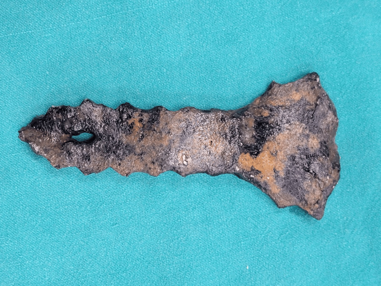 Figure 1
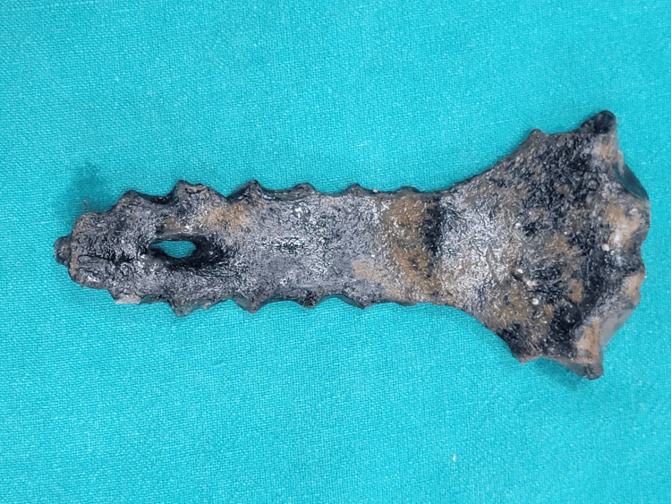 Figure 2
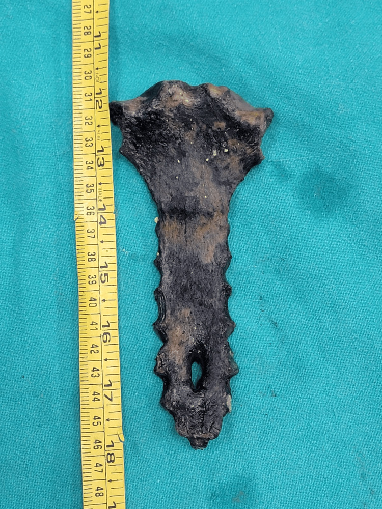 Figure 3
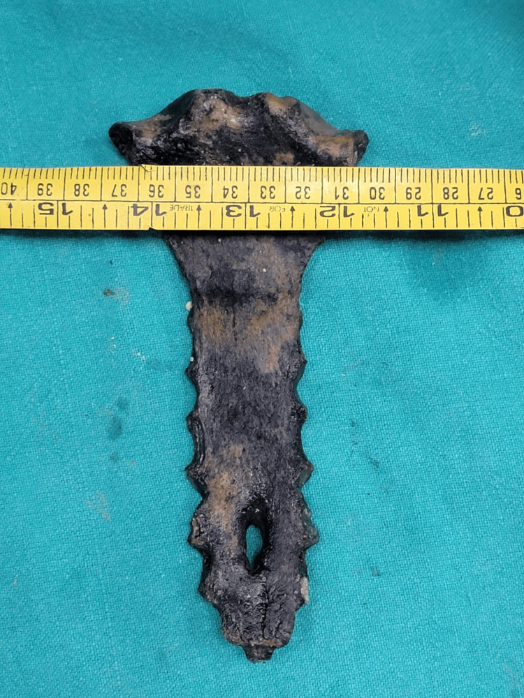 Figure 4
| Feature | Sternal foramen | Gunshot injury |
| Origin | Congenital/developmental | Acquired (traumatic) |
| Margins | Smooth, rounded, well-corticated, often everted | Irregular, jagged, fractured, and absent cortical bone |
| Bevelling | Absent (same dimensions on both sides) | Present (internal for entry, external for exit) |
| Radiating fractures | Absent | Often present, extending from the defect |
| Bone fragmentation | Absent | Often present (comminution) |
| Associated soft tissue damage | Absent | Almost always present (lacerations, hemorrhage, charring) |
| Gunshot residue | Absent | May be present (soot, stippling) at entry |
| Underlying organ damage | Unrelated (unless iatrogenic complication) | Common and severe (heart, lung, vessels) |
| Radiological appearance | Smooth, corticated border, uniform defect | Irregular, fragmented, often with associated foreign bodies |
Peer Reviews
No public reviews on file for this paper yet. If you reviewed it on a platform where reviews are public (OpenReview, ICLR, NeurIPS, ICML), you can paste yours below so the community can read it here.
Videos
No videos yet. Explain this paper in a talk, walkthrough, or lecture? Add one.
Taxonomy
TopicsTraumatic Ocular and Foreign Body Injuries · Trauma Management and Diagnosis · Gun Ownership and Violence Research
Introduction
The human sternum is a pivotal component of the thoracic cage, which originates from multiple ossification centers that fuse during development [1-4]. Anomalies in this fusion process can lead to developmental defects within the sternal body, such as sternal foremen [1-5]. These congenital defects are often asymptomatic but hold significant clinical relevance, such as the risk of iatrogenic injury during invasive cardiothoracic procedures [1,4,6,7].
In forensic medicine, meticulous examination is necessary for identification of sternal defects. The morphological similarities between a sternal foramen and a penetrating traumatic injury, such as a gunshot wound, can be misleading, especially in cases of advanced decomposition or skeletonization [2,3,8]. Misinterpretation in such scenarios can impact the determination of cause and manner of death, leading to erroneous conclusions in medico-legal investigations [2,3,8]. This article presents a forensic case where such a distinction was critical, followed by a comprehensive review of the differentiating characteristics of sternal foramina and gunshot injuries to the sternum.
Case presentation
A tragic incident unfolded when a car with a father and son fell into a river. Days later, the son's body was recovered from a downstream dam, and a post-mortem examination was conducted. However, the father's body remained unaccounted for. Two years after the incident, the submerged car was successfully recovered from the riverbed due to a fall in water level. Inside, amidst the advanced stages of decomposition, was the body of the man. Relatives positively identified the car and the deceased based on the clothing still clinging to the remains.
The body was brought for a post-mortem examination, revealing extreme decomposition. While adipocere was observed over the chest area, indicating prolonged immersion in water under specific conditions, the remaining soft tissues and internal viscera were largely unidentifiable. The bones were carefully extracted for detailed assessment.
During the examination of the skeletal remains, a noticeable anomaly was discovered: a distinct hole in the lower one-third of the sternum (Figure 1). This finding immediately raised a serious suspicion of a gunshot wound to the chest, suggesting the possibility of a projectile injury that may have precipitated the vehicle's plunge into the river. This potential alternate theory underscored the critical need for a precise differentiation. However, circumstantial evidence, absence of any entry or exit wound, and the bullet in-situ pointed towards the congenital nature of the hole.
Detailed examination of the recovered sternum (Figures 1-4) revealed a distinct, oval defect in its lower one-third, measuring 2.2 cm in length and 1 cm in breadth at its inner and outer parts, narrowing to 1 cm in length and 0.4 cm in breadth at its middle.
Anterior surface of the recovered sternum.Anterior view of the recovered sternum from the decomposed remains. The distinct oval defect, identified as a sternal foramen, is visible in the lower one-third of the sternal body. Note the well-corticated margins characteristic of a congenital anomaly.
Posterior surface of the recovered sternum.Posterior view of the recovered sternum. The sternal foramen is visible from this aspect, showing the consistent dimensions of the defect across both the anterior and posterior surfaces, further supporting the absence of bevelling.
Length measurement of the sternal defect.Recovered sternum with a measuring tape, illustrating the length of the sternal defect. The defect measured 2.2 cm in length at its inner and outer parts, constricting to 1 cm in length at its middle. This view also highlights the surrounding intact bone and the absence of radiating fracture lines.
Breadth measurement of the sternal defect.Recovered sternum with a measuring tape, illustrating the breadth of the sternal defect. The defect measured 1 cm in breadth at its inner and outer parts, constricting to 0.4 cm in breadth at its middle. This further demonstrates the uniform nature of the congenital defect.
The margins of the defect were smooth, well-corticated, and regular. The diameter of the defect was consistent across both the anterior and posterior surfaces of the sternum. These findings are consistent with the diagnosis of a sternal foramen. The characteristic features of traumatic impact, such as radiating fracture lines emanating from the defect into the surrounding bone, were absent. Furthermore, bevelling, or the typical conical shape seen in projectile trajectories, was not present. The sternal bone surrounding the anomaly appeared largely intact, lacking the comminution or fragmentation typically associated with a high-energy gunshot wound. While the advanced stage of decomposition precluded soft tissue analysis for gunshot residue, the macroscopic features of the bony defect strongly supported a congenital origin, leading to the conclusion that the anomaly was indeed a sternal foramen rather than a gunshot injury.
Discussion
Embryological development and ossification
Sternal development is a complex process, beginning with the formation and midline fusion of mesenchymal bars during the sixth to tenth weeks of intra-uterine life, followed by chondrogenesis into a cartilaginous rod. Initial fusion and subsequent segmentation into sternebrae occur in a cranio-caudal direction. Ossification commences around the fifth month of gestation. Manubrium and first two sternebrae form from single centers, and the later sternebrae often develop from paired centers, if bar fusion is not complete. Interestingly, the union of these primary ossification centers progresses in the opposite, caudo-cranial direction, with the xiphoid process ossifying much later. The union of primary ossification centers commences after three years of age, and complete fusion of all sternebrae within the mesosternum is usually observed in individuals over 15 years old [2].
Sternal foramina: Developmental anomalies
Sternal foramina are developmental defects resulting from the incomplete fusion of the sternal segments during ossification. The sternum ossifies from six to eight cartilaginous centers, which fuse sequentially from inferior to superior. Failure of fusion, or partial fusion, can lead to various anomalies, including sternal foramina or sternal clefts [1,3,5].
Sternal foramina are most frequently observed in the lower third of the sternal body, particularly at the junction of the fourth and fifth sternebrae, or occasionally in the xiphoid process or manubrium [3,4]. These congenital defects typically present as round or oval defects, varying in size from a few millimeters to over a centimeter in diameter [3-5]. A defining characteristic of their margins is their smooth, well-corticated, and often everted nature, indicative of a naturally formed anatomical structure, with consistent dimensions across both inner and outer surfaces [1,3-5]. These features were crucial in our case, where the observed sternal defect exhibited precisely these smooth, well-corticated margins and uniform dimensions, directly aligning with the morphological characteristics of a sternal foramen and distinguishing it from a traumatic injury. The reported prevalence of sternal foramina varies across different populations and studies, generally ranging from 4% to 10% in living individuals, though higher in skeletal studies (some reports up to 27.4%), and they are generally more common in males [2-4]. While often asymptomatic, the presence of a sternal foramen is clinically significant as it creates a direct pathway to the mediastinum, carrying a considerable risk of complications during procedures involving sternal puncture, such as bone marrow biopsy or acupuncture, potentially leading to pneumothorax, cardiac tamponade, or injury to great vessels [1,4,6,7].
Gunshot injuries to the sternum: Traumatic insults
Gunshot wounds to the sternum are a result of high-velocity projectile impact, causing significant damage to both bone and surrounding soft tissues. The characteristics of the defect depend on multiple factors, including projectile type, velocity, angle of impact, and range of fire [2,8].
Gunshot wounds are always acquired traumatic lesions, not congenital [9]. Their appearance in bone is distinct: the edges of the bony defect are typically irregular, jagged, and often exhibit radiating fracture lines extending from the primary defect, lacking the smooth cortical bone seen in foramina. A critical feature for determining the direction of projectile travel is bevelling, where the entry wound is wider on the inner aspect of the bone compared to the outer entry point, forming a "cone" effect [9]. The impact also commonly causes significant bone fragmentation (comminution), leading to multiple small pieces of bone (shrapnel) [10]. If the projectile exits the sternum, the exit wound is generally larger and more irregular, exhibiting external bevelling [9]. In our case, the sternal defect conspicuously lacked these features; there were no irregular, jagged edges, no radiating fracture lines, no bevelling indicating projectile trajectory, and no evidence of comminution or fragmentation, which strongly argued against a gunshot injury. Associated soft tissue damage is also characteristic of gunshot wounds, including an abrasion collar around the skin entry wound, and for close-range shots, soot and unburnt/partially burnt gunpowder particles (stippling or tattooing) [11]. Extensive tissue damage, hemorrhage, and disruption to underlying muscles and vital organs (heart, lungs, major blood vessels) are almost invariably present [9]. Radiographic imaging or dissection may also reveal retained bullet fragments within the sternal bone or surrounding tissues [9]. Despite the advanced stage of decomposition in our case, the absence of any remnants suggesting an abrasion collar, soot, or stippling, combined with the lack of any projectile fragments within or near the sternal defect, further supported the exclusion of a gunshot wound.
Differentiating sternal foramina from gunshot injuries: A forensic perspective
The ability to accurately distinguish between a sternal foramen and a gunshot injury (Table 1) is paramount in forensic pathology and anthropology, especially in challenging cases like the one presented [2,3,8].
Conclusions
This case illustrates the challenges faced in forensic medicine, particularly when dealing with highly decomposed remains. The initial suspicion of a gunshot injury due to the sternal defect underscores the critical need for a precise differential diagnosis. Through meticulous examination, focusing on the definitive morphological characteristics of the sternal defect -- namely the smooth, corticated margins, the absence of beveling, and the lack of associated traumatic indicators on the skeletal remains and surrounding minimal tissues -- we were able to conclude that the "hole" was indeed a sternal foramen, a congenital anomaly, and not a gunshot wound.
This determination had profound implications, allowing investigators to correctly focus on drowning as the cause of death in the context of the vehicle's plunge into the river. This case reinforces the absolute necessity for forensic experts to possess a deep understanding of anatomical variations and their differentiating features from traumatic injuries, ensuring accuracy in medico-legal investigations and upholding the integrity of justice.
The reference list from the paper itself. Each links out to its DOI / PubMed record.
- 1A comprehensive review of the sternal foramina and its clinical significance Cureus Choi PJ Iwanaga J Tubbs RS 09201710.7759/cureus.1929 PMC 580531929456905 · doi ↗ · pubmed ↗
- 2Development and morphology of the sternal foramen Am J Forensic Med Pathol Cooper PD Stewart JH Mc Cormick WF 34234791988323955610.1097/00000433-198812000-00016 · doi ↗ · pubmed ↗
- 3Morphological approach of the sternal foramen: an anatomic study and a short review of the literature Folia Morphol (Warsz) Gkantsinikoudis N Chaniotakis C Gkasdaris G Georgiou N Kapetanakis S 4844907620172815027210.5603/FM.a 2017.0006 · doi ↗ · pubmed ↗
- 4Prevalence and morphometry of sternal and xiphoid foramen: a meta-analysis on 16,666 subjects Surg Radiol Anat Pasieka P Pasieka PM Komosa A Barnowska A Pękala J Malinowski K Tomaszewski K 6236354520233691841810.1007/s 00276-023-03116-9PMC 10129975 · doi ↗ · pubmed ↗
- 5Anatomical variations of the sternum: sternal foramen and variant xiphoid morphology in dried adult human sternum in Ethiopia F 1000 Res Lema AS 9341220233884561710.12688/f 1000 research.133151.1PMC 11153988 · doi ↗ · pubmed ↗
- 6Midline sternal foramen: CT demonstration J Comput Assist Tomogr Stark P 48949091985398904310.1097/00004728-198505000-00013 · doi ↗ · pubmed ↗
- 7Frequency of sternal variations and anomalies evaluated by MDCTAJR Am J Roentgenol Yekeler E Tunaci M Tunaci A Dursun M Acunas G 95696018620061655456310.2214/AJR.04.1779 · doi ↗ · pubmed ↗
- 8An unusual case of multiple mesosternal foramina J Forensic Sci Mc Cormick WF Flournoy LE Rogers NL Ross AH 706707431998 https://pubmed.ncbi.nlm.nih.gov/9608710/9608710 · pubmed ↗