Images in infectious diseases: milker’s nodule with erythema multiforme after calf bite in a 23-year-old patient
Benjamin T. Schleenvoigt, Christine Kletta, Christine Zollmann, Stefan Hagel, Stefan Glöckner, Eva Krause, Janine Michel, Carlotta Helbig, Andrea Vanegas-Ramirez

Abstract
Genes, proteins, chemicals, diseases, species, mutations and cell lines named across the full text — each resolved to its canonical identifier and authoritative record.
Click any figure to enlarge with its caption.
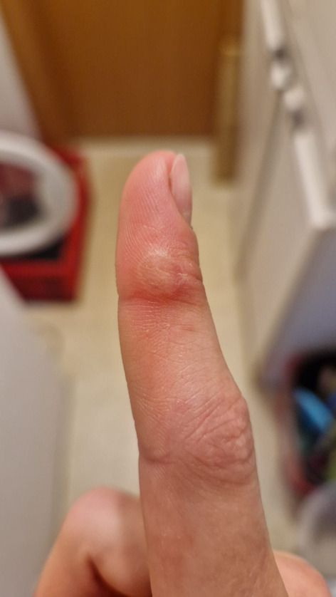 Figure 1
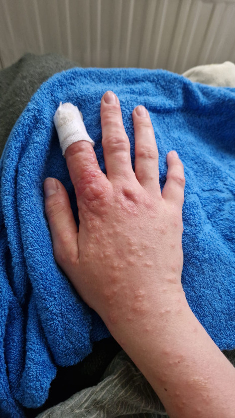 Figure 2
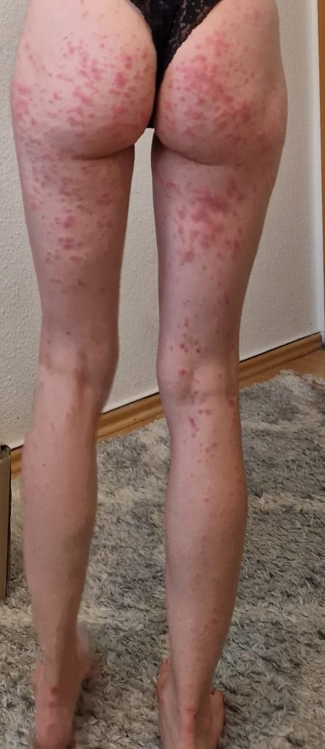 Figure 3
- —Friedrich-Schiller-Universität Jena (1010)
Peer Reviews
No public reviews on file for this paper yet. If you reviewed it on a platform where reviews are public (OpenReview, ICLR, NeurIPS, ICML), you can paste yours below so the community can read it here.
Videos
No videos yet. Explain this paper in a talk, walkthrough, or lecture? Add one.
Taxonomy
TopicsDrug-Induced Adverse Reactions · Eosinophilic Disorders and Syndromes · Chemotherapy-related skin toxicity
A 23-year-old patient presented to the general practitioner´s (GP’s) office with a hard nodule on her right index finger. This had been preceded by a calf bite in the same place 4 weeks earlier. The wound had initially healed within a few days without sequelae. Clinically, the nodule was approximately 1 cm in size, with a whitish margin and central hemorrhagic erythema (Fig. 1).
Fig. 1. Nodule on the right index finger, 4 weeks after the calf bite
The puncture remained dry, and the swab showed no bacterial growth. After starting empirical antibiotic therapy with amoxicillin/clavulanate, a macular pruritic rash occurred, beginning on both distal extremities and spreading to the trunk (Figs. 2 and 3). The patient also reported a general malaise, pain in her limbs and loss of appetite.
Fig. 2. Macular pruritic rash after starting antibiotic treatment
Fig. 3. Macular pruritic rash after starting antibiotic treatment
Amoxicillin/clavulanate was discontinued after 6 days, on the assumption that the rash was drug-related. In view of the previous animal contact, a swab for poxvirus analysis was sent to the Robert Koch Institute, and the PCR test for parapoxviruses returned a positive result [1]. The sample was identified as pseudocowpox virus by Sanger sequencing of the B2L gene. Furthermore, parapoxvirus specific IgM (1:1280) and IgG (1:320) were detected by immunoflourescence assay in a serum sample taken 7 weeks after the bite. In the context of the diagnosis of pseudocowpox, the rash was most likely to be classified as infection-induced erythema multiforme [2].
Topical steroids (mometasone furoate cream 1 mg/g, applied twice daily for two weeks) and systemic steroids (methylprednisolone: 40 mg on day 1, 20 mg for 2 days, 10 mg for 2 days, and 5 mg for 2 days) were administered, complemented by topical antibiotic treatment (Fusidic acid cream) as a preventive measure [3]. Analgesic treatment (Ibuprofen 600 mg) was used as needed. Within the next two weeks, the exanthema gradually subsided, and the nodule on the index finger healed without scarring.
The reference list from the paper itself. Each links out to its DOI / PubMed record.