A Case of Synchronous Bilateral Canalicular Adenoma and Polymorphous Adenocarcinoma of the Minor Salivary Glands of the Upper Lip
Holly Boyes, Rahail Kumar, Mathew Alemkunnapuzha, Mohammed Anabtawi

TL;DR
A 76-year-old woman had four lumps on her upper lip, diagnosed as two polymorphous adenocarcinomas and two canalicular adenomas, showing rare synchronous tumors in minor salivary glands.
Contribution
The paper presents a rare case of synchronous bilateral canalicular adenoma and polymorphous adenocarcinoma in minor salivary glands of the upper lip.
Findings
Two medial lesions were polymorphous adenocarcinomas and two lateral lesions were canalicular adenomas.
The case emphasizes the need for thorough biopsies to identify multiple coexisting salivary gland pathologies.
Patient demographics align with those reported in the literature for similar rare salivary gland tumors.
Abstract
Multiple salivary gland tumours can occur in both major and minor salivary glands. Its histology can be greatly varied and include pleomorphic adenoma, canalicular adenoma, mucoepidermoid carcinoma, or polymorphous adenocarcinoma, amongst other pathologies. A 76-year-old lady was referred on a two-week-wait pathway to our oral and maxillofacial surgery department complaining of a one-year history of painless lumps in her upper lip, which were gradually increasing in size. Examination in the clinic revealed four distinct lumps in the upper lip that were firm on palpation but mobile. Histopathological examination revealed the two medial lesions to be polymorphous adenocarcinomas and the two lateral lesions to be canalicular adenomas. Whilst our case represents uncommon pathologies, our patient’s demographics are typical of others in the literature. This highlights the importance of…
Genes, proteins, chemicals, diseases, species, mutations and cell lines named across the full text — each resolved to its canonical identifier and authoritative record.
Click any figure to enlarge with its caption.
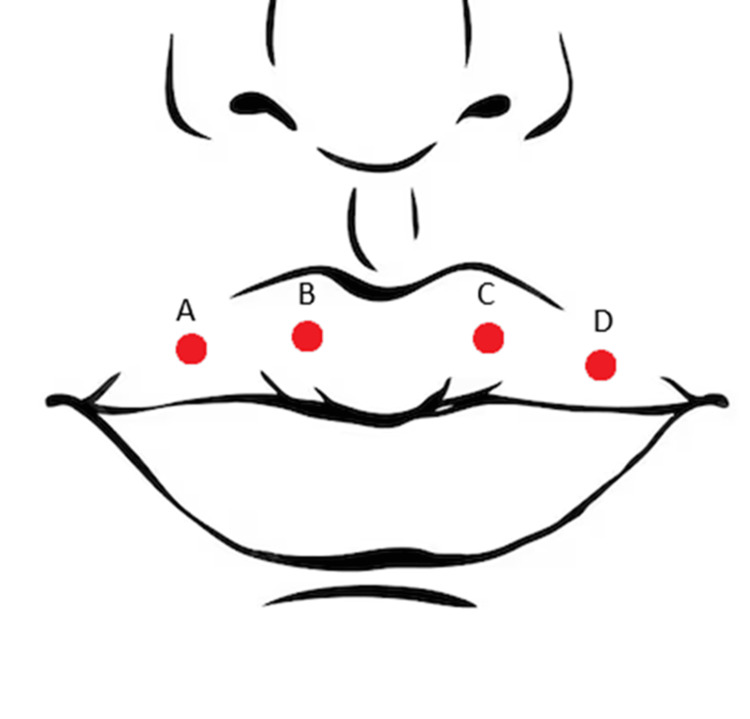 Figure 1
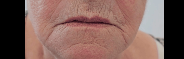 Figure 2
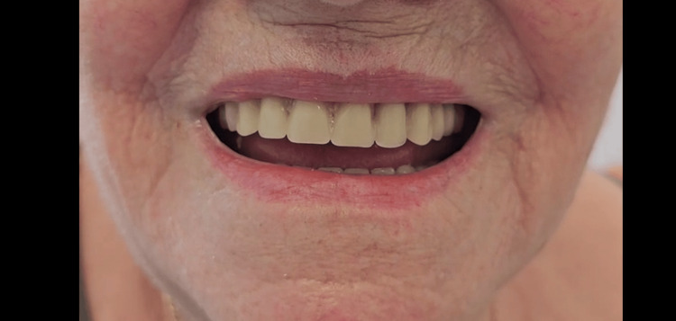 Figure 3
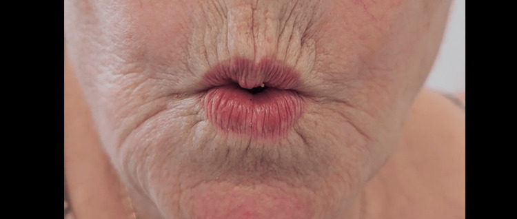 Figure 4
Peer Reviews
No public reviews on file for this paper yet. If you reviewed it on a platform where reviews are public (OpenReview, ICLR, NeurIPS, ICML), you can paste yours below so the community can read it here.
Videos
No videos yet. Explain this paper in a talk, walkthrough, or lecture? Add one.
Taxonomy
TopicsSalivary Gland Tumors Diagnosis and Treatment · Ear and Head Tumors · Cancer and Skin Lesions
Introduction
Salivary gland neoplasms represent a diverse group of tumours that can occur in both major and minor salivary glands [1]. They account for less than 5% of all head and neck neoplasms, and while most arise from the major salivary glands, approximately 10-25% are of minor salivary gland origin, commonly located in the palate, upper lip, and buccal mucosa [2,3]. Though these tumours may occur as isolated lesions, multiple synchronous tumours, especially involving both benign and malignant histology, are notably uncommon.
Tumours of the salivary glands may present with a broad spectrum of histopathological features, ranging from benign entities such as pleomorphic adenoma and canalicular adenoma to malignant forms including mucoepidermoid carcinoma, adenoid cystic carcinoma, and polymorphous adenocarcinoma [3, 4].
Most reported cases of multiple tumours occur in the parotid gland, with synchronous benign and malignant lesions in minor salivary glands being exceedingly rare. A limited number of reports have documented such dual pathologies in the upper lip, including canalicular adenoma and polymorphous adenocarcinoma occurring concurrently [1,5,6].
We present a rare case of bilateral upper lip canalicular adenomas occurring synchronously with polymorphous adenocarcinomas. This case serves to underscore the diagnostic complexity and necessity of complete histological evaluation of all lesions, particularly when multiple lesions are present.
Case presentation
A 76-year-old lady was referred by her dentist on a two-week wait pathway to our oral and maxillofacial surgery department, complaining of a one-year history of painless lumps in her upper lip, which were gradually increasing in size. She denied any bleeding, paraesthesia, taste disturbance, dry mouth, or constitutional symptoms.
Her medical history includes chronic obstructive pulmonary disease, hypertension, osteoarthritis, hypothyroidism, type 2 diabetes mellitus, hypercholesterolaemia, and cataracts. She has an allergy to nuts. Her social history revealed she is an ex-smoker, having stopped at age 74, and a non-drinker.
Examination in the clinic revealed four distinct lumps in the upper lip that were firm on palpation but mobile: (A) in the UR4 region, measuring 8 mm in diameter; (B) in the UR2 region, 4 mm; (C) in the UL2 region, 5 mm; and (D) in the UL4 region, 7 mm. There was no cervical lymphadenopathy, and was edentulous in the upper and lower jaws.
Histopathological examination revealed the following diagnoses (see Figure 1): (A) canalicular adenoma, (B) polymorphous adenocarcinoma of the minor salivary gland, (C) polymorphous adenocarcinoma of the minor salivary gland, and (D) canalicular adenoma.
Schematic illustrating clinical locations of lesions
Following histopathological assessment, she was referred to the regional head and neck cancer service for further investigation and management. An MRI of the neck showed no evidence of nodal disease. She also underwent a staging CT of the chest, which revealed several lung nodules of varying sizes. Her case was discussed at the head and neck multi-disciplinary team (MDT) meeting, and was listed for wide local excision and local reconstruction of the adenocarcinomatous lesions in her upper lip, along with a referral to the local lung MDT regarding her lung nodules.
She underwent wide local excision (5 mm margins) and primary closure of the adenocarcinomatous lip lesions under general anaesthesia, from which she recovered well with a minimal amount of residual numbness. The histopathology showed complete excision of both polymorphous adenocarcinoma (pT1NXM0), and she was scheduled for a routine follow-up.
The lung MDT recommended surveillance CT scans, which showed no concerning findings on follow up and she was discharged from the respiratory department after four months. She was seen by the oral and maxillofacial surgery team every six weeks for a year following her procedure, with four-monthly neck ultrasound scans (all of which were negative for disease). She is currently on four-monthly clinic reviews and progressing well (Figures 2-4).
Post-operative lips at rest
Post-operative lips smiling
Post-operative lips pursed
Discussion
Minor salivary gland tumours present a diagnostic challenge due to their varied histopathology and potential for multiple synchronous lesions. One study analysed 213 cases of minor salivary gland tumours and found 56% were benign tumours (most commonly pleomorphic adenoma, followed by canalicular adenoma) and 44% were malignant (most commonly mucoepidermoid carcinoma, followed by adenoid cystic carcinoma, then polymorphous low-grade adenocarcinoma) [3].
Canalicular adenoma
Canalicular adenomas are benign salivary gland tumours that often present as a slow-growing nodule in the upper lip, more commonly found in women over the age of 50 [7]. They are well-circumscribed and rarely recur after excision. Although typically solitary, multifocal and bilateral presentations have been documented [8-10]. They arise from the terminal ducts of minor salivary glands, are encapsulated, and characterised histologically by columnar or cuboidal cells in a loose, highly vascular stroma [7].
Polymorphous adenocarcinoma
Polymorphous adenocarcinoma most commonly occurs in minor salivary glands of the palate [11]. Morphologically, it can exhibit a variety of growth patterns, including solid, cribriform, tubular, trabecular, fascicular, and papillary patterns [12, 13]. It frequently presents as an asymptomatic, slow-growing mass; it is more commonly seen in women, with a peak incidence in the sixth to seventh decade of life. It commonly presents in the palate; it may be locally destructive and often displays perineural invasion, though distant metastases are rare. Microscopically, the tumour is unencapsulated, infiltrative, with cells arranged in diverse architectural patterns [14].
Clinical significance
Most reported cases of synchronous tumours involve the major glands, especially the parotid [15]. In the literature, only a few cases describe synchronous canalicular adenoma and polymorphous adenocarcinoma, with very limited cases involving bilateral lesions [1, 5, 6]. From a clinical perspective, the identification of one benign lesion should not preclude investigation of adjacent or additional lesions, as these may harbour malignant pathology. This case exemplifies the need for individualised assessment and histopathologic confirmation of each lesion. The patient’s age, sex, and anatomical location align with typical presentations of both canalicular adenoma and polymorphous adenocarcinoma.
Conclusions
Though each tumour entity individually aligns with the known epidemiological and clinical patterns, their concurrent appearance underscores the necessity of detailed clinical, radiological, and pathological assessment. Multiple biopsies should be obtained in cases of multifocal lesions to ensure accurate diagnosis and treatment planning. Clinicians should maintain a high index of suspicion when encountering multiple upper lip lesions, particularly in elderly female patients, as synchronous salivary gland tumours, though rare, carry important diagnostic and management implications.
The reference list from the paper itself. Each links out to its DOI / PubMed record.
- 1Polymorphous low-grade adenocarcinoma of the upper lip with metachronous myoepithelioma of the buccal mucosa Oral Surg Oral Med Oral Pathol Oral Radiol Argyris PP Gopalakrishnan R Pambuccian SE Tosios KI Koutlas IG 08117201410.1016/j.oooo.2013.08.03024268388 · doi ↗ · pubmed ↗
- 2Salivary gland tumours Oral Dis Speight PM Barrett AW 229240820021236310710.1034/j.1601-0825.2002.02870.x · doi ↗ · pubmed ↗
- 3Frequency and distribution pattern of minor salivary gland tumors in a northeastern Chinese population: a retrospective study of 485 patients J Oral Maxillofac Surg Wang XD Meng LJ Hou TT Zheng C Huang SH 81917320152551195810.1016/j.joms.2014.08.019 · doi ↗ · pubmed ↗
- 4WHO Classification of Head and Neck Tumours. 4th ed El-Naggar AK Chan JKC Grandis JR Lyon IARC Press 2017 https://publications.iarc.fr/Book-And-Report-Series/Who-Classification-Of-Tumours/WHO-Classification-Of-Head-And-Neck-Tumours-2017
- 5Synchronous polymorphous adenocarcinoma and canalicular adenoma on the upper lip: an unusual presentation and immunohistochemical analysis Head Neck Pathol Ortega RM Bufalino A Almeida LY 1451491220182876611010.1007/s 12105-017-0838-6PMC 5873492 · doi ↗ · pubmed ↗
- 6An extremely rare case of synchronous low-grade polymorphous adenocarcinoma with canalicular adenoma of the minor salivary gland of the palate Cureus Kasthurirengan S John RS 015202310.7759/cureus.36591 PMC 1012250637095807 · doi ↗ · pubmed ↗
- 7Canalicular adenoma: A systematic review J Craniomaxillofac Surg Peraza AJ Wright J Gómez R 175417584520172889025210.1016/j.jcms.2017.07.020 · doi ↗ · pubmed ↗
- 8Clinical and histological features of multifocal canalicular adenomas of the upper lip Braz Dent J Siqueira CS Fernandes KS Vivas AP Pinto Ddos S Jr de Sousa SC 5425462420132447430110.1590/0103-6440201302264 · doi ↗ · pubmed ↗