Clinical characteristics and prognosis analysis of patients with myocardial infarction with non-obstructive coronary arteries in the Qinghai-Tibet plateau region
Zixu Fan, Zhiyu Wang, Yinghua Wang, Jianwei Ma, Mingyuan Niu, Min Zhang

TL;DR
This study examines the causes and outcomes of heart attacks with non-blocked arteries in patients from the Qinghai-Tibet Plateau region.
Contribution
The study identifies unique clinical features and etiologies of MINOCA in a high-altitude population.
Findings
MINOCA patients had lower BMI and different laboratory markers compared to MI-CAD patients.
Common causes of MINOCA included plaque rupture, artery spasm, and undiagnosed myocarditis.
Electrocardiogram and echocardiography findings differed significantly between the two groups.
Abstract
To investigate the etiology and clinical characteristics of patients with myocardial infarction with nonobstructive coronary arteries (MINOCA) in the Qinghai-Tibet Plateau region. A retrospective analysis was conducted on 82 acute myocardial infarction (AMI) patients who underwent coronary angiography in the Department of Cardiology at Shigatse People's Hospital between December 2020 and December 2021. Patients were divided into two groups based on the results of coronary angiography: the myocardial infarction associated with obstructive coronary artery disease (MI-CAD group, n = 67) and the MINOCA group (n = 15). Etiology, medical history, laboratory findings, and in-hospital adverse events were analyzed for the MINOCA group. Among the 15 MINOCA patients, the primary etiologies included: coronary plaque rupture in 2 cases (13.33%), coronary artery spasm in 2 cases (13.33%), coronary…
Genes, proteins, chemicals, diseases, species, mutations and cell lines named across the full text — each resolved to its canonical identifier and authoritative record.
Click any figure to enlarge with its caption.
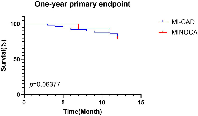 Figure 1
| Characteristic | MINOCA group ( | MI-CAD Group ( | |
|---|---|---|---|
| Male ( | 15 (100.00) | 55 (82.09) | 0.076 |
| Age ( | 53.33 ± 12.88 | 55.00 ± 16.61 | 0.221 |
| BMI (kg/m2, IQR) | 20.3 (20.3–24.22) | 24.5 (23.0–26.0) | 0.020 |
| Smoking history ( | 7 (46.67) | 37 (55.22) | 0.548 |
| History of coronary heart disease ( | 0 (0.00) | 4 (5.97) | 0.332 |
| History of hypertension ( | 8 (53.33) | 29 (43.28) | 0.480 |
| History of diabetes ( | 1 (6.67) | 5 (7.46) | 0.915 |
| History of cerebrovascular accident/TIA ( | 0 (0.00) | 1 (1.49) | 0.634 |
| History of hyperlipidemia ( | 0 (0.00) | 3 (4.48) | 0.404 |
| Variable | MINOCA group ( | MI-CAD Group ( | |
|---|---|---|---|
| Triglycerides (mmol/L, IQR) | 0.95 (0.81–1.30) | 1.19 (0.96–1.80) | 0.396 |
| Total cholesterol (mmol/L, | 3.16 ± 1.61 | 6.88 ± 13.52 | 0.65 |
| High-density lipoprotein cholesterol (mmol/L, | 0.83 ± 0.44 | 0.96 ± 0.69 | 0.568 |
| Low-density lipoprotein cholesterol (mmol/L, IQR) | 1.78 (1.60–3.27) | 2.94 (2.20–3.54) | 0.009 |
| Apolipoprotein A (mmol/L, IQR) | 0.81 (0.58–1.01) | 0.98 (0.79–1.09) | 0.002 |
| Apolipoprotein B (mmol/L, IQR) | 0.66 (0.53–0.88) | 0.74 (0.60–0.87) | 0.427 |
| Lipoprotein (a) (mmol/L, IQR) | 332.00 (132.00–403.00) | 167.00 (90.00–436.00) | 0.496 |
| Blood glucose (mmol/L, IQR) | 5.12 (4.72–8.09) | 6.18 (5.82–7.41) | 0.65 |
| Glycated hemoglobin (%, | 5.11 ± 1.39 | 5.36 ± 1.23 | 0.643 |
| hs -CRP (mmol/L, IQR) | 1.99 (0.50–10.00) | 4.30 (0.96–10.00) | 0.91 |
| hs-cTnI (ng/ml, IQR) | 3.25 (0.43–16.12) | 18.61 (3.21–50) | 0.007 |
| CK-MB (ng/ml, IQR) | 21.90 (3.87–38.39) | 68.80 (16.21–248.60) | 0.007 |
| Variable | MINOCA group ( | MI-CAD Group ( | |
|---|---|---|---|
| Atrial fibrillation ( | 1 (6.67) | 3 (4.48) | 0.722 |
| T wave changes ( | 4 (26.67) | 39 (58.21) | 0.027 |
| ST segment elevation ( | 4 (26.67) | 51 (76.12) | <0.001 |
| Ventricular arrhythmia ( | 0 (0.00) | 3 (4.48) | 0.404 |
| Abnormal ventricular wall contraction activity ( | 1 (6.67) | 49 (73.13) | <0.001 |
| Interventricular septum thickness (mm, IQR) | 9.00 (9.00–9.00) | 12.00 (9.00–13.00) | 0.002 |
| LVEF (%, IQR) | 59.00 (51.00–62.00) | 58.00 (48.00–65.00) | 0.75 |
| Events | MINOCA group ( | MI-CAD Group ( | |
|---|---|---|---|
| Infection ( | 8 (53.33) | 26 (38.80) | 0.302 |
| Deep vein thrombosis ( | 0 (0.00) | 2 (3.00) | 0.498 |
| Heart failure ( | 10 (66.67) | 42 (62.69) | 0.772 |
| Cardiovascular death ( | 0 (0.00) | 1 (1.49) | 0.634 |
| All-cause mortality ( | 0 (0.00) | 1 (1.49) | 0.634 |
- —science and technology project
Peer Reviews
No public reviews on file for this paper yet. If you reviewed it on a platform where reviews are public (OpenReview, ICLR, NeurIPS, ICML), you can paste yours below so the community can read it here.
Videos
No videos yet. Explain this paper in a talk, walkthrough, or lecture? Add one.
Taxonomy
TopicsAcute Myocardial Infarction Research · Cardiac Imaging and Diagnostics · Coronary Interventions and Diagnostics
Introduction
1
According to the Fourth Universal Definition of Myocardial Infarction (MI), MI is characterized by an elevation of cardiac biomarkers—primarily cardiac troponin (cTn)—exceeding the 99th percentile upper reference limit, with dynamic changes and at least one of the following clinical criteria: (1) Ischemic symptoms; (2) New ischemic electrocardiographic changes (e.g., new ST-T changes or new left bundle branch block); (3) Development of pathological Q waves; (4) Imaging evidence of new loss of viable myocardium or new regional wall motion abnormalities (1). Electrocardiographically, MI is classified as ST-elevation MI (STEMI) or non–ST-elevation MI (NSTEMI). Most cases are due to obstructive coronary artery disease (MI-CAD), defined as ≥50% coronary stenosis on angiography. However, about 6%–8% of MIs occur without significant stenosis (MINOCA, myocardial infarction with nonobstructive coronary arteries), identified by <50% stenosis on angiography (2). Despite earlier beliefs, MINOCA patients have a poor prognosis, with a 12-month all-cause mortality of approximately 3.4% (3). Early recognition and management of MINOCA are essential to improve patient outcomes (2).
The Qinghai-Tibet Plateau, often referred to as the “Roof of the World,” is the highest plateau globally, with an average altitude exceeding 4,500 meters (4). The low oxygen environment at this altitude suppresses aerobic metabolism, resulting in reduced energy production and impaired circulatory function, which limits nutrient and energy delivery to tissues (5). To adapt to hypoxic conditions, the body compensates with an increased heart rate and cardiac output (6). Additionally, high-altitude residents have distinct lifestyles and dietary patterns compared to those at lower altitudes (7). These environmental and behavioral factors may contribute to unique cardiovascular disease profiles among high-altitude residents compared to their low-altitude counterparts.
At present, there is no relevant research on MINOCA patients in the Qinghai-Tibet Plateau. We selected MINOCA patients in Shigatse City (average altitude of about 3,800 meters), and summarized their clinical characteristics, diagnosis,treatment and prognosis. We hope to increase the understanding of MINOCA in the plateau area and provide a reference for optimizing clinical treatment strategies and formulating individualized treatment plans for local patients.
Materials and methods
2
General information
2.1
A retrospective analysis was performed on 82 patients who were diagnosed with acute myocardial infarction and underwent coronary angiography in Shigatse People's Hospital from December 2020 to December 2021. Inclusion criteria were: (1) meeting the diagnostic criteria for acute myocardial infarction; (2) age >18 years. Exclusion criteria were: (1) type 3, 4, or 5 myocardial infarction; (2) receiving thrombolytic therapy before coronary angiography; (3) pregnant or lactating women; (4) severe liver and kidney diseases; (5) malignant tumors with an expected survival of less than 1 year. The diagnosis of MINOCA was based on the criteria of the fourth edition of the Universal Definition of Myocardial Infarction (1): (1) meeting the diagnostic criteria for acute myocardial infarction; (2) coronary angiography showed non-obstructive coronary artery disease, that is, no obstructive coronary artery disease (i.e., coronary artery stenosis ≥50%) was found in angiography of any possible infarction-related vessels; (3) non-ischemic troponin elevation was excluded, such as systemic factors (renal failure, pulmonary embolism, severe infection, and stroke) and cardiac factors (heart failure). After screening, 15 patients (18.3%) were included in the MINOCA group and 67 patients (81.7%) were included in the MI-CAD group.
Observation indicators
2.2
The demographic data and medical history information of all the subjects were collected, including the following: (1) Demographics and Lifestyle Factors: age, sex, smoking history, and body mass index (BMI); (2) Medical History: history of coronary artery disease (CAD), hypertension, diabetes mellitus, cerebrovascular events/transient ischemic attacks (TIA), peripheral arterial disease, hyperlipidemia, and other relevant conditions; (3) Biochemical Parameters (within 24 h of admission): lipid profile: triglycerides, total cholesterol, high-density lipoprotein cholesterol (HDL-C), low-density lipoprotein cholesterol (LDL-C), apolipoprotein A-I, apolipoprotein B, and lipoprotein(a); Cardiac Biomarkers: hypersensitive cardiac troponin I (hs-cTnI) and creatine kinase-MB (CK-MB); Other Blood Tests: blood glucose, glycated hemoglobin (HbA1c), and hypersensitive C-reactive protein (hs-CRP); (4) Imaging and Diagnostic Results: Electrocardiogram (ECG) findings, echocardiographic results; (5) In-hospital adverse events: incidence of infections, deep vein thrombosis (DVT), and major adverse cardiovascular events (MACEs) during hospitalization; (6) All patients were followed for one year. The primary endpoint was the composite of MACEs, including all-cause death, myocardial infarction, ischemic stroke, and heart failure.
Statistical analysis methodology
2.3
Data were analyzed using SPSS 26.0 statistical software. For variables conforming to a normal distribution, data were expressed as (mean ± standard deviation). Group comparisons were performed using t-tests. For variables not conforming to a normal distribution, data were presented as P50 (median) with interquartile ranges (P25–P75). Comparisons between groups were conducted using the Wilcoxon rank-sum test. Categorical data were expressed as frequencies (counts and percentages). Relationships between categorical variables were analyzed using the chi-square test (χ^2^). p < 0.05 was considered statistically significant.
Patient and public involvement
2.4
Patients or the public WERE NOT involved in the design, or conduct, or reporting, or dissemination plans of our research.
Results
3
Etiology and risk factors of MINOCA
3.1
Among all 82 patients, 15 had MINOCA, accounting for 18.3% of all patients, which was significantly higher than previous reports, indicating that patients with myocardial infarction in plateau areas may have characteristics different from those in low-altitude areas. In terms of specific causes, there were 2 cases of coronary artery plaque rupture (13.33%), 2 cases of coronary artery spasm (13.33%), 1 case of coronary artery thrombosis and embolism (6.67%), 1 case of type 2 acute myocardial infarction (6.67%), 3 cases of unrecognized myocarditis (20%), and 6 other cases with no clear cause.
The results (Table 1) showed that all 15 patients in the MINOCA group were male, with a significantly higher proportion than that in the MI-CAD group, but no statistical difference was found (100% vs. 82.1%, p = 0.076). At the same time, the BMI of the MINOCA group was lower than that of the MI-CAD group (20.3 kg/m^2^ vs. 24.5 kg/m^2^, p = 0.02). There was no statistical difference in age, BMI, smoking history, coronary heart disease history, hypertension history, diabetes history, cerebrovascular accident/TIA history, and hyperlipidemia history between the two groups.
Laboratory indicators
3.2
The results (Table 2) showed that the low-density lipoprotein cholesterol, apolipoprotein A, hs-cTnI, and CK-MB in the MINOCA group were significantly lower than those in the MI-CAD group (p < 0.01), while there were no statistical differences in triglycerides, total cholesterol, HDL-C, apolipoprotein B, lipoprotein (a), blood glucose, glycosylated hemoglobin, and hs -CRP between the two groups.
ECG and echocardiogram results
3.3
By comparing the ECG characteristics of the two groups of patients (Table 3), we found that the proportion of T wave changes and ST segment elevation in the MINOCA group was lower than that in the MI-CAD group (p < 0.05), and there was no statistical difference in the occurrence of atrial fibrillation and ventricular arrhythmia between the two groups. In the analysis of echocardiography (Table 3), we found that patients in the MI-CAD group were more likely to have abnormal ventricular wall contraction activity (p < 0.001), and their ventricular septum thickness was significantly higher than that in the MINOCA group (p = 0.002). There was no significant difference in the left ventricular ejection fraction (LVEF) between the two groups.
In-hospital adverse events and MACEs
3.4
There was no significant statistical difference between the two groups of patients in terms of infection, deep vein thrombosis, heart failure, cardiovascular death and all-cause death (p > 0.05, Table 4).
At the 1-year follow-up, there was no significant difference in MACE between the MI-CAD group (9 cases) and the MINOCA group (3 cases) (p > 0.05, Figure 1).
One-year MACEs composite endpoint between the MINOCA group and the MI-CAD group.
Discussion
4
Incidence of MINOCA
4.1
Previous studies have reported that MINOCA accounts for about 6%–8% of acute myocardial infarction cases globally (2, 8, 9), and 5.47% to 9.19% in China (10–12). In this study, MINOCA represented 18.3% of cases, significantly higher than reported rates, indicating that MI in the Qinghai-Tibet Plateau may have unique characteristics. The plateau's chronic hypoxic environment leads to compensatory erythrocytosis and various clinical symptoms known as chronic mountain sickness (CMS) (13). Studies have shown that these patients usually have a higher cardiovascular risk, with significantly increased levels of various inflammatory indicators (such as MIP-1β, IL-8, IFN-γ, etc.) and oxidative stress indicator superoxide dismutase (SOD). At the same time, the patient's microcirculation and macrovascular function will also change due to changes in altitude (14). Whether these factors contribute to the high prevalence of MINOCA in this population warrants further investigation.
Risk factors
4.2
Previous studies generally suggest that the proportion of women in MINOCA patients is usually higher than that in MI-CAD patients (1). However, our statistics showed that all 15 MINOCA patients were male, possibly due to the unhealthy lifestyle of local men, exposure to hypoxic environments, and genetic factors. Filippo Zilio et al. explored gender differences in patients with ACS, specifically noting that in many cases, women do not receive timely and adequate diagnosis and treatment (15). In the Qinghai-Tibet Plateau region, women's healthcare seeking rates are even lower, influenced by the social environment and traditional beliefs. Past research reports from China have indicated that among outpatient patients in the Tibet region, there are more males than females, which may also lead to potential research bias (16). Previous studies have found that spontaneous coronary artery dissection (SCAD) and Takotsubo syndrome occur more frequently in women, possibly due to differences in sex hormones and transforming growth factor-β levels. In contrast, myocarditis is more prevalent in men, particularly younger men. Although mortality rates between sexes are similar, further research is needed to clarify the influence of sex on myocarditis outcomes (15, 17).
Consistent with previous studies (18), MINOCA patients had lower BMI and lower levels of blood lipids (LDL-C and apolipoprotein A). In addition, cardiovascular risk factors such as history of hypertension, diabetes, hyperlipidemia, and smoking history vary in different research reports (12, 18). Our statistics found that there were no significant differences between groups in these factors on the plateau. Interestingly, although there was no statistical difference in the age of onset between the two groups of patients, by comparing with previous literature, we found that the average age of patients with myocardial infarction in the Qinghai-Tibet Plateau was significantly lower than that in other areas. Therefore, active health education and cardiovascular screening are needed in plateau areas to enable early detection and intervention, ultimately improving cardiovascular health among residents.
Related inspection results
4.3
The results of this study showed that compared with the MI-CAD group, the MINOCA group had lower levels of hs-cTnI and CK-MB, and fewer abnormal ECG manifestations (including T wave changes and ST segment elevation), suggesting that the level of myocardial damage in MINOCA patients was lower. At the same time, the echocardiogram results of the MINOCA group showed fewer abnormal ventricular wall contraction activity, which was consistent with the performance of myocardial markers and ECG. In addition, previous reports have shown (19, 20) that MINOCA patients usually have higher LVEF, but in our study, there was no significant statistical difference in LVEF between the two groups, possibly due to sample size and regional differences. Notably, ventricular septum thickness was significantly lower in the MINOCA group (p = 0.002), a finding not been reported in previous studies. There was no significant difference in the history of hypertension between the two groups. Whether this difference is due to the years of hypertension or blood pressure control in the two groups of patients is not yet known, and it deserves further study.
Prognostic analysis
4.4
Previous studies have shown that MINOCA patients are more likely to experience in-hospital MACEs compared with MI-CAD patients (11). However, our data showed no significant difference in in-hospital adverse events between the two groups. Possible reasons include: patients had a shorter hospital stay and less chance of complications; the sample size was small and the statistical power was insufficient.
Study limitations
5
It is important to note that limited medical resources and low disease awareness among residents of the Qinghai-Tibet Plateau led to delayed medical consultation for chest pain. As a result, our study included only 15 patients in the MINOCA group, which greatly limited the statistical power of our analysis.
On the other hand, other confounding factors may also influence our findings, such as socioeconomic status, racial disparities, dietary patterns, and other high-altitude-related comorbidities. Lower socioeconomic status is linked to higher coronary heart disease mortality (21), making the economic conditions of the Qinghai-Tibet Plateau an important consideration in interpreting our results. Our patient group included both long-term Tibetan residents and tourists; however, due to the small sample size, we could not conduct detailed subgroup analyses to further assess potential differences between these groups.
In patients with MINOCA, cardiac magnetic resonance (CMR) has been shown to have significant diagnostic and prognostic value, particularly in differentiating myocarditis and Takotsubo syndrome, and is highly recommended by current clinical practice guidelines (22–24). A recent study shows that the use of CMR in contemporary cohorts helps distinguish MINOCA from other cardiac conditions, which is important for guiding patient treatment (25). However, due to limitations in local medical resources, CMR cannot be performed. Thus, myocarditis is diagnosed mainly through medical history and clinical presentation; vasospasm is confirmed by symptom relief after nitroglycerin during angiography; and plaque rupture is identified by Intravascular ultrasound (IVUS) or Optical Coherence Tomography (OCT). As understanding of MINOCA deepens, its complexity is increasingly recognized, and its clinical characteristics and management continue to evolve with updated guidelines. Relevant imaging examinations (including CMR, IVUS, OCT, etc.) play an important role in disease differentiation, guiding treatment, and improving prognosis (17, 25).
Conclusion
6
MINOCA represents a clinical syndrome caused by diverse etiologies, differing significantly from MI due to coronary artery obstruction in terms of risk factors, pathogenesis, clinical presentation, and prognosis. Recognizing and treating MINOCA remains a challenge in clinical practice. In high-altitude environments, the understanding of this condition is further complicated by limited technical resources, the effects of hypoxia, and unique lifestyle factors. These challenges often result in delayed recognition and diagnosis.
This study retrospectively analyzed MINOCA patients at Shigatse People's Hospital, summarizing their clinical characteristics to provide insights into the diagnosis and treatment of such patients in the Qinghai-Tibet Plateau region. Future efforts should focus on enhanced follow-up of these patients and further exploration of the mechanisms underlying MINOCA in high-altitude environments. These steps are essential for improving disease prevention and developing individualized treatment strategies.
The reference list from the paper itself. Each links out to its DOI / PubMed record.
- 1Thygesen K Alpert JS Jaffe AS Chaitman BR Bax JJ Morrow DA Fourth universal definition of myocardial infarction (2018). Circulation. (2018) 138(20):e 618–51. 10.1161/CIR.000000000000061730571511 · doi ↗ · pubmed ↗
- 2Parwani P Kang N Safaeipour M Mamas MA Wei J Gulati M Contemporary diagnosis and management of patients with MINOCA. Curr Cardiol Rep. (2023) 25(6):561–70. 10.1007/s 11886-023-01874-x 37067753 PMC 10188585 · doi ↗ · pubmed ↗
- 3Pasupathy S Lindahl B Litwin P Tavella R Williams MJA Air T Survival in patients with suspected myocardial infarction with nonobstructive coronary arteries: a comprehensive systematic review and meta-analysis from the MINOCA global collaboration. Circ Cardiovasc Qual Outcomes. (2021) 14(11):e 007880. 10.1161/CIRCOUTCOMES.121.00788034784229 · doi ↗ · pubmed ↗
- 4Xu S Li S Yang Y Tan J Lou H Jin W A genome-wide search for signals of high-altitude adaptation in Tibetans. Mol Biol Evol. (2011) 28(2):1003–11. 10.1093/molbev/msq 27720961960 · doi ↗ · pubmed ↗
- 5Lv J Qi P Bai L Yan X Zhang L. Review of the relationship and underlying mechanisms between the Qinghai-Tibet plateau and host intestinal flora. Front Microbiol. (2022) 13:1055632. 10.3389/fmicb.2022.105563236523840 PMC 9745141 · doi ↗ · pubmed ↗
- 6Moore LG Niermeyer S Zamudio S. Human adaptation to high altitude: regional and life-cycle perspectives. Am J Phys Anthropol. (1998) 27(Suppl ):25–64. <25::AID-AJPA 3>3.0.CO;2-L 10.1002/(sici)1096-8644(1998)107:27+<25::aid-ajpa 3>3.0.co;2-l 9881522 · doi ↗ · pubmed ↗
- 7Lan D Ji W Lin B Chen Y Huang C Xiong X Correlations between gut microbiota community structures of Tibetans and geography. Sci Rep. (2017) 7(1):16982. 10.1038/s 41598-017-17194-429209019 PMC 5717229 · doi ↗ · pubmed ↗
- 8Kilic S Aydın GÇoner A Doğan Y Arican ÖzlükÖÇelik Y Prevalence and clinical profile of patients with myocardial infarction with non-obstructive coronary arteries in Turkey (MINOCA-TR): a national multi-center, observational study. Anatol J Cardiol. (2020) 23(3):176–82. 10.14744/Anatol J Cardiol.2019.4680532120362 PMC 7222639 · doi ↗ · pubmed ↗