Urine and occlusion in the pathogenesis of lichen sclerosus: insights from a case of post-urethrectomy remission
Sachini Mendis, Georgios Kravvas, Richard Watchorn, Christopher B Bunker

TL;DR
A man's genital lichen sclerosus completely cleared after surgery to remove his urethra, suggesting that urine exposure plays a key role in causing the condition.
Contribution
This case provides novel evidence that occlusive urine exposure is a critical driver of male genital lichen sclerosus.
Findings
Complete remission of MGLSc occurred after urethrectomy and urine diversion.
Peristomal lichen sclerosus developed, indicating the role of urinary exposure in disease.
Resolution of MGLSc after urethral isolation supports the importance of reducing urine contact.
Abstract
We report the case of a 58-year-old uncircumcised man with male genital lichen sclerosus (MGLSc) achieving complete remission following urethrectomy and ileal conduit formation for urothelial carcinoma. The patient presented with typical clinical features of MGLSc. Initial treatment with ultrapotent topical corticosteroids provided only moderate improvement. Circumcision was planned but delayed due to the diagnosis of bladder and ureteral carcinomas. Postsurgery, urine was diverted through an ileal stoma, and the urethral meatus was sealed. Remarkably, despite cessation of topical treatments, the patient’s penile MGLSc showed complete resolution, with no residual active disease over a 4-year follow-up period. However, peristomal lichen sclerosus developed, emphasizing the role of occlusive urinary exposure in disease pathogenesis. This case supports the hypothesis that occlusive…
Genes, proteins, chemicals, diseases, species, mutations and cell lines named across the full text — each resolved to its canonical identifier and authoritative record.
Click any figure to enlarge with its caption.
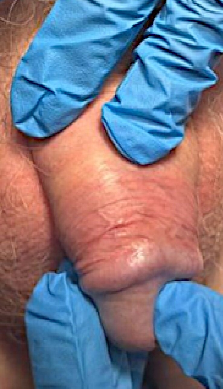 Figure 1
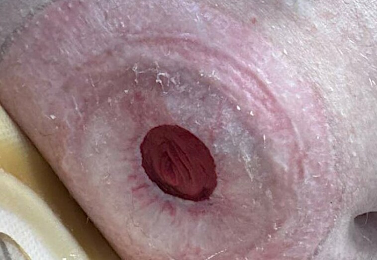 Figure 2
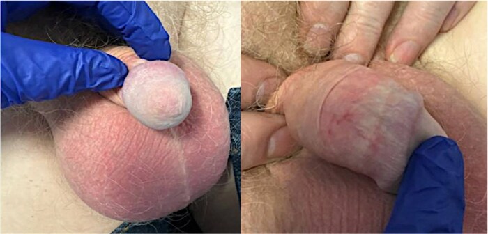 Figure 3
Peer Reviews
No public reviews on file for this paper yet. If you reviewed it on a platform where reviews are public (OpenReview, ICLR, NeurIPS, ICML), you can paste yours below so the community can read it here.
Videos
No videos yet. Explain this paper in a talk, walkthrough, or lecture? Add one.
Taxonomy
TopicsGenital Health and Disease · Urological Disorders and Treatments · Urologic and reproductive health conditions
Case presentation
We report a case of male genital lichen sclerosus (MGLSc) where clinical remission was achieved after complete urethrectomy and ileal conduit formation.
A 58-year-old uncircumcised male patient who formerly smoked presented to our tertiary male genital dermatology clinic in 2018 with penile erythema, itching and foreskin tightness. On examination, findings included an etiolated glans, trans-coronal adhesions, and subtle erythematous-to-violaceous macular inflammation of the ventral frenulum and coronal sulcus (Figure 1). A firm clinical diagnosis of MGLSc was established. Initial management consisted of soap avoidance, skin barrier preparations and a 4-week course of clobetasol propionate ointment. Despite consistent adherence, the treatment achieved only moderate improvement and failed to provide a cure, leading to the recommendation of circumcision.
Active male genital lichen sclerosus prior to urethrectomy. Clinical photograph illustrating male genital lichen sclerosus (MGLSc) with active inflammation and chronic architectural changes prior to the urethrectomy. The image demonstrates areas of lichenoid inflammation, waxy pallor (sclerosis), loss of the coronal sulcus and constrictive posthitis with a prominent sclerotic band.
However, before circumcision could be performed, the patient was diagnosed with carcinoma of the bladder and right ureter (G3 pT1). He underwent robotic radical cystoprostatectomy, right nephroureterectomy, pelvic lymph node dissection and ileal conduit formation. The urine outflow was diverted to the ileal stoma, and the urethral meatus was sealed. Postsurgery, the patient discontinued the use of topical corticosteroids and barrier preparations for the glans and foreskin.
Two months after surgery, erythematous and scarring changes were noted around the stoma site (Figure 2). A clinical diagnosis of peristomal lichen sclerosus (LSc) was made, and treatment with barrier preparations and topical corticosteroids was initiated with good results. Since then, no residual signs of active MGLSc on the penis could be elicited, despite the patient remaining uncircumcised and having ceased topical treatments (Figure 3).
Development of lichen sclerosus (LSc) around the stoma site following urethrectomy. Clinical photograph demonstrating erythematous and scarring changes characteristic of LSc surrounding the stoma site. These features developed following urethrectomy and underscore the irritant effects of urine, a key factor in the pathophysiology of LSc.
Remission of active male genital lichen sclerosus (MGLSc) following urethrectomy. Combined clinical photographs illustrating the remission of active lichen sclerosus (LSc) from the penis following urethrectomy. The images demonstrate features of burned-out LSc, including etiolation of the glans and residual scarring. While all signs of active inflammation have resolved, erythematous changes persist due to chronic LSc-driven telangiectasia. The cessation of inflammation highlights the removal of urine, a key factor in the pathogenesis of LSc, achieved through urethrectomy. Note the obliteration of the urethral meatus secondary to the removal of the urethra.
The patient has been under annual surveillance since, and the penile LSc remains quiescent.
Discussion
LSc is an acquired, chronic, inflammatory skin disorder that predominantly affects the anogenital region. In male patients, it typically presents as waxy white, often atrophic patches and plaques. Purpura, telangiectasias or ecchymotic changes may also occur in the affected areas. Other common features include constrictive posthitis, lichenoid or zoonoid inflammation, thinning, and sclerosis of the skin, which can result in scarring, loss of elasticity, adhesions and architectural changes, such as the obliteration of the coronal sulcus. The frenulum is frequently affected, becoming fibrotic or, in some cases, resorbed.^1–3^
If left untreated or inadequately managed, MGLSc can lead to significant complications, including phimosis; it may involve the urethra causing stenosis; it is associated with differentiated penile intraepithelial neoplasia (dPeIN) and frank squamous carcinoma of the penis (PeSCC).^2,4,5^ More recently, emerging evidence has also suggested a potential link between MGLSc and the development of genital melanoma.^6–8^
MGLSc predominantly affects uncircumcised men and is strongly associated with postvoiding micro incontinence.^9^ It is proposed that in these individuals, after the foreskin is repositioned following urination, small droplets of residual urine leak from the urethral meatus and accumulate between the apposed mucosal surfaces of the glans and prepuce. This occlusive environment, combined with as-yet-undefined epithelial susceptibility factors, contributes to chronic inflammation and subsequent sclerosis.^10–12^
The presence of hypospadias, or interventions that can lead to postvoiding incompetence of the naviculomeatal valve, such as urological procedures and genital piercing, pose a recognized risk factor. Obesity is another important risk factor, not only as a systemic driver but also as a local mechanical contributor to the pathogenesis of MGLSc. Importantly, in circumcised men, obesity can lead to a ‘buried penis’ and ‘pseudoforeskin’ formation, where excess suprapubic fat and redundant penile skin form folds that recreate the occlusive environment seen in uncircumcised men. These represent the necessary conditions for urine trapping and prolonged tissue exposure, facilitating chronic irritation and inflammation.^6,8^
A growing body of evidence strongly supports the hypothesis that MGLSc is primarily driven by occlusive exposure to urine, and this case further strengthens that argument. Key evidence underpinning this hypothesis includes
(i)the anatomical distribution of LSc lesions, which aligns with areas of occlusive contact with urine;(ii)the high prevalence of post-micturition dribbling in men with MGLSc;(iii)the characteristic resolution of MGLSc following circumcision;(iv)the anatomical abnormalities of the naviculomeatal fossa that are frequently seen in men with MGLSc; these impair its function as a low-pressure valve, thereby allowing urine leakage into the potential balanopreputial space;(v)the increased risk of LSc following urological instrumentation or genital piercing; as these procedures may similarly impair the function of the naviculomeatal fossa as a low-pressure valve, promoting urine leakage into the balanopreputial space;(vi)the sparing of the perianal region in MGLSc, unlike female genital LSc, due to the protective shielding provided by the foreskin and scrotum against urinary exposure; and(vii)the recurrence of MGLSc in circumcised men when a pseudo- or neo-foreskin forms post-circumcision, associated with obesity; and(viii)molecular evidence pointing to epithelial stress as the initiating event.^2,10,11,13,14^
Peristomal LSc has been increasingly recognized as a distinct manifestation of LSc, likely driven by chronic occlusion and exposure to irritants around the stoma site. Shim et al. reported cases of LSc developing in association with perineal urethrostomy, attributing its pathogenesis to the persistent contact of urine with peristomal skin, exacerbated by occlusion from stoma appliances.^15^ Similarly, Weng and Charles-Holmes described cases of LSc affecting colostomy sites, suggesting that the combination of mechanical irritation, occlusion and exposure to faecal material could act as triggers for disease development.^16^ Finally, Al-Niaimi and Lyon further highlighted the pivotal role of occlusion and urinary exposure in peristomal LSc, documenting cases where stoma appliances and chronic urinary contact created a localized environment conducive to inflammation and sclerosis.^17^
This case highlights the role of urinary occlusion in the pathogenesis of MGLSc and demonstrates a unique instance of remission following urethrectomy and ileal conduit formation.
The reference list from the paper itself. Each links out to its DOI / PubMed record.
- 1Bunker CB, Porter WM. Male genital dermatology. In: Rook’s Textbook of Dermatology (Griffiths C, Barker J, Bleiker T, Chalmers R, Creamer D, eds). Chichester: Wiley-Blackwell, 2016; 111.1–111.41.
- 2Kravvas G, Shim TN, Doiron PR et al The diagnosis and management of male genital lichen sclerosus: a retrospective review of 301 patients. J Eur Acad Dermatol Venereol 2018; 32:91–5.28750140 10.1111/jdv.14488 · doi ↗ · pubmed ↗
- 3Bunker C . Male Genital Skin Disease. London: Saunders, 2004.
- 4Kravvas G, Ge L, Ng J et al The management of penile intraepithelial neoplasia (Pe IN): clinical and histological features and treatment of 345 patients and a review of the literature. J Dermatolog Treat 2022; 33:1047–62.32705920 10.1080/09546634.2020.1800574 · doi ↗ · pubmed ↗
- 5James ML, Kravvas G, Lallas A, Bunker CB. The clinical and dermatoscopic features of penile pigmentation in men with genital lichen sclerosus. Skin Health Dis 2024; 4:e 435.39355751 10.1002/ski 2.435PMC 11442078 · doi ↗ · pubmed ↗
- 6Mastoraki E, Kravvas G, Dear K et al Primary vulval melanoma and genital lichen sclerosus. Skin Health Dis 2024; 4:e 411.39104656 10.1002/ski 2.411PMC 11297432 · doi ↗ · pubmed ↗
- 7Dear K, Kravvas G, Sim S et al Primary penile melanoma and genital lichen sclerosus. Skin Health Dis 2023; 3:e 274.38047263 10.1002/ski 2.274PMC 10690690 · doi ↗ · pubmed ↗
- 8Sim SJY, Dear K, Mastoraki E et al Genital lichen sclerosus and melanoma; a systematic review. Skin Health Dis 2022; 3:e 198.37013116 10.1002/ski 2.198PMC 10066758 · doi ↗ · pubmed ↗