Bright Zinc Probes with Thiomorpholine Monoxide Auxochromes for Imaging Insulin Secretion
Rundong Yu, Junwei Zhang, Xiaohong Peng, Zhixing Chen

TL;DR
Scientists developed new zinc probes that improve imaging of insulin secretion in mouse islets with minimal harm.
Contribution
The novel PKZnBR probes have enhanced fluorescence and reduced phototoxicity for long-term insulin secretion imaging.
Findings
PKZnBR probes have fluorescence quantum yields up to seven times higher than PKZnRs due to TICT state inhibition.
PKZnBR-3 shows a high turn-on ratio (134) and 340 nM affinity for zinc, suitable for ex vivo insulin secretion studies.
The probes enable long-term, low-toxicity imaging of zinc and insulin corelease in mouse islets.
Abstract
Zinc biology significantly impacts human physiological and pathological processes, especially for β-cell endocrinology. In 2021, our group reported the PKZnR family with minimal phototoxicity and μM affinities for monitoring Zn2+/insulin corelease during vesicular secretory events on β-cells (Zhang et al. Angew. Chem. Int. Ed. 2021, 60 (49), 25846−25855 10.1002/anie.20210951034423531). Here, we synthesized a series of Zn2+ probes (PKZnBR) featuring thiomorpholine monoxide auxochromes with K d values ranging from 160 nM to 94 μM. By inhibiting the twisted intramolecular charge transfer (TICT) state, the fluorescence quantum yields of PKZnBRs can be effectively increased to ∼7 times those of PKZnRs. A privileged candidate, PKZnBR-3, has a high turn-on ratio (∼134), appropriate affinity (340 nM), and excellent hydrophilicity, making it a powerful tool for long-term ex vivo recording of…
Genes, proteins, chemicals, diseases, species, mutations and cell lines named across the full text — each resolved to its canonical identifier and authoritative record.
Click any figure to enlarge with its caption.
 1
1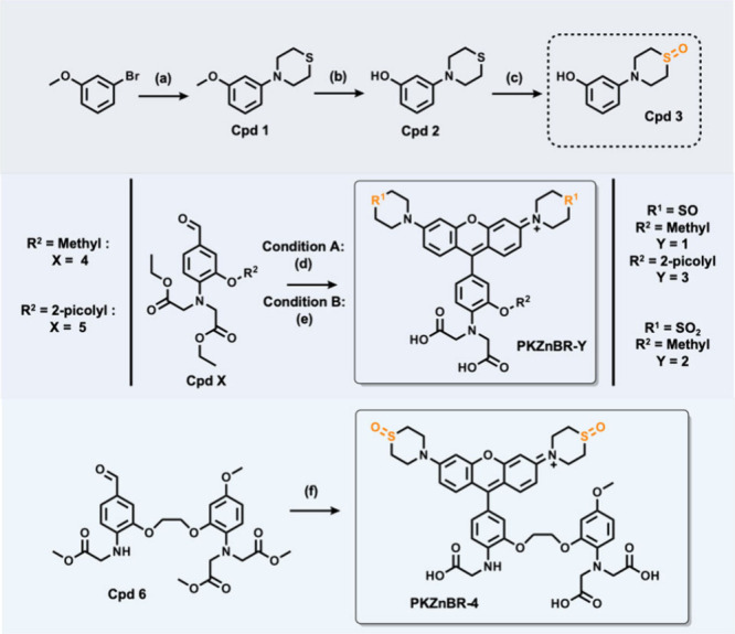 1
1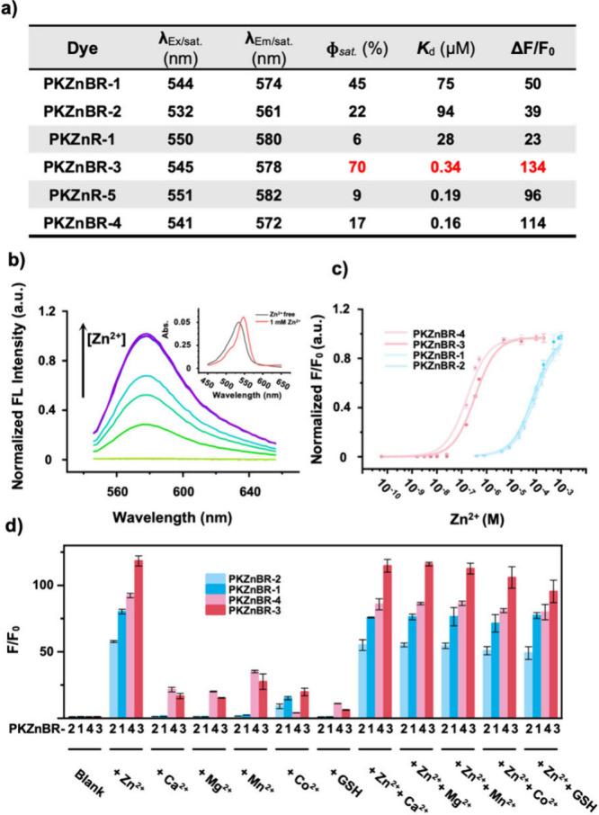 2
2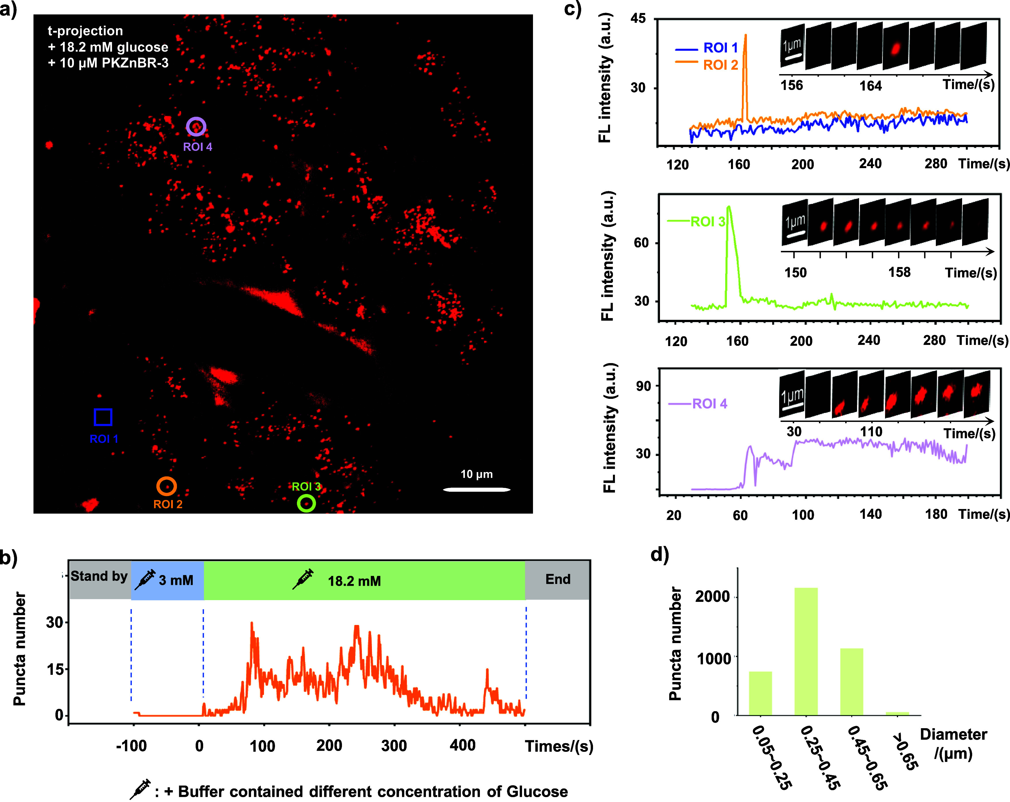 3
3- —National Natural Science Foundation of China10.13039/501100001809
- —Natural Science Foundation of Beijing Municipality10.13039/501100005089
- —National Key Research and Development Program of China10.13039/501100012166
Peer Reviews
No public reviews on file for this paper yet. If you reviewed it on a platform where reviews are public (OpenReview, ICLR, NeurIPS, ICML), you can paste yours below so the community can read it here.
Videos
No videos yet. Explain this paper in a talk, walkthrough, or lecture? Add one.
Taxonomy
TopicsLipid Membrane Structure and Behavior · Molecular Sensors and Ion Detection · Advanced biosensing and bioanalysis techniques
Introduction
Zinc (Zn^2+^) impacts human health through its involvement in metabolic and immune processes. The concentration of Zn^2+^ exhibits high cellular heterogeneity and rapid dynamics, making fluorescent probes an irreplaceable tool for studying Zn^2+^ biology.? Recently, the coevolution of fluorescent probes, microscopy, and image algorithms allowed us to obtain high-resolution spatiotemporal information at cellular or subcellular levels. ?,? Small-molecule ion probes have unique advantages in expanding the cutting edge in Zn^2+^ biology, as they can provide better photophysical properties in brightness and photostability than fluorescent protein and produce fluorescence signals with high spatiotemporal dynamics in living cells and tissues without genetic manipulation. ?−? ? ?
The application of small-molecule-based probes in Zn^2+^ biology can be tracked to the 1960s when 8-hydroxyquinoline was used to quantitatively analyze Zn^2+^ in human serum or urine.? Two decades later, another quinoline-based Zn^2+^ probe TSQ was synthesized with enhanced photophysical properties.? Upon the half-century’s development, large quantities of small-molecule-based Zn^2+^ probes have been reported and can be divided into three types according to their activation mechanism. The first type is ICT (Intramolecular Charge Transfer)-based probes, such as the original version of FuraZin and IndoZin in the UV range and newly improved NBD-TPE, etc. in the visible region, featured on the ratiometric response of Zn^2+^. ?−? ? ? ? ? ? Another working mode known as FRET (Fluorescence Resonance Energy Transfer) bred a series of probes such as CZ-1, CPBT, etc. ?−? ? ? FRET-based Zn^2+^ probes were designed with paired fluorophores coupled with a short linker to ensure a higher FRET efficiency. The above two types of ratiomatric and intensiometric probes can be used for absolute quantification of Zn^2+^ concentration. Intensity-based probes usually sense Zn^2+^ through the PET (Photoinduced Electron Transfer) mechanism. ?,? For example, the fluorescein-based QZ probes and ZP probes were reported in the 2000s. Both of them exhibited high brightness and visible emission spectra. ?−? ? Meanwhile, derived from the DPA (2,2′-dipicolylamine) chelating group, the ZnAF probes also featured excellent selectivity and strong binding affinity toward Zn^2+^. ?−? ? In addition, another series of Zn^2+^ probes such as RhodZin and FluoZin were commercialized during the same period. ?,? Their structures are featured on a modified Ca^2+^ chelator BAPTA (1,2-bis(2-aminophenoxy)ethane-N,N,N′,N′-tetraacetic acid). These types of molecules can also be combined with a self-labeling protein tag to achieve precise subcellular localization. ?,? To date, chemists continue to evolve fluorophores and chelating groups to achieve higher brightness, faster kinetics, better selectivity, and tunable binding affinities (K d) for the growing Zn^2+^ biological research.
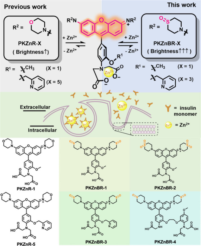
The remarkably high concentration of Zn^2+^ in insulin secretory granules (∼20 mM) suggests a special biological process. Insulin is vitally important for glucose homeostasis by lowering blood sugar levels. The crystal structure of insulin suggested that the coexistence of Zn^2+^ could stabilize the insulin hexamer structure.? Basically, insulin is synthesized and stored in pancreatic β-cells and released as a Zn^2+^-insulin complex when blood glucose rises. ?,? During the diffusion of these complexes, the located transient concentration of Zn^2+^ could arrive at the level of micromole per liter.? Thus, a plausible design for sensing insulin release is to detect the corelease of Zn^2+^ with fluorescent probes. In the 2010s, a series of fluorescein-based Zn^2+^ probes such as ZIMIRs and ZIGIR revealed Zn^2+^ dynamics within islets with high spatiotemporal resolution. ?−? ? ? Recently, our group reported a family of red- and far-red-emitting rhodamine-based Zn^2+^ probes called PKZnR/FRs with minimized phototoxicity, which enabled long-term 4D imaging of islet β-cell secretion due to their excellent biocompatibility and appropriate affinities for Zn^2+^.? These excellent properties are derived from the hydrophilic morpholino auxochrome, which reduced the phototoxicity by hindering nonspecific binding and adjustable chelating groups. We also noticed that the morpholino auxochrome limited their brightness by leaving the saturated quantum yields of PKZnRs under 10%. We hypothesized that the limited quantum yield resulted from the “Twisted Intramolecular Charge Transfer (TICT)” effect of rhodamine-based fluorescent probes. The early solution to avoid the TICT state is to reduce the steric hindrance effect of auxochromes. Although this strategy can significantly enhance the brightness of fluorophores, it often leads to an increase in its hydrophobicity. In 2019 and 2020, Xiao and Guo groups reported a series of new auxochromes that contained strong electron withdrawing groups (quaternary piperazine or thiomorpholine 1,2-dioxide) to inhibit the TICT state. ?,? Inspired by these pioneering works, we assumed that replacing the morpholine moiety of the PKZnRs by a strong electron withdrawing group might significantly upgrade the brightness and hydrophilicity of probes for monitoring Zn^2+^/insulin corelease in islets.
In this study, we designed and synthesized a brand new class of PK Zinc Bright Red (PKZnBR) family with 4 candidates featuring the thiomorpholine-monoxide/1,1-dioxide auxochromes (Figure). Their quantum yields were generally increased ∼7 times over the old generation PKZnR probes. Better turn-on ratio (up to 134), appropriate binding affinity (10^–7^ to 10^–4^ M), and water solubility make them powerful tools for ex vivo recording of insulin secretion compared to existing Zn^2+^ probes (Figure S1).
A new generation PK Zinc Bright Red dye (PKZnBR) featuring the thiomorpholine-monoxide auxochrome moiety exhibits brighter fluorescence signals in visualizing Zn2+/insulin corelease events.
Experimental Section
Tests
of Photophysical Properties
Details of the photophysical tests are given in the Supporting Information.
Synthesis of PKZnBR-1–4
The synthesis routes of PKZnBR-1–4 are shown in Scheme. Experimental details are given in the Supporting Information. For the synthesis of Cpd 3, the commercially available 3-bromoanisole was fused with a thiomorpholine moiety through the Buchwald–Hartwig coupling reaction and oxidation. The aldehyde intermediates (Cpd 4–6) were synthesized according to the previous work, and the conditions of key Friedel–Crafts reactions between the aldehyde intermediates and Cpd 3 were generally identical. Finally, the ester protecting groups were removed by a saponification reaction to obtain the final products. Their characterization results are as follows.
Syntheses of PKZnBR-1–4
PKZnBR-1
^1^H NMR (400 MHz, DMSO-d 6) δ 7.61 (d, J = 9.6 Hz, 2H), 7.46 (dd, J = 9.7 Hz, 2.3 Hz, 2H), 7.34 (d, J = 2.3 Hz, 2H), 7.10 (d, J = 1.9 Hz, 1H), 7.04 (dd, J = 8.3 Hz, 1.9 Hz,1H), 6.87 (d, J = 8.3 Hz, 1H), 4.30 (d, J = 14.9 Hz, 4H), 4.18–4.12 (m, 8H), 3.75 (s, 3H), 3.07–3.00 (m, 4H), 2.88 (d, J = 13.6 Hz, 4H).
^13^C NMR (101 MHz, DMSO-d 6) δ 172.1, 157.94, 157.89, 155.8, 149.4, 141.4, 132.5, 123.8, 122.3, 116.3, 115.2, 114.8, 113.5, 97.8, 56.1, 54.0, 44.6, 39.2.
HRMS(ESI) calcd for C_32_H_34_N_3_O_8_S_2_ ^+^ [M]^+^ 652.1782, found 652.1785.
PKZnBR-2
^1^H NMR (400 MHz, DMSO-d 6) δ 7.67 (d, J = 9.6 Hz, 2H), 7.50 (dd, J = 9.6 Hz, 2.2 Hz, 2H), 7.41 (d, J = 2.2 Hz, 2H), 7.12 (d, J = 1.8 Hz, 1H), 7.06 (dd, J = 8.3 Hz, 1.8 Hz, 1H), 6.88 (d, J = 8.3 Hz, 1H), 4.25 (bs, 8H), 4.17 (s, 4H), 3.76 (s, 3H), 3.34 (bs, 8H).
^13^C NMR (101 MHz, DMSO-d 6) δ 172.1, 159.0, 157.9, 155.9, 149.3, 141.6, 132.6, 124.1, 122.1, 116.3, 115.7, 114.9, 114.0, 98.5, 56.1, 54.0, 51.0, 45.8.
HRMS(ESI) calcd for C_32_H_34_N_3_O_10_S_2_ ^+^ [M]^+^ 684.1680, found 684.1677.
PKZnBR-3
^1^H NMR (400 MHz, DMF-d 7) δ 8.60–8.59 (m, 1H), 7.93 (td, J = 7.7 Hz, 1.6 Hz, 1H), 7.67 (d, J = 7.9 Hz, 1H), 7.59 (d, J = 9.6 Hz, 2H), 7.50 (dd, J = 9.6 Hz, 2H), 7.45–7.42 (m, 3H), 7.30 (d, J = 1.7 Hz, 1H), 7.19 (dd, J = 8.4 Hz, 1.8 Hz, 1H), 7.13 (d, J = 8.3 Hz, 1H), 5.37 (s, 2H), 4.50 (s, 2H), 4.45 (bs, 6H), 4.36–4.29 (m, 4H), 3.26–3.20 (m, 4H), 2.98 (d, J = 13.6 Hz, 4H).
^13^C NMR (400 MHz, DMF-d 7) δ 173.4, 163.4, 159.4, 157.8, 157.3, 150.4, 149.7, 143.3, 138.3, 133.6, 125.6, 124.2, 124.0, 123.1, 118.6, 117.7, 116.2, 114.9, 99.1, 72.6, 55.0, 46.2, 40.4.
HRMS(ESI) calcd for C_37_H_37_N_4_O_8_S_2_ ^+^ [M]^+^ 729.2047, found 729.2042.
PKZnBR-4
^1^H NMR (600 MHz, DMSO-d 6) δ 7.71 (d, J = 9.6 Hz, 2H), 7.41 (dd, J = 9.7 Hz, 2.2 Hz, 2H), 7.30 (d, J = 2.2 Hz, 2H), 7.14 (d, J = 1.5 Hz, 1H), 7.06 (dd, J = 8.1 Hz, 1.5 Hz, 2H), 6.78 (d, J = 8.9 Hz, 1H), 6.74 (d, J = 8.4 Hz, 1H), 6.60 (d, J = 2.9 Hz, 1H), 6.45 (dd, J = 8.7 Hz, 2.7 Hz, 1H), 4.39 (s, 4H), 4.26 (d, J = 14.6 Hz, 4H), 4.14 (t, J = 13.2 Hz, 4H), 4.02 (s, 2H), 3.96 (s, 4H), 3.68 (s, 3H), 3.05–3.01 (m, 4H), 2.88 (d, J = 13.3 Hz, 4H).
^13^C NMR (151 MHz, DMSO-d 6) δ: 172.5, 171.9, 158.6, 157.8, 155.6, 154.9, 151.0, 145.0, 140.8, 132.8, 132.7, 125.4, 120.3, 118.2, 115.0, 113.5, 113.4, 109.1, 105.2, 101.9, 97.8, 67.14, 67.06, 55.3, 53.8, 44.6, 44.1, 40.0.
HRMS(ESI)calculated for C_42_H_45_N_4_O_12_S_2_ ^+^ [M]^+^ 861.2470, found 861.2475.
Imaging of Glucose-Stimulated Exocytosis
of Insulin Vesicles
To image insulin granules exocytosis, islets were cultured on a 35 mm glass bottom confocal dish (Cellvis, D35-14-1-N) for 24 h and then washed twice and bathed in prewarmed KRBB solution containing 125 mM NaCl, 5.9 mM KCl, 2.4 mM CaCl_2_, 1.2 mM MgCl_2_, 1 mM l-Glutamine, 25 mM HEPES, 3 mM glucose, 0.1% bovine serum albumin, and 10 μM Zn^2+^-probe for ∼15 min to silent the β-cells’ activity. Next, stimulated islets with a KRBB solution containing designated glucose and 10 μM Zn^2+^-dyes were imaged. All fluorescence images were acquired with a spinning-disc confocal microscope based on a CSU-X1 Yokogawa head mounted on an inverted IX-81 Olympus microscope. Images were acquired by a 60× (NA1.35, Olympus) oil immersion objective lens and at a sampling rate of ∼1 Hz. Finally, 10 μM FM4-64 (Invitrogen, T3166) was applied to label the plasma membrane of islet cells after islets were stimulated by glucose.
Results and Discussion
Probe
Design
We choose thiomorpholine monoxide/thiomorpholine-1,1-dioxide to replace morpholino auxochromes in the fluorophore section and expected the following benefits: (1) They have strong electron-withdrawing effects that can prevent the formation of TICT states, increasing the brightness and turn-on ratio of probes. (2) They have extremely high water solubility that can minimize nonspecific staining of probes, allowing them to stay outside the cell membrane with minimal phototoxicity. (3) Two different electron-withdrawing auxochromes can fine-tune the spectrum and binding affinity of the probes. PKZnBR-1 and -2 feature 2-methoxyaniline-N,N-diacetate chelator while PKZnBR-3 installs 2-pyridylmethyl on the side chain to obtain a higher affinity and more specific selectivity. We also introduced a BAPTA-like chelating group into PKZnBR-4, which had been reported to have more coordinating atoms and higher binding affinity.
Characterization
of PKZnBR-1–4 in Vitro
With PKZnBR-1–4 in hand, we tested their photophysical properties in vitro (Figures and S2 to S5), including Zn^2+^ dependent change in absorption, fluorescence (Figureb), titration (Figurec), and selectivity against other divalent cations and biological active species. Compared to the previous PKZnRs probes, the corresponding PKZnBR probes displayed a blue shift on their absorption and emission spectra for 6 or 18 nm (Figurea), mainly because the sulfone group can reduce the electron-donating ability of the chromophores. Consistent with our expectations, the introduction of electron withdrawing auxochromes significantly improved the brightness of probes. To be specific, PKZnBR-1 obtained a higher brightness than PKZnBR-2 and PKZnR-1 (45% vs 22% and 6%), and the turn-on ratio of PKZnBR-1 (indicated by ΔF/F 0) is also higher than the others(50 vs 39 and 23), which indicates that the formation of the TICT state and probes’ photophysical properties do not precisely match a linear relationship on some occasions. It is worth noting that the introduction of thiomorpholine monoxide/thiomorpholine-1,1-dioxide also reduced the binding affinity of the probes. We hypothesized that it was attributed to the sulfur oxides’ electronic withdrawing effect, which might decrease the electronic density of the carboxyl group and nitrogen atom of the chelating group. Summarizing the above data, we found that probes containing thiomorpholine monoxide autochrome feature higher quantum yields, turn-on ratios, binding affinities, and red-shifted spectra than those of thiomorpholine-1,1-dioxide auxochrome. Thus, we applied this auxochrome in the design of PKZnBR-3 and -4. PKZnBR-3 is the brightest probe in the PKZnBR family, since its quantum yields can be 70%, which is 6.7 times higher than that of PKZnR-5 with the same chelating group (Figurea). Furthermore, it is also a hydrophilic candidate, which will enormously reduce phototoxicity by minimizing nonspecific binding in tissue-level insulin-Zn^2+^ imaging. PKZnBR-4 has the highest affinity within the PKZnBR family by following the design of RhodZin-3. Although a 160 nM-level K d was accomplished, the afforded PKZnBR-4 exhibited a lower saturated quantum yield than that of PKZnBR-3. Counting on their higher affinities, PKZnBR-3 and -4 should have the capability to detect Zn^2+^/insulin corelease in living systems with different secretion abilities.
Characterizations of PKZnBR-1–4 in vitro. (a) Photophysical properties of PKZnBRs (λEx/sat. and λEm/sat.: the maximum of excitation and emission wavelength when the probe was saturated by 1 mM Zn2+; ϕsat.: fluorescence quantum yield when the probe was saturated by 1 mM Zn2+; K d: dissociation constant). (b) Normalized fluorescence intensity of PKZnBR-3 (1 μM) in buffers containing different concentrations of Zn2+. (c) Normalized PKZnBRs’ (1 μM) binding curve fitted from individual emission maxima. For PKZnBR-2, a light blue color; for PKZnBR-4, a light red color; for PKZnBR-1, a deep blue color; for PKZnBR-3, a deep red color. (d) Selectivity and cross-talk trials. Zn2+, Ca2+, Mg2+, Mn2+, and Co2+ (1 mM) and GSH (10 mM) were added to PKZnBRs (1 μM). For PKZnBR-2, light blue column; for PKZnBR-4, light red column; for PKZnBR-1, deep blue column,; for PKZnBR-3, deep red column. Measurements were performed in HEPES buffer (100 mM HEPES, pH = 7.4, I (NaNO3) = 0.1, <0.5% DMSO as cosolvent), which contained different concentrations of Zn2+. For the concentration of free Zn2+ < 100 nM, 10 mM NTA was added to control the free Zn2+ concentration. 10 μM TPEN was added to ensure the free Zn2+ condition. Excitation/emission wavelength pairs of corresponding tests were set at 480/562 nm (for PKZnBR-2), 496/572 nm (for PKZnBR-4), 496/578 nm (for PKZnBR-3), and 495/573 nm (for PKZnBR-1) separately. Error bars denote SD; n = 3.
In the test of selectivity, PKZnBR-1 and PKZnBR-2 showed a negligible fluorescence response to the metal ions from the first and second main groups, which exactly matched what we had expected (Figured). Furthermore, transition metal ions, such as Mn^2+^ and Co^2+^, were also trialed. The results indicated that Co^2+^ might slightly disturb Zn^2+^ recognition but Mn^2+^ almost did not. Besides, glutathione (GSH) was examined for roughly evaluating the interference resulting from biological electrophilic species, and it was proven to be ignorable in Zn^2+^ sensing events. PKZnBR-3 and PKZnBR-4 showed minor crosstalk effects on the listed metal ions and GSH, supposing an increase of coordination number might induce misrecognition chances. The above experiments demonstrate that the PKZnBR family has strong selectivity and robustness for Zn^2+^ against other metal ions or some important biologically active species.
Due to its superior brightness, turn-on ratio, and appropriate affinity, we selected PKZnBR-3 for monitoring the corelease of insulin and Zn^2+^ in intact mouse islets. ?,? Upon stimulation with 18.2 mM glucose at 37 °C, we observed the abrupt emergence of dozens of brightly fluorescent puncta across the islet, indicative of physiologically relevant exocytosis of insulin granules (Figurea and Movie S1). The high hydrophilicity of PKZnBR-3 enabled a nonphototoxic recording of Zn^2+^/insulin corelease for over 500 s, free from nonspecific staining (Figurea). The long-term recording revealed that, (1) within the intact islets, a significant increase in fluorescent puncta occurred approximately 1 min after glucose stimulation, lasting for about 5 min before gradually diminishing (Figureb). This biphasic pattern of insulin secretion by β-cells, with an initial robust phase followed by a subdued subsequent phase under high glucose stimulation, ?,? was clearly reflected by PKZnBR-3, demonstrating its reliability in capturing the dynamics of insulin secretion. (2) Analysis of individual fusion events identified three distinct modes of fusion in pancreatic islet β-cells.? The first, termed “full fusion”, involves rapid and complete fusion of vesicles with the plasma membrane, primarily occurring within 1 s (ROI 2 in Figurec). The second mode, “short-lived fusion”, likely involves granules fusing with the cell membrane and gradually dissolving their insulin crystals (ROI 3 in Figurec). The third, “long-lived fusion”, might involve secretory vesicles fusing with the plasma membrane through a small pore to release their contents (ROI 4 in Figurec). Notably, the diameter of these fluorescent puncta ranged from 0.2 to 0.5 μm (Figured). Furthermore, numerous diffuse signals were observed in the intercellular space as the stimulation continued. Given that the concentration of insulin-bound Zn^2+^ within β-cell vesicles can reach millimolar levels, ?,? the release of bound Zn^2+^ upon secretion to the extracellular environment increases extracellular Zn^2+^ concentration, resulting in these diffuse signals. These data confirm that PKZnBR-3, with its enhanced photophysical and chemical properties, is a potent sensor for ex vivo insulin recording.
PKZnBR-3-powered visualization of three types of Zn2+/insulin fusion modes in living mouse islet. (a) The t-projection of fluorescence signals detected by PKZnBR-3 under a high concentration of glucose stimulation (18.2 mM). Scale bar: 10 μm. ROI: Region of interest. (b) The number of Zn2+/insulin coreleasing puncta over time during continued stimulation. Puncta was extracted from ImageJ through a built-in particle analysis function. (c) The fluorescence intensity curves over time within selected regions of interest (ROIs) (the inset montages show consecutive image series of corresponding ROIs at 2 s (ROI 1–3) or 10 s (ROI 4)/frame. Scale bar = 1 μm). (d) Distribution of diameters of fluorescent dots.
Conclusions
In summary, we introduced thiomorpholine monoxide auxochrome into Zn^2+^ probes and obtained a brand new class of Zn^2+^ probes (PKZnBR) featuring higher brightness (∼7×), better turn-on ratio (up to 134), and improved hydrophilicity than the previous version (PKZnR). From a chemical perspective, this work indicated the significant advantages of introducing modern auxochrome engineering strategies into the design of ion probes. From a biological perspective, our work expanded the tool kit of extracellular Zn^2+^ probes for long-term monitoring of insulin secretion with minimal phototoxicity, highlighting the potential impact of biocompatible sensors on β-cell endocrinology.
Supplementary Material
The reference list from the paper itself. Each links out to its DOI / PubMed record.
- 1Liu M.Zhang J.Chen Z.Emerging Trends in Fluorescence Bioimaging of Divalent Metal Cations Using Small-Molecule Indicators Chem. -Eur. J.202228 e 20220058710.1002/chem.20220058735608008 · doi ↗ · pubmed ↗
- 2Rust M. J.Bates M.Zhuang X.Sub-diffraction-limit imaging by stochastic optical reconstruction microscopy (STORM)Nat. Methods 2006379379610.1038/nmeth 92916896339 PMC 2700296 · doi ↗ · pubmed ↗
- 3Betzig E.Patterson G. H.Sougrat R.Lindwasser O. W.Olenych S.Bonifacino J. S.Davidson M. W.Lippincott-Schwartz J.Hess H. F.Imaging Intracellular Fluorescent Proteins at Nanometer Resolution Science 200631357931642164510.1126/science.112734416902090 · doi ↗ · pubmed ↗
- 4Carter K. P.Young A. M.Palmer A. E.Fluorescent Sensors for Measuring Metal Ions in Living Systems Chem. Rev.201411484564460110.1021/cr 400546 e 24588137 PMC 4096685 · doi ↗ · pubmed ↗
- 5Chang C. J.Introduction: Fluorescent Probes in Biology Chem. Rev.202412421116391164010.1021/acs.chemrev.4c 0055239533871 PMC 11849401 · doi ↗ · pubmed ↗
- 6Grover K.Koblova A.Pezacki A. T.Chang C. J.New E. J.Small-Molecule Fluorescent Probes for Binding- and Activity-Based Sensing of Redox-Active Biological Metals Chem. Rev.202412495846592910.1021/acs.chemrev.3c 0081938657175 PMC 11485196 · doi ↗ · pubmed ↗
- 7Chen Y.Bai Y.Han Z.He W.Guo Z.Photoluminescence imaging of Zn 2+ in living systems Chem. Soc. Rev.2015444517454610.1039/C 5CS 00005 J 25747236 · doi ↗ · pubmed ↗
- 8Houck J. C.Mahanand D.Fluorometric Determination of Zinc in Biologic Fluids Clin. Chem.196814161110.1093/clinchem/14.1.6 · doi ↗