Commentary On: Three Calcium Hydroxylapatite–Based Dermal Fillers Marketed in Mexico: Comparison of Particle Size and Shape Using Electron Microscopy
Nabil Fakih‐Gomez, Jonathan Kadouch, Cristina Muñoz‐Gonzalez

Abstract
Genes, proteins, chemicals, diseases, species, mutations and cell lines named across the full text — each resolved to its canonical identifier and authoritative record.
Click any figure to enlarge with its caption.
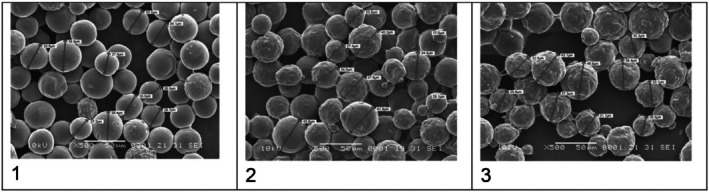 Figure 1
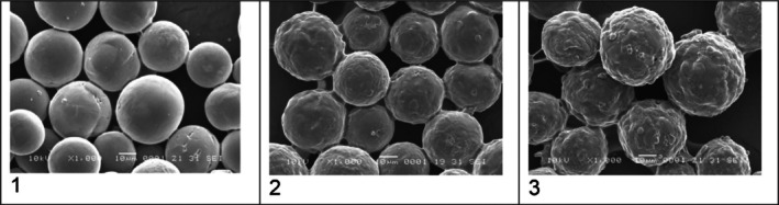 Figure 2
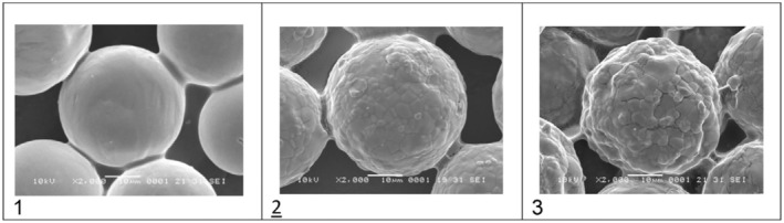 Figure 3
Peer Reviews
No public reviews on file for this paper yet. If you reviewed it on a platform where reviews are public (OpenReview, ICLR, NeurIPS, ICML), you can paste yours below so the community can read it here.
Videos
No videos yet. Explain this paper in a talk, walkthrough, or lecture? Add one.
Taxonomy
TopicsFacial Rejuvenation and Surgery Techniques · Body Contouring and Surgery · Dermatologic Treatments and Research
We read with great interest the article titled “Three Calcium Hydrofxylapatite‐Based Dermal Fillers Marketed in Mexico: Comparison of Particle Size and Shape Using Electron Microscopy” by Gilberto A. Sánchez Rico and Silvia Beatriz Andrade Canto, recently published in the Journal of Cosmetic Dermatology, 2015;24(3):e70100. doi: 10.1111/jocd.70100. While we appreciate the effort to explore calcium hydroxylapatite (CaHA) particle morphology, we would like to respectfully express several scientific concerns regarding the methodology and conclusions drawn.
First, the authors' decision to avoid washing and centrifugation—standard steps for isolating CaHA particles—raises questions about data validity. They claim to have avoided centrifugation to prevent microsphere fragmentation; however, previously validated protocols have demonstrated that Radiesse (Merz Aesthetics, Frankfurt, Germany) microspheres remain morphologically intact following multiple high‐speed centrifugation cycles (5000 rpm for 5 min, repeated five times), with no evidence of damage [1]. Moreover, studies such as those by de Moraes Nobre et al. and Kunzler et al. successfully performed centrifugation (up to 10 000 rpm) without microsphere fragmentation [1, 2]. In contrast, scanning electron microscopy (SEM) images of HarmonyCa (Allergan Aesthetics, Irvine, CA, USA) microspheres showed broken, irregular, and fragmented microspheres, with polymer deposits in the fissures [1].
Second, the analysis presented in the article is not representative. Only one image per filler and one particle per image were used for morphological assessment, despite millions of microspheres being present in each syringe. In contrast, other studies quantified 150+ particles per sample to ensure statistical robustness. This selective imaging raises the possibility of cherry‐picking favorable data. Further, the authors fail to disclose their conflicts of interest, with the first author directly reporting that he is a trainer and speaker for Allergan Medical Institute on social media. Therefore, the lack of transparent disclosure and potential bias toward Allergan may have resulted in selective image analysis with bulk SEM imaging.
Third, the claim that HarmonyCa microspheres are “uniform and spherical” is inconsistent with the authors' own micrographs. Figure 1 (500×) and Figure 2 (1000×) clearly show fractured, irregular particles, contradicting the assertion in Figure 3's caption. Prior studies using SEM have also identified broken, hollow, and anisomorphic HarmonyCa particles, including variability in surface texture.
Fourth, the lack of a discussion section precludes scientific interpretation. The authors fail to reconcile their findings with a large body of literature showing that Radiesse microspheres are uniformly smooth and spherical, with limited phagocytosable material [1, 3, 4]. By omitting comparative discussion or methodological limitations, the study misses an opportunity for constructive scientific dialogue.
Fifth, the authors express vague concerns about Radiesse's biocompatibility, yet this contradicts published evidence. Multiple studies have shown Radiesse to be immunologically inert, producing limited macrophage activation and lower phagocytosable content relative to PLLA and other CaHA‐based materials [5, 6]. Histological studies support its role in collagen I and III stimulation, fibroblast activation, and low granuloma risk.
Sixth, the quantitative analysis appears flawed. The graphs comparing particle sizes include ≤ 10 particles per filler—insufficient for any statistically meaningful conclusion, especially given the natural variability across batches and syringe sites. We support the effort to better characterize dermal fillers, but accurate, reproducible, and statistically sound methods are essential. We encourage future studies to align with standardized SEM protocols, use representative sampling, and transparently report limitations. We appreciate the opportunity to share these comments and hope they contribute constructively to the scientific discourse in the field of cosmetic dermatology.
Ethics Statement
The authors have nothing to report.
Consent
The authors have nothing to report.
Conflicts of Interest
The authors N.F.‐G., J.K, C.M.‐G. are consultants for Merz Aesthetics (Frankfurt, Germany).
The reference list from the paper itself. Each links out to its DOI / PubMed record.
- 1C. Kunzler , C. Hartmann , B. Nowag , et al., “Comparison of Physicochemical Characteristics and Biostimulatory Functions in Two Calcium Hydroxyapatite‐Based Dermal Fillers,” Journal of Drugs in Dermatology 22, no. 9 (2023): 910–916.37683069 10.36849/JDD.7684 · doi ↗ · pubmed ↗
- 2M. de Moraes Nobre , M. A. A. Fraga , V. Dal Coll , et al., “Characterization of Particles From Collagen Bio‐Stimulating Materials Used in Facial Filling Procedures,” Microscopy Research and Technique 88, no. 6 (2025): 1935–1944, 10.1002/jemt.24837.39995085 · doi ↗ · pubmed ↗
- 3S. B. Aguilera , A. Mc Carthy , S. Khalifian , Z. P. Lorenc , K. Goldie , and W. G. Chernoff , “The Role of Calcium Hydroxylapatite (Radiesse) as a Regenerative Aesthetic Treatment: A Narrative Review,” Aesthetic Surgery Journal 43, no. 10 (2023): 1063–1090.37635437 10.1093/asj/sjad 173PMC 11025388 · doi ↗ · pubmed ↗
- 4H. Oh , S. Lee , J. Na , and J. H. Kim , “Comparative Evaluation of Safety and Efficacy of a Novel Hyaluronic Acid‐Polynucleotide/Poly‐L‐Lactic Acid Composite Dermal Filler,” Aesthetic Plastic Surgery 45, no. 4 (2021): 1792–1801.33876290 10.1007/s 00266-021-02295-3 · doi ↗ · pubmed ↗
- 5G. Lemperle , V. Morhenn , and U. Charrier , “Human Histology and Persistence of Various Injectable Filler Substances for Soft Tissue Augmentation,” Aesthetic Plastic Surgery 44, no. 4 (2020): 1348–1360.32766911 10.1007/s 00266-020-01827-7 · doi ↗ · pubmed ↗
- 6R. Mazzuco , C. Evangelista , D. O. Gobbato , and L. M. de Almeida , “Clinical and Histological Comparative Outcomes After Injections of Poly‐L‐Lactic Acid and Calcium Hydroxyapatite in Arms: A Split Side Study,” Journal of Cosmetic Dermatology 21, no. 12 (2022): 6727–6733.36098704 10.1111/jocd.15356 · doi ↗ · pubmed ↗