Design and implementation of a low-cost gimbal-based angular ultrasound gantry for optimal tissue slice selection using deep learning
Abhishek Kumar, Akshay S. Menon, Divyansh Sharma, Raviteja Sista, Debdoot Sheet

TL;DR
This paper presents a low-cost angular ultrasound gantry system with deep learning to automate optimal tissue slice selection for tumor diagnosis, improving accuracy and reducing human error.
Contribution
A novel angular ultrasound gantry system integrated with deep learning for automated, accurate tissue slice selection.
Findings
The angular gantry system achieved 98% accuracy in selecting optimal tissue slices.
The system reduces time, resources, and human error in tumor diagnosis and treatment planning.
The angular design captures more comprehensive tumor geometry compared to linear gantries.
Abstract
Ultrasound (US) is a widely popular imaging technique for the diagnosis of tumors and associated soft tissue pathology. Traditionally, excised tumor masses are manually sliced for microscopic examination, which is a resource-intensive, time-consuming process, and prone to human error. The proposed work addresses these challenges by developing a cost-effective US gantry system integrated with a deep learning algorithm to automate the tissue slice selection process. This system scans the entire tumor and by integrating a deep learning algorithm predicts the optimal slice to assist its preparation for microscopic analysis. Automating this process reduces the time and resources required while minimizing the risk of human error. Optimal tissue slice reduces sampling associated uncertainty in diagnosis and treatment planning. Thereby determining tumor grade and type, and helping to reduce the…
Genes, proteins, chemicals, diseases, species, mutations and cell lines named across the full text — each resolved to its canonical identifier and authoritative record.
Click any figure to enlarge with its caption.
 Figure 1
 Figure 2
 Figure 3
 Figure 4
 Figure 5
 Figure 6
 Figure 7
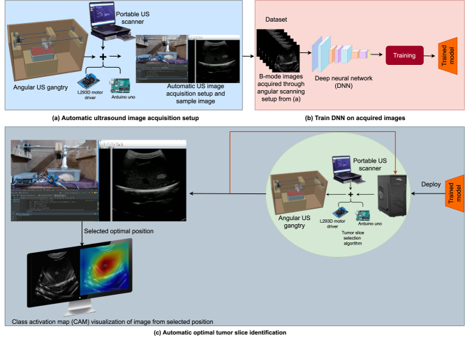 Figure 8
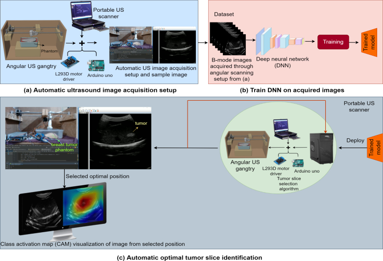 Figure 9
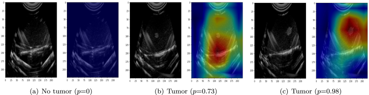 Figure 10
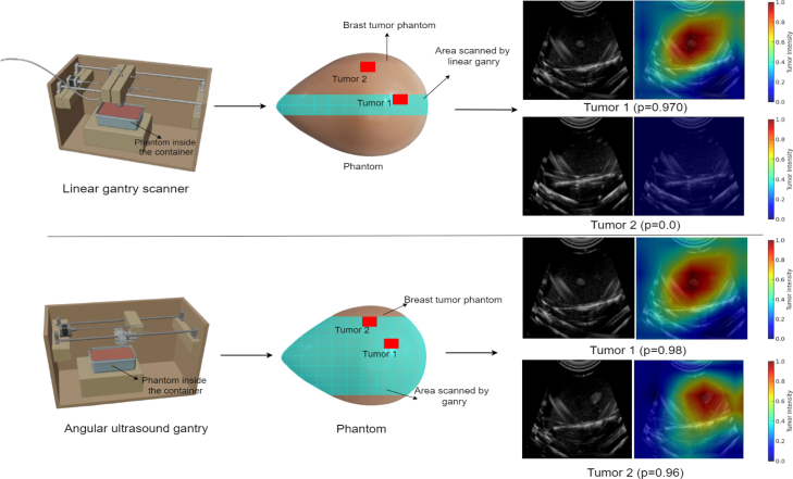 Figure 11
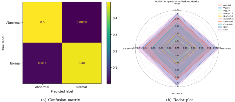 Figure 12
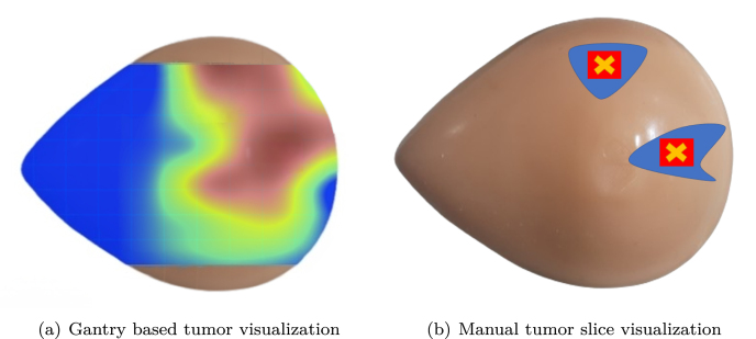 Figure 13
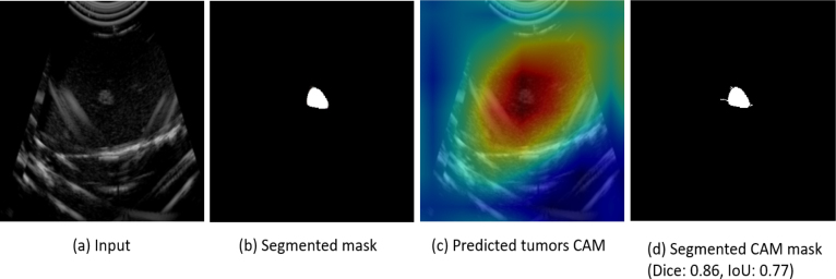 Figure 14
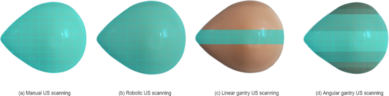 Figure 15
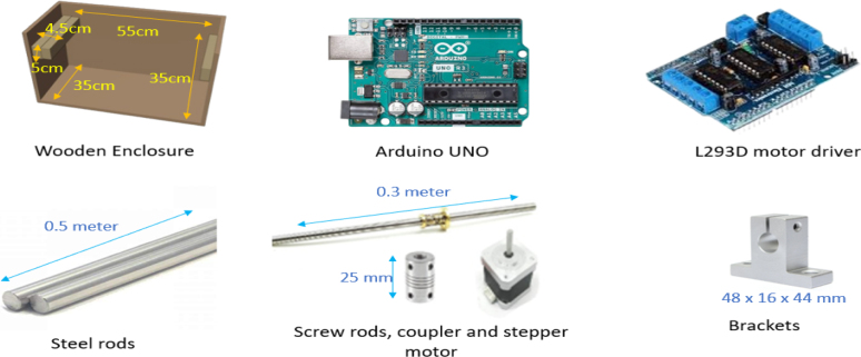 Figure 16
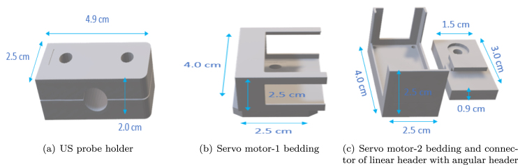 Figure 17
 Figure 18
 Figure 19
 Figure 20
 Figure 21
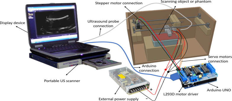 Figure 22
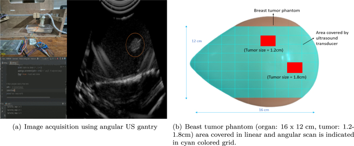 Figure 23
Peer Reviews
No public reviews on file for this paper yet. If you reviewed it on a platform where reviews are public (OpenReview, ICLR, NeurIPS, ICML), you can paste yours below so the community can read it here.
Videos
No videos yet. Explain this paper in a talk, walkthrough, or lecture? Add one.
Taxonomy
TopicsMedical Imaging and Analysis