Spatiotemporal fluctuations in fluorescence intensity of rhodamine phalloidin–labeled actin filaments
Kenta Toshino, Yosuke Yamazaki, Shunsuke Ando, Ryuichi Kaneda, Kazunori Ono, Takahiro Suzuki, Saku T. Kijima, Taro Q.P. Uyeda

TL;DR
This study reveals that the uneven fluorescence in actin filaments labeled with rhodamine phalloidin is due to nonuniform binding and structural changes influenced by phosphate and oxygen.
Contribution
The paper identifies two mechanisms for nonuniform phalloidin binding and dynamic fluorescence fluctuations in actin filaments.
Findings
Fluorescence inhomogeneities arise from nonuniform phalloidin binding density, not fluorophore quantum yield.
Inorganic phosphate suppresses fluorescence inhomogeneities and the correlation between Rh and Alexa488 intensities.
Temporal fluorescence fluctuations depend on glucose, glucose oxidase, and are suppressed by Trolox.
Abstract
Phalloidin (Ph) is widely used for fluorescent labeling of actin filaments. We observed ADP–actin filaments labeled with rhodamine phalloidin (RhPh) or Alexa488–Ph in vitro and discovered that the fluorescence intensities along the filaments showed a mottled pattern of bright and dark regions. Filaments labeled with substoichiometric RhPh exhibited more significant fluorescence inhomogeneities than those labeled with excess RhPh. Because the quantum yield of Alexa488 fluorescence is hardly affected by the environment, we concluded that the inhomogeneities arise from nonuniform Ph binding density rather than locally inhomogeneous quantum yield of the fluorophores. Simulations assuming random RhPh binding alone partially produced fluorescence inhomogeneities, but the degree of inhomogeneities was significantly smaller than the experimental results. Furthermore, filaments colabeled with…
Genes, proteins, chemicals, diseases, species, mutations and cell lines named across the full text — each resolved to its canonical identifier and authoritative record.
Click any figure to enlarge with its caption.
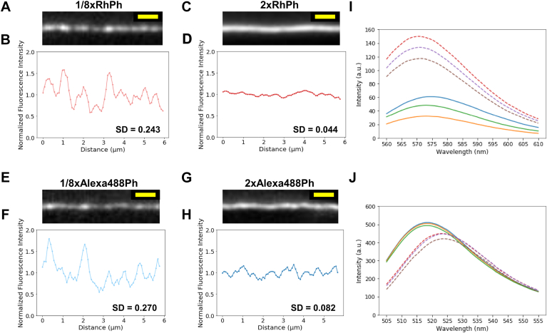 Figure 1
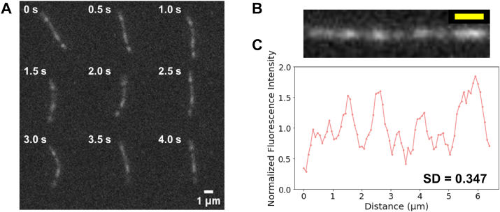 Figure 2
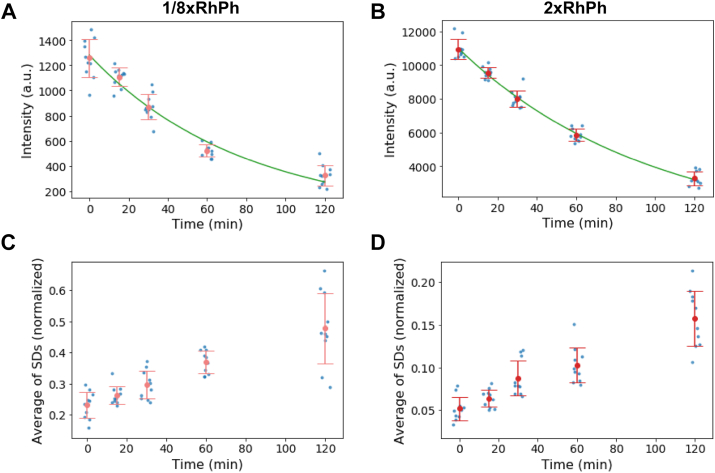 Figure 3
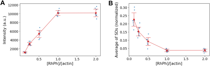 Figure 4
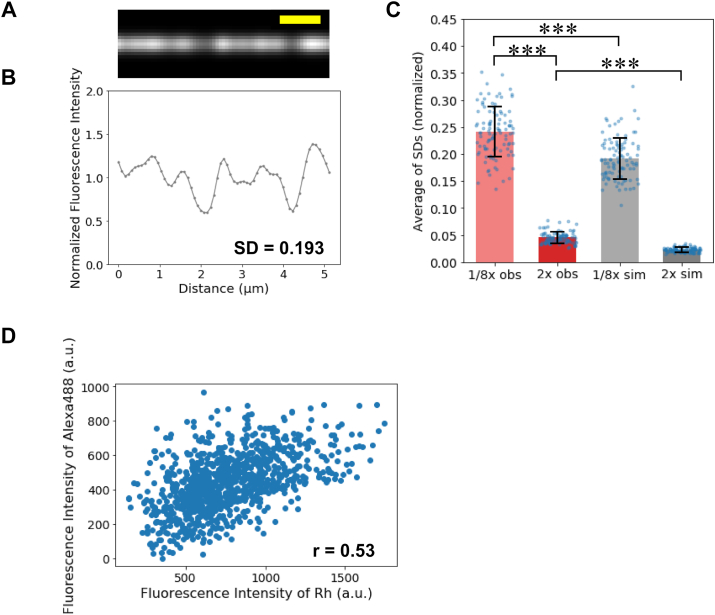 Figure 5
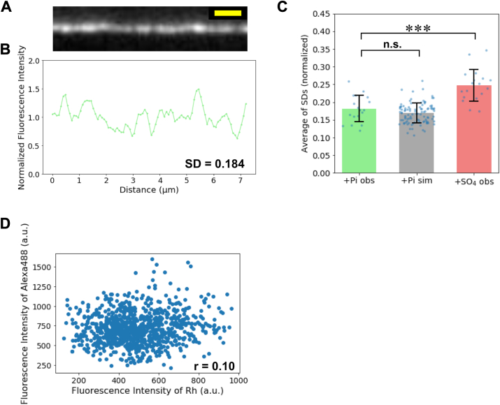 Figure 6
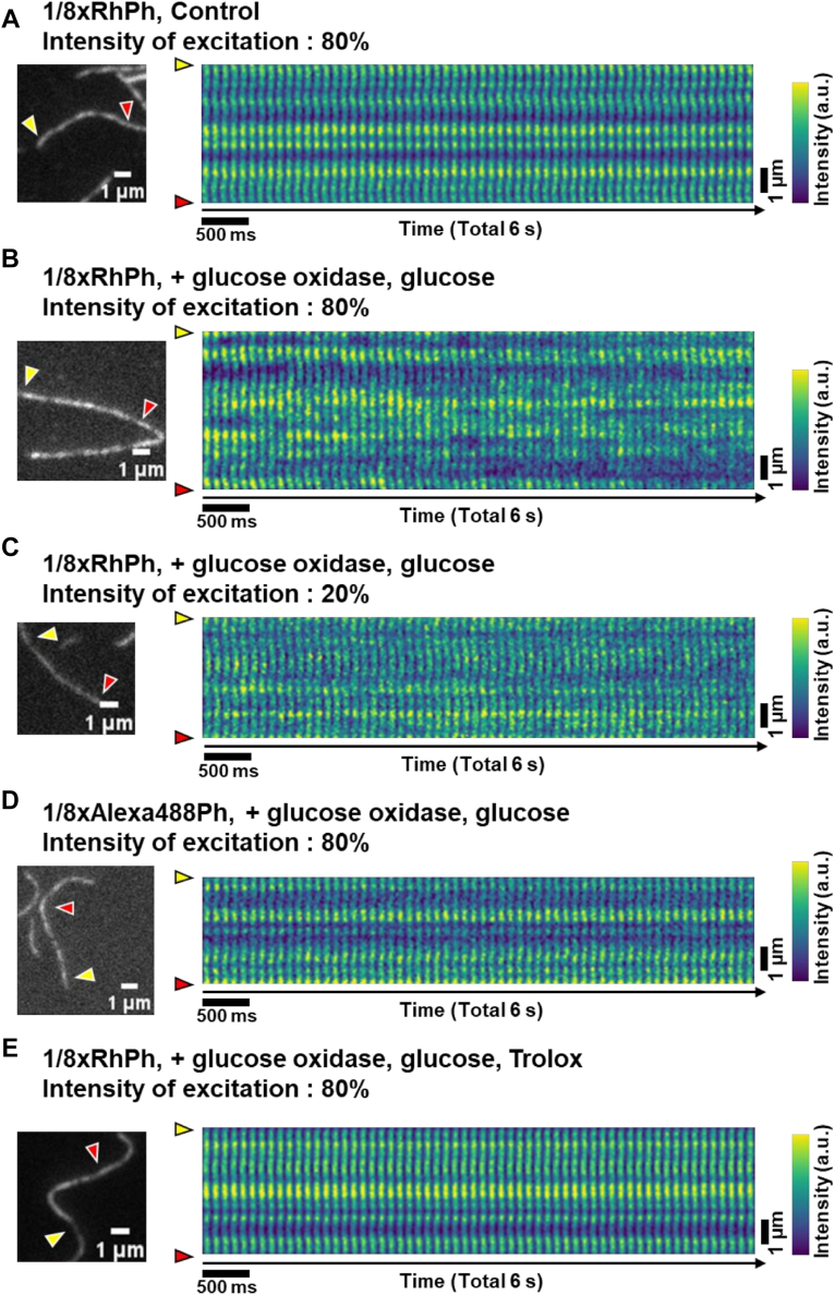 Figure 7
Peer Reviews
No public reviews on file for this paper yet. If you reviewed it on a platform where reviews are public (OpenReview, ICLR, NeurIPS, ICML), you can paste yours below so the community can read it here.
Videos
No videos yet. Explain this paper in a talk, walkthrough, or lecture? Add one.
Taxonomy
TopicsAdvanced Fluorescence Microscopy Techniques · bioluminescence and chemiluminescence research · Photoreceptor and optogenetics research